|
Rectouterine Folds
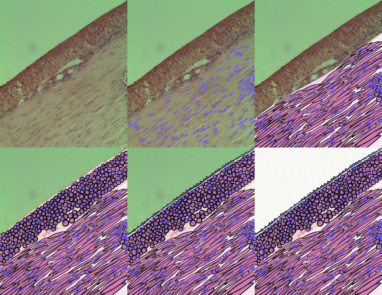
The rectouterine folds, (sacrogenital folds) are folds of the rectouterine pouch. They contain a considerable amount of fibrous tissue and smooth muscle fibers which are attached to the front of the sacrum and constitute the uterosacral ligament The uterosacral ligaments (or rectouterine ligaments) belong to the major ligaments of uterus. Structure The rectouterine folds contain a considerable amount of fibrous tissue and non-striped muscular fibers which are attached to the front of the ...s. References External links * - "The Female Pelvis: Rectouterine" Pelvis {{Anatomy-stub ... [...More Info...] [...Related Items...] OR: [Wikipedia] [Google] [Baidu] |
Rectouterine Pouch
The rectouterine pouch (recto-uterine pouch), pouch of Douglas, or rectovaginal pouch is the extension of the peritoneum between the rectum and the posterior wall of the uterus in the human female. Its anterior boundary is formed by the posterior fornix of the vagina. Structure In women, the rectouterine pouch is the deepest point of the peritoneal cavity. It lies posterior to the uterus and anterior to the rectum. (The pouch on the other side of the uterus is the vesicouterine pouch.) It is near the posterior fornix of the vagina. It is normal to have approximately 1 to 3 ml (or mL) of fluid in the rectouterine pouch throughout the menstrual cycle. After ovulation there is between 4 and 5 ml of fluid in the rectouterine pouch. In men, the region corresponding to the rectouterine pouch is the rectovesical pouch, which lies between the urinary bladder and rectum. Clinical significance The rectouterine pouch, being the lowest part of the peritoneal cavity in a woman at supine p ... [...More Info...] [...Related Items...] OR: [Wikipedia] [Google] [Baidu] |
Smooth Muscle
Smooth muscle is an involuntary non-striated muscle, so-called because it has no sarcomeres and therefore no striations (''bands'' or ''stripes''). It is divided into two subgroups, single-unit and multiunit smooth muscle. Within single-unit muscle, the whole bundle or sheet of smooth muscle cells contracts as a syncytium. Smooth muscle is found in the walls of hollow organs, including the stomach, intestines, bladder and uterus; in the walls of passageways, such as blood, and lymph vessels, and in the tracts of the respiratory, urinary, and reproductive systems. In the eyes, the ciliary muscles, a type of smooth muscle, dilate and contract the iris and alter the shape of the lens. In the skin, smooth muscle cells such as those of the arrector pili cause hair to stand erect in response to cold temperature or fear. Structure Gross anatomy Smooth muscle is grouped into two types: single-unit smooth muscle, also known as visceral smooth muscle, and multiunit smooth muscle. ... [...More Info...] [...Related Items...] OR: [Wikipedia] [Google] [Baidu] |
Sacrum
The sacrum (plural: ''sacra'' or ''sacrums''), in human anatomy, is a large, triangular bone at the base of the spine that forms by the fusing of the sacral vertebrae (S1S5) between ages 18 and 30. The sacrum situates at the upper, back part of the pelvic cavity, between the two wings of the pelvis. It forms joints with four other bones. The two projections at the sides of the sacrum are called the alae (wings), and articulate with the ilium at the L-shaped sacroiliac joints. The upper part of the sacrum connects with the last lumbar vertebra (L5), and its lower part with the coccyx (tailbone) via the sacral and coccygeal cornua. The sacrum has three different surfaces which are shaped to accommodate surrounding pelvic structures. Overall it is concave (curved upon itself). The base of the sacrum, the broadest and uppermost part, is tilted forward as the sacral promontory internally. The central part is curved outward toward the posterior, allowing greater room for the pel ... [...More Info...] [...Related Items...] OR: [Wikipedia] [Google] [Baidu] |
Uterosacral Ligament
The uterosacral ligaments (or rectouterine ligaments) belong to the major ligaments of uterus. Structure The rectouterine folds contain a considerable amount of fibrous tissue and non-striped muscular fibers which are attached to the front of the sacrum and constitute the uterosacral ligaments.* Netter, Atlas of Human Anatomy, 371 (4th Edition) These ligaments travel from the uterus to the anterior aspect of the sacrum. The pelvic splanchnic nerves Pelvic splanchnic nerves or nervi erigentes are splanchnic nerves that arise from sacral spinal nerves S2, S3, S4 to provide parasympathetic innervation to the organs of the pelvic cavity. Structure The pelvic splanchnic nerves arise from t ... run on top of the ligament. References {{Authority control Ligaments Pelvis Uterus ... [...More Info...] [...Related Items...] OR: [Wikipedia] [Google] [Baidu] |

