|
Quadriceps Tendon
In human anatomy, the quadriceps tendon works with the quadriceps muscle to extend the leg. All four parts of the quadriceps muscle attach to the shin via the patella (knee cap), where the quadriceps tendon becomes the patellar ligament. It attaches the quadriceps to the top of the patella, which in turn is connected to the shin from its bottom by the patellar ligament. A tendon connects muscle to bone, while a ligament connects bone to bone.Saladin, Kenneth S. Anatomy & Physiology: The Unity of Form and Function. 6th ed. New York: McGraw-Hill, 2012. Print. Injuries are common to this tendon, with tears, either partial or complete, being the most common. If the quadriceps tendon is completely torn, surgery will be required to regain function of the knee."Patellar Tendon Tear." OrthoInfo - AAOS. American Academy of Orthopaedic Surgeons, Aug. 2009. Web. 07 Dec. 2014. Without the quadriceps tendon, the knee cannot extend. Often, when the tendon is completely torn, part of the kneec ... [...More Info...] [...Related Items...] OR: [Wikipedia] [Google] [Baidu] |
Knee
In humans and other primates, the knee joins the thigh with the leg and consists of two joints: one between the femur and tibia (tibiofemoral joint), and one between the femur and patella (patellofemoral joint). It is the largest joint in the human body. The knee is a modified hinge joint, which permits flexion and extension as well as slight internal and external rotation. The knee is vulnerable to injury and to the development of osteoarthritis. It is often termed a ''compound joint'' having tibiofemoral and patellofemoral components. (The fibular collateral ligament is often considered with tibiofemoral components.) Structure The knee is a modified hinge joint, a type of synovial joint, which is composed of three functional compartments: the patellofemoral articulation, consisting of the patella, or "kneecap", and the patellar groove on the front of the femur through which it slides; and the medial and lateral tibiofemoral articulations linking the femur, or thigh bone ... [...More Info...] [...Related Items...] OR: [Wikipedia] [Google] [Baidu] |
Human Anatomy
The human body is the structure of a human being. It is composed of many different types of cells that together create tissues and subsequently organ systems. They ensure homeostasis and the viability of the human body. It comprises a head, hair, neck, trunk (which includes the thorax and abdomen), arms and hands, legs and feet. The study of the human body involves anatomy, physiology, histology and embryology. The body varies anatomically in known ways. Physiology focuses on the systems and organs of the human body and their functions. Many systems and mechanisms interact in order to maintain homeostasis, with safe levels of substances such as sugar and oxygen in the blood. The body is studied by health professionals, physiologists, anatomists, and by artists to assist them in their work. Composition The human body is composed of elements including hydrogen, oxygen, carbon, calcium and phosphorus. These elements reside in trillions of cells and non-cellula ... [...More Info...] [...Related Items...] OR: [Wikipedia] [Google] [Baidu] |
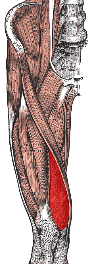
Quadriceps Muscle
The quadriceps femoris muscle (, also called the quadriceps extensor, quadriceps or quads) is a large muscle group that includes the four prevailing muscles on the front of the thigh. It is the sole extensor muscle of the knee, forming a large fleshy mass which covers the front and sides of the femur. The name derives . Structure Parts The quadriceps femoris muscle is subdivided into four separate muscles (the 'heads'), with the first superficial to the other three over the femur (from the trochanters to the condyles): *The rectus femoris muscle occupies the middle of the thigh, covering most of the other three quadriceps muscles. It originates on the ilium. It is named for its straight course. *The vastus lateralis muscle is on the ''lateral side'' of the femur (i.e. on the outer side of the thigh). *The vastus medialis muscle is on the ''medial side'' of the femur (i.e. on the inner part thigh). *The vastus intermedius muscle lies between vastus lateralis and vastus mediali ... [...More Info...] [...Related Items...] OR: [Wikipedia] [Google] [Baidu] |
Tuberosity Of The Tibia
The tuberosity of the tibia or tibial tuberosity or tibial tubercle is an elevation on the proximal, anterior aspect of the tibia, just below where the anterior surfaces of the lateral and medial tibial condyles end. Structure The tuberosity of the tibia gives attachment to the patellar ligament, which attaches to the patella from where the suprapatellar ligament forms the distal tendon of the quadriceps femoris muscles. The quadriceps muscles consist of the rectus femoris, vastus lateralis, vastus medialis, and vastus intermedius. These quadriceps muscles are innervated by the femoral nerve. KneeHipPain (1998) The tibial tuberosity thus forms the terminal part of the large structure that acts as a lever to extend the knee-joint and prevents the knee from collapsing when the foot strikes the ground. The tw ... [...More Info...] [...Related Items...] OR: [Wikipedia] [Google] [Baidu] |
Patella
The patella, also known as the kneecap, is a flat, rounded triangular bone which articulates with the femur (thigh bone) and covers and protects the anterior articular surface of the knee joint. The patella is found in many tetrapods, such as mice, cats, birds and dogs, but not in whales, or most reptiles. In humans, the patella is the largest sesamoid bone (i.e., embedded within a tendon or a muscle) in the body. Babies are born with a patella of soft cartilage which begins to ossify into bone at about four years of age. Structure The patella is a sesamoid bone roughly triangular in shape, with the apex of the patella facing downwards. The apex is the most inferior (lowest) part of the patella. It is pointed in shape, and gives attachment to the patellar ligament. The front and back surfaces are joined by a thin margin and towards centre by a thicker margin. The tendon of the quadriceps femoris muscle attaches to the base of the patella., with the vastus intermedius muscle ... [...More Info...] [...Related Items...] OR: [Wikipedia] [Google] [Baidu] |
Patellar Ligament
The patellar tendon is the distal portion of the common tendon of the quadriceps femoris, which is continued from the patella to the tibial tuberosity. It is also sometimes called the patellar ligament as it forms a bone to bone connection when the patella is fully ossified. Structure The patellar tendon is a strong, flat ligament, which originates on the apex of the patella distally and adjoining margins of the patella and the rough depression on its posterior surface; below, it inserts on the tuberosity of the tibia; its superficial fibers are continuous over the front of the patella with those of the tendon of the quadriceps femoris. It is about 4.5 cm long in adults (range from 3 to 6 cm). The medial and lateral portions of the quadriceps tendon pass down on either side of the patella to be inserted into the upper extremity of the tibia on either side of the tuberosity; these portions merge into the capsule, as stated above, forming the medial and lateral patellar retina ... [...More Info...] [...Related Items...] OR: [Wikipedia] [Google] [Baidu] |
Quadriceps Tendon Rupture
A quadriceps tendon rupture is a tear of the tendon that runs from the quadriceps muscle to the top of the knee cap. Signs and symptoms Symptoms are pain and the inability to extend the knee against resistance. A gap can often be palpated at the tendon's normal location. Diagnosis The diagnosis is usually made clinically, but ultrasound or MRI can be used if there is any doubt. Image:Quadriceps Ruptur Roe1.jpg, Quadriceps tendon rupture in plain X-ray Image:Quadriceps Ruptur Roe2.jpg, Quadriceps tendon rupture in plain X-ray: Incomplete rupture with haematoma in tendon. Image:Quadriceps Ruptur Roe3.jpg, Quadriceps tendon rupture in plain X-ray Image:Patellarsehenruptur Quadrizepssehnenruptur Roe.jpg, X-ray of a tear of the patellar tendon. On the left: The kneecap is pulled up. On the right: Significant dent in the soft tissue above the kneecap. Image:Quadrizepssehnenruptur.jpg, Operative image: 1. Kneecap 2. upper patella pole with drill holes 3. Stump of the quadriceps tendo ... [...More Info...] [...Related Items...] OR: [Wikipedia] [Google] [Baidu] |
Patellofemoral Pain Syndrome
Patellofemoral pain syndrome (PFPS; not to be confused with jumper's knee) is knee pain as a result of problems between the kneecap and the femur. The pain is generally in the front of the knee and comes on gradually. Pain may worsen with sitting, excessive use, or climbing and descending stairs. While the exact cause is unclear, it is believed to be due to overuse. Risk factors include trauma, increased training, and a weak quadriceps muscle. It is particularly common among runners. The diagnosis is generally based on the symptoms and examination. If pushing the kneecap into the femur increases the pain, the diagnosis is more likely. Treatment typically involves rest and rehabilitation with a Physical Therapist. Runners may need to switch to activities such as cycling or swimming. Insoles may help some people. Symptoms may last for years despite treatment. Patellofemoral pain syndrome is the most common cause of knee pain, affecting more than 20% of young adults. It occurs ... [...More Info...] [...Related Items...] OR: [Wikipedia] [Google] [Baidu] |


