|
Queckenstedt's Maneuver
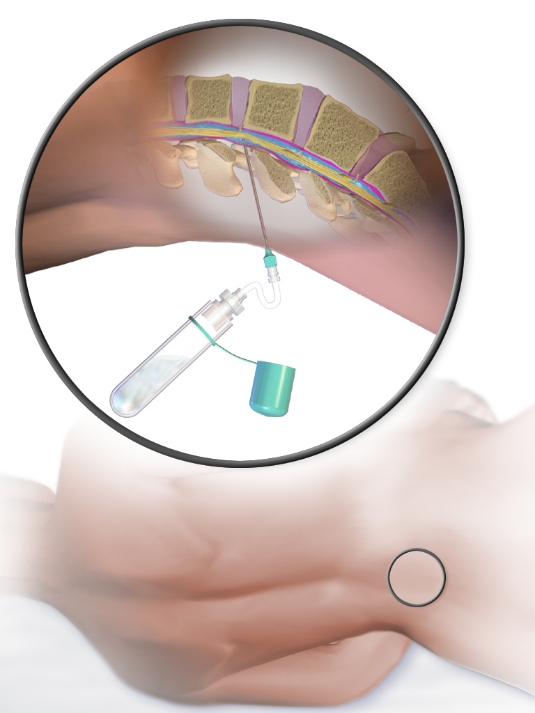
Queckenstedt's maneuver is a clinical test, formerly used for diagnosing spinal stenosis. The test is performed by placing the patient in the lateral decubitus position, thereafter the clinician performs a lumbar puncture. The opening pressure is measured. Then, the clinician's assistant compresses both jugular veins (if increased intracranial pressure is not suspected then one may exert pressure on both external jugular veins but usually pressure is first exerted on the abdomen, this pressure causes an engorgement of spinal veins and in turn rapidly increases cerebrospinal fluid pressure), which leads to a rise in the intracranial pressure. Given normal anatomy, the intracranial pressure will be reflected as a rapidly rising pressure measured from the lumbar needle, within 10–12 seconds. If there is a stenosis in the spine, there will be a damped, delayed response in the lumbar pressure, thus a positive Queckenstedt's maneuver. Nowadays this test has been made mostly superflu ... [...More Info...] [...Related Items...] OR: [Wikipedia] [Google] [Baidu] |
Spinal Stenosis
Spinal stenosis is an abnormal narrowing of the spinal canal or neural foramen that results in pressure on the spinal cord or nerve roots. Symptoms may include pain, numbness, or weakness in the arms or legs. Symptoms are typically gradual in onset and improve with leaning forward. Severe symptoms may include loss of bladder control, loss of bowel control, or sexual dysfunction. Causes may include osteoarthritis, rheumatoid arthritis, spinal tumors, trauma, Paget's disease of the bone, scoliosis, spondylolisthesis, and the genetic condition achondroplasia. It can be classified by the part of the spine affected into cervical, thoracic, and lumbar stenosis. Lumbar stenosis is the most common, followed by cervical stenosis. Diagnosis is generally based on symptoms and medical imaging. Treatment may involve medications, bracing, or surgery. Medications may include NSAIDs, acetaminophen, or steroid injections. Stretching and strengthening exercises may also be useful. Limiting ... [...More Info...] [...Related Items...] OR: [Wikipedia] [Google] [Baidu] |
Lumbar Puncture
Lumbar puncture (LP), also known as a spinal tap, is a medical procedure in which a needle is inserted into the spinal canal, most commonly to collect cerebrospinal fluid (CSF) for diagnostic testing. The main reason for a lumbar puncture is to help diagnose diseases of the central nervous system, including the brain and spine. Examples of these conditions include meningitis and subarachnoid hemorrhage. It may also be used therapeutically in some conditions. Increased intracranial pressure (pressure in the skull) is a contraindication, due to risk of brain matter being compressed and pushed toward the spine. Sometimes, lumbar puncture cannot be performed safely (for example due to a bleeding diathesis, severe bleeding tendency). It is regarded as a safe procedure, but post-dural-puncture headache is a common side effect if a small atraumatic needle is not used. The procedure is typically performed under local anesthesia using a aseptic technique, sterile technique. A hypodermic ... [...More Info...] [...Related Items...] OR: [Wikipedia] [Google] [Baidu] |
Jugular Veins
The jugular veins are veins that take deoxygenated blood from the head back to the heart via the superior vena cava. The internal jugular vein descends next to the internal carotid artery and continues posteriorly to the sternocleidomastoid muscle. Structure and Function There are two sets of jugular veins: external and internal. The left and right external jugular veins drain into the subclavian veins. The internal jugular veins join with the subclavian veins more medially to form the brachiocephalic veins. Finally, the left and right brachiocephalic veins join to form the superior vena cava, which delivers deoxygenated blood to the right atrium of the heart. The Jugular veins help carry blood from the heart to and from the brain. An average human brain weighs about 3 pounds, and gets about 15%-20% of the blood that the heart pumps out. It is important for the brain to get enough blood for many reasons. The jugular ... [...More Info...] [...Related Items...] OR: [Wikipedia] [Google] [Baidu] |
Intracranial Pressure
Intracranial pressure (ICP) is the pressure exerted by fluids such as cerebrospinal fluid (CSF) inside the skull and on the brain tissue. ICP is measured in millimeters of mercury (mmHg) and at rest, is normally 7–15 Millimeter of mercury, mmHg for a Supine position, supine adult. The body has various mechanisms by which it keeps the ICP stable, with CSF pressures varying by about 1 mmHg in normal adults through shifts in production and absorption of CSF. Changes in ICP are attributed to volume changes in one or more of the constituents contained in the cranium. CSF pressure has been shown to be influenced by abrupt changes in intrathoracic pressure during coughing (which is induced by contraction of the diaphragm and abdominal wall muscles, the latter of which also increases intra-abdominal pressure), the valsalva maneuver, and communication with the vasculature (venous and arterial systems). Intracranial hypertension (IH), also called increased ICP (IICP) or raised intracrani ... [...More Info...] [...Related Items...] OR: [Wikipedia] [Google] [Baidu] |
Cerebrospinal Fluid
Cerebrospinal fluid (CSF) is a clear, colorless body fluid found within the tissue that surrounds the brain and spinal cord of all vertebrates. CSF is produced by specialised ependymal cells in the choroid plexus of the ventricles of the brain, and absorbed in the arachnoid granulations. There is about 125 mL of CSF at any one time, and about 500 mL is generated every day. CSF acts as a shock absorber, cushion or buffer, providing basic mechanical and immunological protection to the brain inside the skull. CSF also serves a vital function in the cerebral autoregulation of cerebral blood flow. CSF occupies the subarachnoid space (between the arachnoid mater and the pia mater) and the ventricular system around and inside the brain and spinal cord. It fills the ventricles of the brain, cisterns, and sulci, as well as the central canal of the spinal cord. There is also a connection from the subarachnoid space to the bony labyrinth of the inner ear via the perilymphat ... [...More Info...] [...Related Items...] OR: [Wikipedia] [Google] [Baidu] |
Stenosis
A stenosis (from Ancient Greek στενός, "narrow") is an abnormal narrowing in a blood vessel or other tubular organ or structure such as foramina and canals. It is also sometimes called a stricture (as in urethral stricture). ''Stricture'' as a term is usually used when narrowing is caused by contraction of smooth muscle (e.g. achalasia, prinzmetal angina); ''stenosis'' is usually used when narrowing is caused by lesion that reduces the space of lumen (e.g. atherosclerosis). The term coarctation is another synonym, but is commonly used only in the context of aortic coarctation. Restenosis is the recurrence of stenosis after a procedure. Types The resulting syndrome depends on the structure affected. Examples of vascular stenotic lesions include: * Intermittent claudication (peripheral artery stenosis) * Angina ( coronary artery stenosis) * Carotid artery stenosis which predispose to (strokes and transient ischaemic episodes) * Renal artery stenosis The types of sten ... [...More Info...] [...Related Items...] OR: [Wikipedia] [Google] [Baidu] |
Computed Tomography
A computed tomography scan (CT scan; formerly called computed axial tomography scan or CAT scan) is a medical imaging technique used to obtain detailed internal images of the body. The personnel that perform CT scans are called radiographers or radiology technologists. CT scanners use a rotating X-ray tube and a row of detectors placed in a gantry to measure X-ray attenuations by different tissues inside the body. The multiple X-ray measurements taken from different angles are then processed on a computer using tomographic reconstruction algorithms to produce tomographic (cross-sectional) images (virtual "slices") of a body. CT scans can be used in patients with metallic implants or pacemakers, for whom magnetic resonance imaging (MRI) is contraindicated. Since its development in the 1970s, CT scanning has proven to be a versatile imaging technique. While CT is most prominently used in medical diagnosis, it can also be used to form images of non-living objects. The 1979 Nob ... [...More Info...] [...Related Items...] OR: [Wikipedia] [Google] [Baidu] |
Hans Heinrich Georg Queckenstedt
Hans Heinrich Georg Queckenstedt (1876 in Leipzig-Reudnitz – 8 November 1918 in Bertrix) was a German neurologist remembered for describing Queckenstedt's phenomenon. He graduated from the University of Leipzig in 1900, having studied under Emil Kraepelin. He worked under Sigbert Josef Maria Ganser, and gained his doctorate in 1904. He worked in Rostock, and was habilitated as Privatdozent in 1913. He studied cerebrospinal fluid dynamics, noting the fluctuation of pressure with respiration. This led to experiments with the Valsalva manoeuvre and jugular vein The jugular veins are veins that take deoxygenated blood from the head back to the heart via the superior vena cava. The internal jugular vein descends next to the internal carotid artery and continues posteriorly to the sternocleidomastoid ... pressure from which his eponymous test was published. He took part in the First World War as a medical officer and died shortly before the armistice due to an accident.E ... [...More Info...] [...Related Items...] OR: [Wikipedia] [Google] [Baidu] |
Tobey–Ayer Test
The Tobey–Ayer test is used for lateral sinus thrombosis by monitoring cerebrospinal fluid pressure during a lumbar puncture. No increase of cerebrospinal fluid pressure during compression of the internal jugular vein on the affected side, and an exaggerated response on the patent side, is suggestive of lateral sinus thrombosis. History Tobey–Ayer test was the first specific test for lateral sinus thrombosis. It was created by Tobey, G. L. and Ayer, J. B. in 1925 when they first introduced modifications to the Queckenstedt's maneuver Queckenstedt's maneuver is a clinical test, formerly used for diagnosing spinal stenosis. The test is performed by placing the patient in the lateral decubitus position, thereafter the clinician performs a lumbar puncture. The opening pressure ... test used at the time to diagnose obstruction to spinal cerebrospinal fluid flow. References Medical tests {{med-sign-stub ... [...More Info...] [...Related Items...] OR: [Wikipedia] [Google] [Baidu] |


