|
Quadrate Ligament
In human anatomy, the quadrate ligament or ligament of Denucé is one of the ligaments of the proximal radioulnar joint in the upper forearm. Structure The quadrate ligament is a fibrous band attached to the inferior border of the radial notch on the ulna and to the neck of the radius In classical geometry, a radius ( : radii) of a circle or sphere is any of the line segments from its center to its perimeter, and in more modern usage, it is also their length. The name comes from the latin ''radius'', meaning ray but also the .... Its borders are strengthened by fibers from the upper border of the annular ligament. The ligament is long, wide, and thick. Function The quadrate ligament reinforces the inferior part of the capsule of the elbow joint and contributes to joint stability by securing the proximal radius against the radial notch and by restricting excessive supination (10–20° restriction) and, to a lesser degree, pronation (5–8°). History The quadrate liga ... [...More Info...] [...Related Items...] OR: [Wikipedia] [Google] [Baidu] |
Ligament
A ligament is the fibrous connective tissue that connects bones to other bones. It is also known as ''articular ligament'', ''articular larua'', ''fibrous ligament'', or ''true ligament''. Other ligaments in the body include the: * Peritoneal ligament: a fold of peritoneum or other membranes. * Fetal remnant ligament: the remnants of a fetal tubular structure. * Periodontal ligament: a group of fibers that attach the cementum of teeth to the surrounding alveolar bone. Ligaments are similar to tendons and fasciae as they are all made of connective tissue. The differences among them are in the connections that they make: ligaments connect one bone to another bone, tendons connect muscle to bone, and fasciae connect muscles to other muscles. These are all found in the skeletal system of the human body. Ligaments cannot usually be regenerated naturally; however, there are periodontal ligament stem cells located near the periodontal ligament which are involved in the adult reg ... [...More Info...] [...Related Items...] OR: [Wikipedia] [Google] [Baidu] |
Proximal Radioulnar Joint
The proximal radioulnar articulation, also known as the proximal radioulnar joint (PRUJ), is a synovial pivot joint between the circumference of the head of the radius and the ring formed by the radial notch of the ulna and the annular ligament. Structure The proximal radioulnar joint is a synovial pivot joint. It occurs between the circumference of the head of the radius and the ring formed by the radial notch of the ulna and the annular ligament. The interosseous membrane of the forearm and the annular ligament stabilise the joint. A number of nerves run close to the proximal radioulnar joint, including: *median nerve *musculocutaneous nerve * radial nerve See also * Distal radioulnar articulation The distal radioulnar articulation (also known as the distal radioulnar joint, or inferior radioulnar joint) is a synovial pivot joint between the two bones in the forearm; the radius and ulna. It is one of two joints between the radius and ulna ... * Supination Refere ... [...More Info...] [...Related Items...] OR: [Wikipedia] [Google] [Baidu] |
Forearm
The forearm is the region of the upper limb between the elbow and the wrist. The term forearm is used in anatomy to distinguish it from the arm, a word which is most often used to describe the entire appendage of the upper limb, but which in anatomy, technically, means only the region of the upper arm, whereas the lower "arm" is called the forearm. It is homologous to the region of the leg that lies between the knee and the ankle joints, the crus. The forearm contains two long bones, the radius and the ulna, forming the two radioulnar joints. The interosseous membrane connects these bones. Ultimately, the forearm is covered by skin, the anterior surface usually being less hairy than the posterior surface. The forearm contains many muscles, including the flexors and extensors of the wrist, flexors and extensors of the digits, a flexor of the elbow (brachioradialis), and pronators and supinators that turn the hand to face down or upwards, respectively. In cross-section, ... [...More Info...] [...Related Items...] OR: [Wikipedia] [Google] [Baidu] |
Radial Notch
The radial notch of the ulna (lesser sigmoid cavity) is a narrow, oblong, articular depression on the lateral side of the coronoid process; it receives the circumferential articular surface of the head of the radius. It is concave from before backward, and its prominent extremities serve for the attachment of the annular ligament. Additional images File:Gray333.png, Annular ligament of radius, from above. References External links * *elbow/elbowbones/bones3at the Dartmouth Medical School The Geisel School of Medicine at Dartmouth is the graduate medical school of Dartmouth College in Hanover, New Hampshire. The fourth oldest medical school in the United States, it was founded in 1797 by New England physician Nathan Smith. It is o ...'s Department of Anatomy Upper limb anatomy Ulna {{musculoskeletal-stub ... [...More Info...] [...Related Items...] OR: [Wikipedia] [Google] [Baidu] |
Ulna
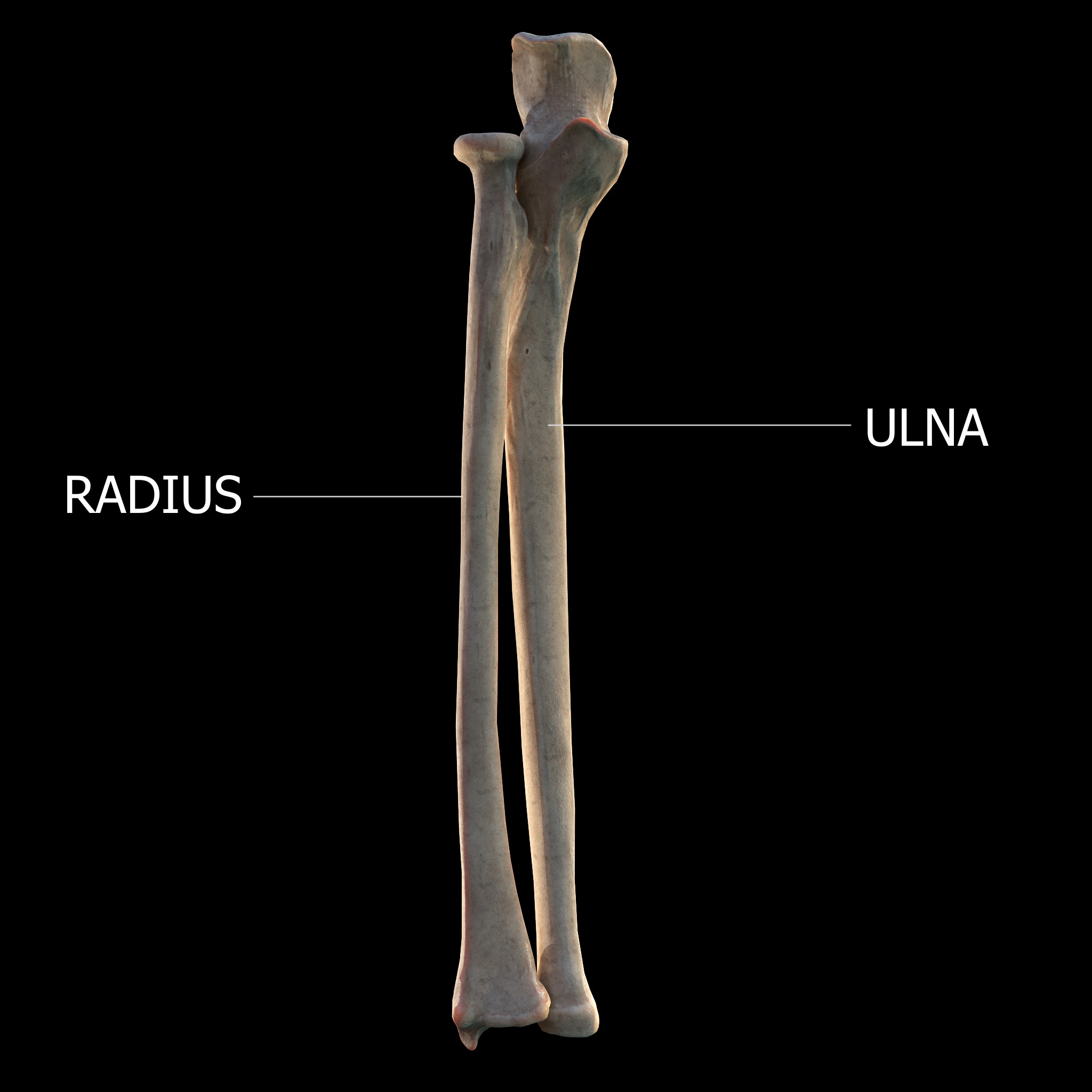
The ulna (''pl''. ulnae or ulnas) is a long bone found in the forearm that stretches from the elbow to the smallest finger, and when in anatomical position, is found on the medial side of the forearm. That is, the ulna is on the same side of the forearm as the little finger. It runs parallel to the radius, the other long bone in the forearm. The ulna is usually slightly longer than the radius, but the radius is thicker. Therefore, the radius is considered to be the larger of the two. Structure The ulna is a long bone found in the forearm that stretches from the elbow to the smallest finger, and when in anatomical position, is found on the medial side of the forearm. It is broader close to the elbow, and narrows as it approaches the wrist. Close to the elbow, the ulna has a bony process, the olecranon process, a hook-like structure that fits into the olecranon fossa of the humerus. This prevents hyperextension and forms a hinge joint with the trochlea of the humerus. Ther ... [...More Info...] [...Related Items...] OR: [Wikipedia] [Google] [Baidu] |
Radius (bone)
The radius or radial bone is one of the two large bones of the forearm, the other being the ulna. It extends from the lateral side of the elbow to the thumb side of the wrist and runs parallel to the ulna. The ulna is usually slightly longer than the radius, but the radius is thicker. Therefore the radius is considered to be the larger of the two. It is a long bone, prism-shaped and slightly curved longitudinally. The radius is part of two joints: the elbow and the wrist. At the elbow, it joins with the capitulum of the humerus, and in a separate region, with the ulna at the radial notch. At the wrist, the radius forms a joint with the ulna bone. The corresponding bone in the lower leg is the fibula. Structure The long narrow medullary cavity is enclosed in a strong wall of compact bone. It is thickest along the interosseous border and thinnest at the extremities, same over the cup-shaped articular surface (fovea) of the head. The trabeculae of the spongy ti ... [...More Info...] [...Related Items...] OR: [Wikipedia] [Google] [Baidu] |
Anular Ligament Of Radius
The annular ligament (orbicular ligament) is a strong band of fibers that encircles the head of the radius, and retains it in contact with the radial notch of the ulna.''Gray's Anatomy'' (1918), see infobox Per '' Terminologia Anatomica 1998'', the spelling is "anular", but the spelling "annular" is frequently encountered. Indeed, the most recent version of ''Terminologia Anatomica'' (2019) uses "annular" as the preferred English spelling. Anatomy The annular ligament is attached by both its ends to the anterior and posterior margins of the radial notch of the ulna, together with which it forms the articular surface that surrounds the head and neck of the radius. The ligament is strong and well defined, yet its flexibility permits the slightly oval head of the radius to rotate freely during pronation and supination. The head of the radius is wider than the bone's neck, and, because the annular ligament embraces both, the radial head is "trapped" inside the ligament which thus a ... [...More Info...] [...Related Items...] OR: [Wikipedia] [Google] [Baidu] |
Supination
Motion, the process of movement, is described using specific anatomical terms. Motion includes movement of organs, joints, limbs, and specific sections of the body. The terminology used describes this motion according to its direction relative to the anatomical position of the body parts involved. Anatomists and others use a unified set of terms to describe most of the movements, although other, more specialized terms are necessary for describing unique movements such as those of the hands, feet, and eyes. In general, motion is classified according to the anatomical plane it occurs in. ''Flexion'' and ''extension'' are examples of ''angular'' motions, in which two axes of a joint are brought closer together or moved further apart. ''Rotational'' motion may occur at other joints, for example the shoulder, and are described as ''internal'' or ''external''. Other terms, such as ''elevation'' and ''depression'', describe movement above or below the horizontal plane. Many anatom ... [...More Info...] [...Related Items...] OR: [Wikipedia] [Google] [Baidu] |
Pronation
Motion, the process of movement, is described using specific anatomical terms. Motion includes movement of organs, joints, limbs, and specific sections of the body. The terminology used describes this motion according to its direction relative to the anatomical position of the body parts involved. Anatomists and others use a unified set of terms to describe most of the movements, although other, more specialized terms are necessary for describing unique movements such as those of the hands, feet, and eyes. In general, motion is classified according to the anatomical plane it occurs in. ''Flexion'' and ''extension'' are examples of ''angular'' motions, in which two axes of a joint are brought closer together or moved further apart. ''Rotational'' motion may occur at other joints, for example the shoulder, and are described as ''internal'' or ''external''. Other terms, such as ''elevation'' and ''depression'', describe movement above or below the horizontal plane. Many anatom ... [...More Info...] [...Related Items...] OR: [Wikipedia] [Google] [Baidu] |
Nomina Anatomica
''Nomina Anatomica'' (''NA'') was the international standard on human anatomic terminology from 1895 until it was replaced by ''Terminologia Anatomica'' in 1998. In the late nineteenth century some 30,000 terms for various body parts were in use. The same structures were described by different names, depending (among other things) on the anatomist's school and national tradition. Vernacular translations of Latin and Greek, as well as various eponymous terms, were barriers to effective international communication. There was disagreement and confusion among anatomists regarding anatomical terminology. Editions The first and last entries in the following table are not NA editions, but they are included for the sake of continuity. Although these early editions were authorized by different bodies, they are sometimes considered part of the same series. Before these codes of terminology, approved at anatomists congresses, the usage of anatomical terms was based on authoritative work ... [...More Info...] [...Related Items...] OR: [Wikipedia] [Google] [Baidu] |


