|
Qianzhousaurus Sinensis
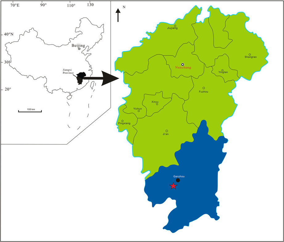
''Qianzhousaurus'' (meaning "Qianzhou lizard") is a genus of tyrannosaurid dinosaurs that lived in Asia during the Late Cretaceous period. There is currently only one species named, the type species ''Qianzhousaurus sinensis'' which is a member of the tribe Alioramini within the group, and most closely related to ''Alioramus'', another alioramin. History of discovery The holotype specimen, GM F10004, was unearthed in southern China, Ganzhou, at the Nanxiong Formation in the summer of 2010 during the construction of an industrial park and it was first described by paleontologists Junchang Lü, Laiping Yi, Stephen L. Brusatte, Ling Yang, Hua Li and Liu Chen in the journal ''Nature Communications'' in 2014. The genus is known from a partial sub-adult individual consisting of a nearly complete skull with the lower jaws missing all teeth (lost during fossilization), 9 cervical vertebrae, 3 dorsal vertebrae, 18 caudal vertebrae, both scapulocoracoids, partial ilia, and the l ... [...More Info...] [...Related Items...] OR: [Wikipedia] [Google] [Baidu] |
Late Cretaceous
The Late Cretaceous (100.5–66 Ma) is the younger of two epochs into which the Cretaceous Period is divided in the geologic time scale. Rock strata from this epoch form the Upper Cretaceous Series. The Cretaceous is named after ''creta'', the Latin word for the white limestone known as chalk. The chalk of northern France and the white cliffs of south-eastern England date from the Cretaceous Period. Climate During the Late Cretaceous, the climate was warmer than present, although throughout the period a cooling trend is evident. The tropics became restricted to equatorial regions and northern latitudes experienced markedly more seasonal climatic conditions. Geography Due to plate tectonics, the Americas were gradually moving westward, causing the Atlantic Ocean to expand. The Western Interior Seaway divided North America into eastern and western halves; Appalachia and Laramidia. India maintained a northward course towards Asia. In the Southern Hemisphere, Australia and Ant ... [...More Info...] [...Related Items...] OR: [Wikipedia] [Google] [Baidu] |
Ling Yang (paleontologist)
Yang Ling (, born May 24, 1968 in Beijing) is a male Chinese sports shooter. He won both the 1996 Summer Olympics and the 2000 Olympic Games in 10 metre running target 10 meter running target is one of the ISSF shooting events, shot with an airgun at a target that moves sideways. The target is pulled across a two meter wide aisle at the range of 10 meters from the firing point. The target is pulled at either of ..., being the only shooter to successfully defend an Olympic title in that event. Olympic results Other major performances *1994 National Champions Tournament - 1st 10m moving target 30+30 *1994 National Qualification Tournament - 1st team, 2nd 10m moving target 30+30 *1994 National Moving Target Championships - 1st 10m moving target mixed speed & 2nd 10m moving target standard speed *1996 World Championships - 1st 50m moving target team & 2nd 25m standard pistol 60 shots team Records References China Daily 1968 births Living people Chinese male spo ... [...More Info...] [...Related Items...] OR: [Wikipedia] [Google] [Baidu] |
Calcaneum
In humans and many other primates, the calcaneus (; from the Latin ''calcaneus'' or ''calcaneum'', meaning heel) or heel bone is a bone of the tarsus of the foot which constitutes the heel. In some other animals, it is the point of the hock. Structure In humans, the calcaneus is the largest of the tarsal bones and the largest bone of the foot. Its long axis is pointed forwards and laterally. The talus bone, calcaneus, and navicular bone are considered the proximal row of tarsal bones. In the calcaneus, several important structures can be distinguished:Platzer (2004), p 216 There is a large calcaneal tuberosity located posteriorly on plantar surface with medial and lateral tubercles on its surface. Besides, there is another peroneal tubecle on its lateral surface. On its lower edge on either side are its lateral and medial processes (serving as the origins of the abductor hallucis and abductor digiti minimi). The Achilles tendon is inserted into a roughened area on its superior ... [...More Info...] [...Related Items...] OR: [Wikipedia] [Google] [Baidu] |
Talus Bone
The talus (; Latin for ankle or ankle bone), talus bone, astragalus (), or ankle bone is one of the group of foot bones known as the tarsus. The tarsus forms the lower part of the ankle joint. It transmits the entire weight of the body from the lower legs to the foot.Platzer (2004), p 216 The talus has joints with the two bones of the lower leg, the tibia and thinner fibula. These leg bones have two prominences (the lateral and medial malleoli) that articulate with the talus. At the foot end, within the tarsus, the talus articulates with the calcaneus (heel bone) below, and with the curved navicular bone in front; together, these foot articulations form the ball-and-socket-shaped talocalcaneonavicular joint. The talus is the second largest of the tarsal bones; it is also one of the bones in the human body with the highest percentage of its surface area covered by articular cartilage. It is also unusual in that it has a retrograde blood supply, i.e. arterial blood enters the ... [...More Info...] [...Related Items...] OR: [Wikipedia] [Google] [Baidu] |
Fibula
The fibula or calf bone is a leg bone on the lateral side of the tibia, to which it is connected above and below. It is the smaller of the two bones and, in proportion to its length, the most slender of all the long bones. Its upper extremity is small, placed toward the back of the head of the tibia, below the knee joint and excluded from the formation of this joint. Its lower extremity inclines a little forward, so as to be on a plane anterior to that of the upper end; it projects below the tibia and forms the lateral part of the ankle joint. Structure The bone has the following components: * Lateral malleolus * Interosseous membrane connecting the fibula to the tibia, forming a syndesmosis joint * The superior tibiofibular articulation is an arthrodial joint between the lateral condyle of the tibia and the head of the fibula. * The inferior tibiofibular articulation (tibiofibular syndesmosis) is formed by the rough, convex surface of the medial side of the lower end of the f ... [...More Info...] [...Related Items...] OR: [Wikipedia] [Google] [Baidu] |
Tibia
The tibia (; ), also known as the shinbone or shankbone, is the larger, stronger, and anterior (frontal) of the two bones in the leg below the knee in vertebrates (the other being the fibula, behind and to the outside of the tibia); it connects the knee with the ankle. The tibia is found on the medial side of the leg next to the fibula and closer to the median plane. The tibia is connected to the fibula by the interosseous membrane of leg, forming a type of fibrous joint called a syndesmosis with very little movement. The tibia is named for the flute ''tibia''. It is the second largest bone in the human body, after the femur. The leg bones are the strongest long bones as they support the rest of the body. Structure In human anatomy, the tibia is the second largest bone next to the femur. As in other vertebrates the tibia is one of two bones in the lower leg, the other being the fibula, and is a component of the knee and ankle joints. The ossification or formation of the bone ... [...More Info...] [...Related Items...] OR: [Wikipedia] [Google] [Baidu] |
Femur
The femur (; ), or thigh bone, is the proximal bone of the hindlimb in tetrapod vertebrates. The head of the femur articulates with the acetabulum in the pelvic bone forming the hip joint, while the distal part of the femur articulates with the tibia (shinbone) and patella (kneecap), forming the knee joint. By most measures the two (left and right) femurs are the strongest bones of the body, and in humans, the largest and thickest. Structure The femur is the only bone in the upper leg. The two femurs converge medially toward the knees, where they articulate with the proximal ends of the tibiae. The angle of convergence of the femora is a major factor in determining the femoral-tibial angle. Human females have thicker pelvic bones, causing their femora to converge more than in males. In the condition ''genu valgum'' (knock knee) the femurs converge so much that the knees touch one another. The opposite extreme is ''genu varum'' (bow-leggedness). In the general populatio ... [...More Info...] [...Related Items...] OR: [Wikipedia] [Google] [Baidu] |
Ilium (bone)
The ilium () (plural ilia) is the uppermost and largest part of the hip bone, and appears in most vertebrates including mammals and birds, but not bony fish. All reptiles have an ilium except snakes, although some snake species have a tiny bone which is considered to be an ilium. The ilium of the human is divisible into two parts, the body and the wing; the separation is indicated on the top surface by a curved line, the arcuate line, and on the external surface by the margin of the acetabulum. The name comes from the Latin (''ile'', ''ilis''), meaning "groin" or "flank". Structure The ilium consists of the body and wing. Together with the ischium and pubis, to which the ilium is connected, these form the pelvic bone, with only a faint line indicating the place of union. The body ( la, corpus) forms less than two-fifths of the acetabulum; and also forms part of the acetabular fossa. The internal surface of the body is part of the wall of the lesser pelvis and gives ... [...More Info...] [...Related Items...] OR: [Wikipedia] [Google] [Baidu] |
Scapulocoracoid
The scapulocoracoid is the unit of the pectoral girdle that contains the coracoid and scapula. The coracoid itself is a beak-shaped bone that is commonly found in most vertebrates with a few exceptions. The scapula is commonly known as the ''shoulder blade''. The humerus is linked to the body via the scapula, and the clavicle is connected to the sternum via the scapula as well. Theria Theria (; Greek: , wild beast) is a subclass of mammals amongst the Theriiformes. Theria includes the eutherians (including the placental mammals) and the metatherians (including the marsupials) but excludes the egg-laying monotremes. Ch ...n mammals lack a scapulocoracoid. References * Vertebrates Comparative Anatomy, Function, Evolution by Kenneth V. Kardong. Page 325. Vertebrate anatomy {{Vertebrate anatomy-stub ... [...More Info...] [...Related Items...] OR: [Wikipedia] [Google] [Baidu] |
Dorsal Vertebrae
In vertebrates, thoracic vertebrae compose the middle segment of the vertebral column, between the cervical vertebrae and the lumbar vertebrae. In humans, there are twelve thoracic vertebrae and they are intermediate in size between the cervical and lumbar vertebrae; they increase in size going towards the lumbar vertebrae, with the lower ones being much larger than the upper. They are distinguished by the presence of facets on the sides of the bodies for articulation with the heads of the ribs, as well as facets on the transverse processes of all, except the eleventh and twelfth, for articulation with the tubercles of the ribs. By convention, the human thoracic vertebrae are numbered T1–T12, with the first one (T1) located closest to the skull and the others going down the spine toward the lumbar region. General characteristics These are the general characteristics of the second through eighth thoracic vertebrae. The first and ninth through twelfth vertebrae contain certain ... [...More Info...] [...Related Items...] OR: [Wikipedia] [Google] [Baidu] |
Cervical Vertebrae
In tetrapods, cervical vertebrae (singular: vertebra) are the vertebrae of the neck, immediately below the skull. Truncal vertebrae (divided into thoracic and lumbar vertebrae in mammals) lie caudal (toward the tail) of cervical vertebrae. In sauropsid species, the cervical vertebrae bear cervical ribs. In lizards and saurischian dinosaurs, the cervical ribs are large; in birds, they are small and completely fused to the vertebrae. The vertebral transverse processes of mammals are homologous to the cervical ribs of other amniotes. Most mammals have seven cervical vertebrae, with the only three known exceptions being the manatee with six, the two-toed sloth with five or six, and the three-toed sloth with nine. In humans, cervical vertebrae are the smallest of the true vertebrae and can be readily distinguished from those of the thoracic or lumbar regions by the presence of a foramen (hole) in each transverse process, through which the vertebral artery, vertebral veins, an ... [...More Info...] [...Related Items...] OR: [Wikipedia] [Google] [Baidu] |
Teeth
A tooth ( : teeth) is a hard, calcified structure found in the jaws (or mouths) of many vertebrates and used to break down food. Some animals, particularly carnivores and omnivores, also use teeth to help with capturing or wounding prey, tearing food, for defensive purposes, to intimidate other animals often including their own, or to carry prey or their young. The roots of teeth are covered by gums. Teeth are not made of bone, but rather of multiple tissues of varying density and hardness that originate from the embryonic germ layer, the ectoderm. The general structure of teeth is similar across the vertebrates, although there is considerable variation in their form and position. The teeth of mammals have deep roots, and this pattern is also found in some fish, and in crocodilians. In most teleost fish, however, the teeth are attached to the outer surface of the bone, while in lizards they are attached to the inner surface of the jaw by one side. In cartilaginous fish, s ... [...More Info...] [...Related Items...] OR: [Wikipedia] [Google] [Baidu] |


