|
Pulsus Alternans
Pulsus alternans is a physical finding with arterial pulse waveform showing alternating strong and weak beats. It is almost always indicative of left ventricular systolic impairment, and carries a poor prognosis. Pathophysiology One explanation is that in left ventricular dysfunction, the ejection fraction will decrease significantly, causing reduction in stroke volume, hence causing an increase in end-diastolic volume. As a result, during the next cycle of systolic phase, the myocardial muscle will be stretched more than usual and as a result there will be an increase in myocardial contraction, related to the Frank–Starling physiology of the heart. This results, in turn, in a stronger systolic pulse. There may initially be a tachycardia as a compensatory mechanism to try to maintain cardiac output. Other explanation is due to the heterogeneity of the refractory period between the healthy and diseased myocardial cells. Diagnosis Begin by palpating the radial or femoral ... [...More Info...] [...Related Items...] OR: [Wikipedia] [Google] [Baidu] |
Cardiology
Cardiology () is a branch of medicine that deals with disorders of the heart and the cardiovascular system. The field includes medical diagnosis and treatment of congenital heart defects, coronary artery disease, heart failure, valvular heart disease and electrophysiology. Physicians who specialize in this field of medicine are called cardiologists, a specialty of internal medicine. Pediatric cardiologists are pediatricians who specialize in cardiology. Physicians who specialize in cardiac surgery are called cardiothoracic surgeons or cardiac surgeons, a specialty of general surgery. Specializations All cardiologists study the disorders of the heart, but the study of adult and child heart disorders each require different training pathways. Therefore, an adult cardiologist (often simply called "cardiologist") is inadequately trained to take care of children, and pediatric cardiologists are not trained to treat adult heart disease. Surgical aspects are not included in ... [...More Info...] [...Related Items...] OR: [Wikipedia] [Google] [Baidu] |
Heart Failure
Heart failure (HF), also known as congestive heart failure (CHF), is a syndrome, a group of signs and symptoms caused by an impairment of the heart's blood pumping function. Symptoms typically include shortness of breath, excessive fatigue, and leg swelling. The shortness of breath may occur with exertion or while lying down, and may wake people up during the night. Chest pain, including angina, is not usually caused by heart failure, but may occur if the heart failure was caused by a heart attack. The severity of the heart failure is measured by the severity of symptoms during exercise. Other conditions that may have symptoms similar to heart failure include obesity, kidney failure, liver disease, anemia, and thyroid disease. Common causes of heart failure include coronary artery disease, heart attack, high blood pressure, atrial fibrillation, valvular heart disease, excessive alcohol consumption, infection, and cardiomyopathy. These cause heart failure by alterin ... [...More Info...] [...Related Items...] OR: [Wikipedia] [Google] [Baidu] |
Ejection Fraction
An ejection fraction (EF) is the volumetric fraction (or portion of the total) of fluid (usually blood) ejected from a chamber (usually the heart) with each contraction (or heartbeat). It can refer to the cardiac atrium, ventricle, gall bladder, or leg veins, although if unspecified it usually refers to the left ventricle of the heart. EF is widely used as a measure of the pumping efficiency of the heart and is used to classify heart failure types. It is also used as an indicator of the severity of heart failure, although it has recognized limitations. The EF of the left heart, known as the left ventricular ejection fraction (LVEF), is calculated by dividing the volume of blood pumped from the left ventricle per beat ( stroke volume) by the volume of blood collected in the left ventricle at the end of diastolic filling ( end-diastolic volume). LVEF is an indicator of the effectiveness of pumping into the systemic circulation. The EF of the right heart, or right ventricular ejecti ... [...More Info...] [...Related Items...] OR: [Wikipedia] [Google] [Baidu] |
Stroke Volume
In cardiovascular physiology, stroke volume (SV) is the volume of blood pumped from the left ventricle per beat. Stroke volume is calculated using measurements of ventricle volumes from an echocardiogram and subtracting the volume of the blood in the ventricle at the end of a beat (called end-systolic volume) from the volume of blood just prior to the beat (called end-diastolic volume). The term ''stroke volume'' can apply to each of the two ventricles of the heart, although it usually refers to the left ventricle. The stroke volumes for each ventricle are generally equal, both being approximately 70 mL in a healthy 70-kg man. Stroke volume is an important determinant of cardiac output, which is the product of stroke volume and heart rate, and is also used to calculate ejection fraction, which is stroke volume divided by end-diastolic volume. Because stroke volume decreases in certain conditions and disease states, stroke volume itself correlates with cardiac function. Calcula ... [...More Info...] [...Related Items...] OR: [Wikipedia] [Google] [Baidu] |
End-diastolic Volume
In cardiovascular physiology, end-diastolic volume (EDV) is the volume of blood in the right or left ventricle at end of filling in diastole which is ammount of blood present in ventricle at the end of diastole systole. Because greater EDVs cause greater distention of the ventricle, ''EDV'' is often used synonymously with '' preload'', which refers to the length of the sarcomeres in cardiac muscle Cardiac muscle (also called heart muscle, myocardium, cardiomyocytes and cardiac myocytes) is one of three types of vertebrate muscle tissues, with the other two being skeletal muscle and smooth muscle. It is an involuntary, striated muscle th ... prior to contraction (systole). An increase in EDV increases the preload on the heart and, through the Frank-Starling mechanism of the heart, increases the amount of blood ejected from the ventricle during systole ( stroke volume). __TOC__ Sample values The right ventricular end-diastolic volume (RVEDV) ranges between 100 and 160 mL. Th ... [...More Info...] [...Related Items...] OR: [Wikipedia] [Google] [Baidu] |
Systole (medicine)
Systole ( ) is the part of the cardiac cycle during which some chambers of the heart contract after refilling with blood. The term originates, via New Latin, from Ancient Greek (''sustolē''), from (''sustéllein'' 'to contract'; from ''sun'' 'together' + ''stéllein'' 'to send'), and is similar to the use of the English term ''to squeeze''. The mammalian heart has four chambers: the left atrium above the left ventricle (lighter pink, see graphic), which two are connected through the mitral (or bicuspid) valve; and the right atrium above the right ventricle (lighter blue), connected through the tricuspid valve. The atria are the receiving blood chambers for the circulation of blood and the ventricles are the discharging chambers. In late ventricular diastole, the atrial chambers contract and send blood to the larger, lower ventricle chambers. This flow fills the ventricles with blood, and the resulting pressure closes the valves to the atria. The ventricles now perfor ... [...More Info...] [...Related Items...] OR: [Wikipedia] [Google] [Baidu] |
Myocardium
Cardiac muscle (also called heart muscle, myocardium, cardiomyocytes and cardiac myocytes) is one of three types of vertebrate muscle tissues, with the other two being skeletal muscle and smooth muscle. It is an involuntary, striated muscle that constitutes the main tissue of the wall of the heart. The cardiac muscle (myocardium) forms a thick middle layer between the outer layer of the heart wall (the pericardium) and the inner layer (the endocardium), with blood supplied via the coronary circulation. It is composed of individual cardiac muscle cells joined by intercalated discs, and encased by collagen fibers and other substances that form the extracellular matrix. Cardiac muscle contracts in a similar manner to skeletal muscle, although with some important differences. Electrical stimulation in the form of a cardiac action potential triggers the release of calcium from the cell's internal calcium store, the sarcoplasmic reticulum. The rise in calcium causes the cell' ... [...More Info...] [...Related Items...] OR: [Wikipedia] [Google] [Baidu] |
Refractory Period (physiology)
Refractoriness is the fundamental property of any object of autowave nature (especially excitable medium) not to respond on stimuli, if the object stays in the specific ''refractory state''. In common sense, refractory period is the characteristic recovery time, a period that is associated with the motion of the image point on the left branch of the isocline \dot = 0 (for more details, see also Reaction–diffusion and Parabolic partial differential equation). In physiology, a refractory period is a period of time during which an organ or cell is incapable of repeating a particular action, or (more precisely) the amount of time it takes for an excitable membrane to be ready for a second stimulus once it returns to its resting state following an excitation. It most commonly refers to electrically excitable muscle cells or neurons. Absolute refractory period corresponds to depolarization and repolarization, whereas relative refractory period corresponds to hyperpolarization ... [...More Info...] [...Related Items...] OR: [Wikipedia] [Google] [Baidu] |
Arterial Waveform
An artery (plural arteries) () is a blood vessel in humans and most animals that takes blood away from the heart to one or more parts of the body (tissues, lungs, brain etc.). Most arteries carry oxygenated blood; the two exceptions are the pulmonary and the umbilical arteries, which carry deoxygenated blood to the organs that oxygenate it (lungs and placenta, respectively). The effective arterial blood volume is that extracellular fluid which fills the arterial system. The arteries are part of the circulatory system, that is responsible for the delivery of oxygen and nutrients to all cells, as well as the removal of carbon dioxide and waste products, the maintenance of optimum blood pH, and the circulation of proteins and cells of the immune system. Arteries contrast with veins, which carry blood back towards the heart. Structure The anatomy of arteries can be separated into gross anatomy, at the macroscopic level, and microanatomy, which must be studied with a microsc ... [...More Info...] [...Related Items...] OR: [Wikipedia] [Google] [Baidu] |
Korotkoff Sounds
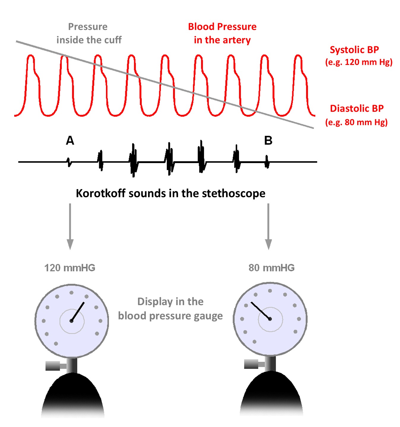
Korotkoff sounds are the sounds that medical personnel listen for when they are taking blood pressure using a non-invasive procedure. They are named after Nikolai Korotkov, a Russian physician who discovered them in 1905, when he was working at the Imperial Medical Academy in St. Petersburg, the Russian Empire. Description The sounds heard during the measurement of blood pressure are not the same as the heart sounds heard during chest auscultation that are due to vibrations inside the ventricles associated with the snapping shut of the valves. If a stethoscope is placed over the brachial artery in the antecubital fossa in a normal person (without arterial disease), no sound should be audible. As the heart beats, these pulses are transmitted smoothly via laminar (non-turbulent) blood flow throughout the arteries, and no sound is produced. Similarly, if the cuff of a sphygmomanometer is placed around a patient's upper arm and inflated to a pressure above the patient's systolic ... [...More Info...] [...Related Items...] OR: [Wikipedia] [Google] [Baidu] |
Sons And Lovers
''Sons and Lovers'' is a 1913 novel by the English writer D. H. Lawrence. It traces emotional conflicts through the protagonist, Paul Morel, and his suffocating relationships with a demanding mother and two very different lovers, which exert complex influences on the development of his manhood. The novel was originally published by Gerald Duckworth and Company Ltd., London, and Mitchell Kennerley Publishers, New York. While the novel initially received a lukewarm critical reception, along with allegations of obscenity, it is today regarded as a masterpiece by many critics and is often regarded as Lawrence's finest achievement. It tells us more about Lawrence's life and his phases, as his first was when he lost his mother in 1910 to whom he was particularly attached. And it was from then that he met Frieda Richthofen, and around this time that he began conceiving his two other great novels, '' The Rainbow'' and ''Women In Love'', which had more sexual emphasis and maturity. Deve ... [...More Info...] [...Related Items...] OR: [Wikipedia] [Google] [Baidu] |


