|
Pronator Teres Syndrome
Pronator teres syndrome is a compression neuropathy of the median nerve at the elbow. It is rare compared to compression at the wrist (carpal tunnel syndrome) or isolated injury of the anterior interosseous branch of the median nerve (anterior interosseous syndrome). Symptoms Compression of the median nerve in the region of the elbow or proximal part of the forearm can cause pain and/or numbness in the distribution of the distal median nerve, and weakness of the muscles innervated by the anterior interosseous nerve: the flexor pollicis longus ("FPL"), the flexor digitorum profundus of the index finger ("FDP IF"), and the pronator quadratus ("PQ"). The pain tends to be at the wrist joint, in the distribution of the terminal branch of the anterior interosseous nerve, and is exacerbated by sustained pronation (i.e., wrist down). The weakness of the FPL and FDP IF is painless, but causes people to "drop things" and have a sense of loss of dexterity. Pinching with the wrist flexed ma ... [...More Info...] [...Related Items...] OR: [Wikipedia] [Google] [Baidu] |
Median Nerve
The median nerve is a nerve in humans and other animals in the upper limb. It is one of the five main nerves originating from the brachial plexus. The median nerve originates from the lateral and medial cords of the brachial plexus, and has contributions from ventral roots of C5-C7 (lateral cord) and C8 and T1 (medial cord). The median nerve is the only nerve that passes through the carpal tunnel. Carpal tunnel syndrome is the disability that results from the median nerve being pressed in the carpal tunnel. Structure The median nerve arises from the branches from lateral and medial cords of the brachial plexus, courses through the anterior part of arm, forearm, and hand, and terminates by supplying the muscles of the hand. Arm After receiving inputs from both the lateral and medial cords of the brachial plexus, the median nerve enters the arm from the axilla at the inferior margin of the teres major muscle. It then passes vertically down and courses lateral to the brachial a ... [...More Info...] [...Related Items...] OR: [Wikipedia] [Google] [Baidu] |
Carpal Tunnel Syndrome
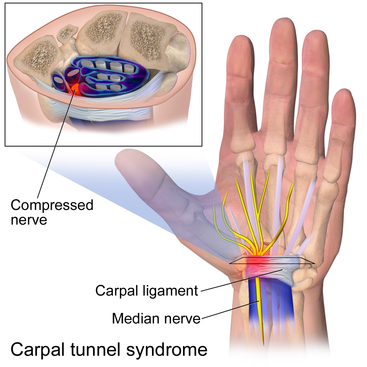
Carpal tunnel syndrome (CTS) is the collection of symptoms and signs associated with median neuropathy at the carpal tunnel. Most CTS is related to idiopathic An idiopathic disease is any disease with an unknown cause or mechanism of apparent spontaneous origin. From Greek ἴδιος ''idios'' "one's own" and πάθος ''pathos'' "suffering", ''idiopathy'' means approximately "a disease of its own kin ... compression of the median nerve as it travels through the wrist at the carpal tunnel (IMNCT). Idiopathic means that there is no other disease process contributing to pressure on the nerve. As with most structural issues, it occurs in both hands, and the strongest risk factor is genetics. Other conditions can cause CTS such as wrist fracture or rheumatoid arthritis. After fracture, swelling, bleeding, and deformity compress the median nerve. With rheumatoid arthritis, the enlarged synovial lining of the tendons causes compression. The main symptoms are numbness and Pares ... [...More Info...] [...Related Items...] OR: [Wikipedia] [Google] [Baidu] |
Anterior Interosseous Syndrome
Anterior interosseous syndrome is a medical condition in which damage to the anterior interosseous nerve (AIN), a distal motor and sensory branch of the median nerve, classically with severe weakness of the pincer movement of the thumb and index finger, and can cause transient pain in the wrist (the terminal, sensory branch of the AIN innervates the bones of the carpal tunnel). Most cases of AIN syndrome are now thought to be due to a transient neuritis, although compression of the AIN in the forearm is a risk, such as pressure on the forearm from immobilization after shoulder surgery. Trauma to the median nerve or around the proximal median nerve have also been reported as causes of AIN syndrome. Although there is still controversy among upper extremity surgeons, AIN syndrome is now regarded as a neuritis (inflammation of the nerve) in most cases; this is similar to Parsonage–Turner syndrome. Although the exact etiology is unknown, there is evidence that it is caused by an ... [...More Info...] [...Related Items...] OR: [Wikipedia] [Google] [Baidu] |
Flexor Pollicis Longus
The flexor pollicis longus (; FPL, Latin ''flexor'', bender; ''pollicis'', of the thumb; ''longus'', long) is a muscle in the forearm and hand that flexes the thumb. It lies in the same plane as the flexor digitorum profundus. This muscle is unique to humans, being either rudimentary or absent in other primates. A meta-analysis indicated accessory flexor pollicis longus is present in around 48% of the population. Human anatomy Origin and insertion It arises from the grooved anterior (side of palm) surface of the body of the radius, extending from immediately below the radial tuberosity and oblique line to within a short distance of the pronator quadratus muscle.Gray 1918, ''Flexor Pollicis Longus'', paras 20, 25 An occasionally present accessory long head of the flexor pollicis longus muscle is called 'Gantzer's muscle'. It may cause compression of the anterior interosseous nerve. It arises also from the adjacent part of the interosseous membrane of the forearm, and genera ... [...More Info...] [...Related Items...] OR: [Wikipedia] [Google] [Baidu] |
Flexor Digitorum Profundus
The flexor digitorum profundus is a muscle in the forearm of humans that flexes the fingers (also known as digits). It is considered an extrinsic hand muscle because it acts on the hand while its muscle belly is located in the forearm. Together the flexor pollicis longus, pronator quadratus, and flexor digitorum profundus form the deep layer of ventral forearm muscles.Platzer 2004, p 162 The muscle is named . Structure Flexor digitorum profundus originates in the upper 3/4 of the anterior and medial surfaces of the ulna, interosseous membrane and deep fascia of the forearm. The muscle fans out into four tendons (one to each of the second to fifth fingers) to the palmar base of the distal phalanx. Along with the flexor digitorum superficialis, it has long tendons that run down the arm and through the carpal tunnel and attach to the palmar side of the phalanges of the fingers. Flexor digitorum profundus lies deep to the superficialis, but it attaches more distally. There ... [...More Info...] [...Related Items...] OR: [Wikipedia] [Google] [Baidu] |
Pronator Quadratus
Pronator quadratus is a square-shaped muscle on the distal forearm that acts to pronate (turn so the palm faces downwards) the hand. Structure Its fibres run perpendicular to the direction of the arm, running from the most distal quarter of the anterior ulna to the distal quarter of the radius. It has two heads: the superficial head originates from the anterior distal aspect of the diaphysis (shaft) of the ulna and inserts into the anterior distal diaphysis of the radius, as well as its anterior metaphysis. The deep head has the same origin, but inserts proximal to the ulnar notch. It is the only muscle that attaches only to the ulna at one end and the radius at the other end. Arterial blood comes via the anterior interosseous artery. Innervation Pronator quadratus muscle is innervated by the anterior interosseous nerve, a branch of the median nerve. Function When pronator quadratus contracts, it pulls the lateral side of the radius towards the ulna, thus pronating the hand ... [...More Info...] [...Related Items...] OR: [Wikipedia] [Google] [Baidu] |
Bicipital Aponeurosis
The bicipital aponeurosis (also known as lacertus fibrosus) is a broad aponeurosis of the biceps brachii, which is located in the cubital fossa of the elbow. It separates superficial from deep structures in much of the fossa. Structure The bicipital aponeurosis originates from the distal insertion of the biceps brachii, and inserts into the deep fascia of the forearm. The biceps tendon inserts on the radial tuberosity, and the bicipital aponeurosis lies medially to it. It reinforces the cubital fossa, helping to protect the brachial artery and the ulnar nerve running underneath. Variations Some individuals (about 3% of the population) have a ''superficial ulnar artery'' that runs superficially to the bicipital aponeurosis instead of underneath it. These individuals are at risk for accidental injury to the ulnar artery during venipuncture. Clinical significance The bicipital aponeurosis is superficial to the brachial artery and the median nerve, but deep to the median cubit ... [...More Info...] [...Related Items...] OR: [Wikipedia] [Google] [Baidu] |
Cubital Fossa
The cubital fossa, chelidon, or elbow pit, is the triangular area on the anterior side of the upper limb between the arm and forearm of a human or other hominid animals. It lies anteriorly to the elbow (Latin ) when in standard anatomical position. Boundaries * superior (proximal) boundary – an imaginary horizontal line connecting the medial epicondyle of the humerus to the lateral epicondyle of the humerus * medial (ulnar) boundary – lateral border of pronator teres muscle originating from the medial epicondyle of the humerus. * lateral (radial) boundary – medial border of brachioradialis muscle originating from the lateral supraepicondylar ridge of the humerus. * apex – it is directed inferiorly, and is formed by the meeting point of the lateral and medial boundaries * superficial boundary (roof) – skin, superficial fascia containing the median cubital vein, the lateral cutaneous nerve of the forearm and the medial cutaneous nerve of the forearm, deep fascia reinforce ... [...More Info...] [...Related Items...] OR: [Wikipedia] [Google] [Baidu] |
Pronator Teres
The pronator teres is a muscle (located mainly in the forearm) that, along with the pronator quadratus, serves to pronate the forearm (turning it so that the palm faces posteriorly when from the anatomical position). It has two attachments, to the medial humeral supracondylar ridge and the ulnar tuberosity, and inserts near the middle of the radius. Structure The pronator teres has two heads—humeral and ulnar. * The humeral head, the larger and more superficial, arises from the medial supracondylar ridge immediately superior to the medial epicondyle of the humerus, and from the common flexor tendon (which arises from the medial epicondyle). * The ulnar head (or ulnar tuberosity) is a thin fasciculus, which arises from the medial side of the coronoid process of the ulna, and joins the preceding at an acute angle. The median nerve enters the forearm between the two heads of the muscle, and is separated from the ulnar artery by the ulnar head. The muscle passes obliquely acros ... [...More Info...] [...Related Items...] OR: [Wikipedia] [Google] [Baidu] |
Flexor Digitorum Superficialis
Flexor digitorum superficialis (''flexor digitorum sublimis'') is an extrinsic flexor muscle of the fingers at the proximal interphalangeal joints. It is in the anterior compartment of the forearm. It is sometimes considered to be the deepest part of the superficial layer of this compartment, and sometimes considered to be a distinct, "intermediate layer" of this compartment. It is relatively common for the Flexor digitorum superficialis to be missing from the little finger, bilaterally and unilaterally, which can cause problems when diagnosing a little finger injury. Structure The muscle has two classically described heads – the humeroulnar and radial – and it is between these heads that the median nerve and ulnar artery pass. The ulnar collateral ligament of elbow joint gives its origin to part of this muscle. Four long tendons come off this muscle near the wrist and travel through the carpal tunnel formed by the flexor retinaculum. These tendons, along with those of fle ... [...More Info...] [...Related Items...] OR: [Wikipedia] [Google] [Baidu] |
Carpal Tunnel
In the human body, the carpal tunnel or carpal canal is the passageway on the palmar side of the wrist that connects the forearm to the hand. The tunnel is bounded by the bones of the wrist and flexor retinaculum from connective tissue. Normally several tendons from the flexor group of forearm muscles and the median nerve pass through it. There are described cases of variable median artery occurrence. When any of the nine long flexor tendons passing through the narrow carpal canal swell or degenerate, the narrowing of the canal may result in the median nerve becoming entrapped or compressed, a common medical condition known as carpal tunnel syndrome (CTS). Structure The carpal bones that make up the wrist form an arch which is convex on the dorsal side of the hand and concave on the palmar side. The groove on the palmar side, the ''sulcus carpi'', is covered by the flexor retinaculum, a sheath of tough connective tissue, thus forming the carpal tunnel. On the side of ... [...More Info...] [...Related Items...] OR: [Wikipedia] [Google] [Baidu] |
Anterior Interosseous Nerve
The anterior interosseous nerve (volar interosseous nerve) is a branch of the median nerve that supplies the deep muscles on the anterior of the forearm, except the ulnar (medial) half of the flexor digitorum profundus. Its nerve roots come from C8 and T1. It accompanies the anterior interosseous artery along the anterior of the interosseous membrane of the forearm, in the interval between the flexor pollicis longus and flexor digitorum profundus, supplying the whole of the former and (most commonly) the radial half of the latter, and ending below in the pronator quadratus and wrist joint. Note that the median nerve supplies all flexor muscles of the forearm except for the ulnar half of flexor digitorum profundus and the flexor carpi ulnaris, which is a superficial muscle of the forearm. Innervation The anterior interosseous nerve classically innervates 2.5 muscles: which are deep muscles of the forearm * flexor pollicis longus * pronator quadratus * the radial (lateral) half of ... [...More Info...] [...Related Items...] OR: [Wikipedia] [Google] [Baidu] |


