|
Presacral Fascia
The presacral fascia lines the anterior aspect of the sacrum, enclosing the sacral vessels and nerves. It continues anteriorly as the pelvic parietal fascia, covering the entire pelvic cavity. The presacral fascia is limited postero-inferiorly, as it fuses with the mesorectal fascia, lying above the levator ani muscle, at the level of the anorectal junction. These two fascias have been erroneously confused, though they are in fact, separate anatomical entities. The colloquial term, among colo-rectal surgeons, for this inter-fascial plane, is known as the holy plane of dissection first coined by Bill Heald. During rectal surgery and mesorectum excision, dissection along the avascular alveolar plane between these two fascias, facilitates a straightforward dissection and preserves the sacral vessels and hypogastric nerves. Waldeyer's fascia (a.k.a. rectosacral fascia) originates from the presacral parietal fascia at the S2 to S4 level fusing with the rectal visceral fascia at th ... [...More Info...] [...Related Items...] OR: [Wikipedia] [Google] [Baidu] |
Sacrum
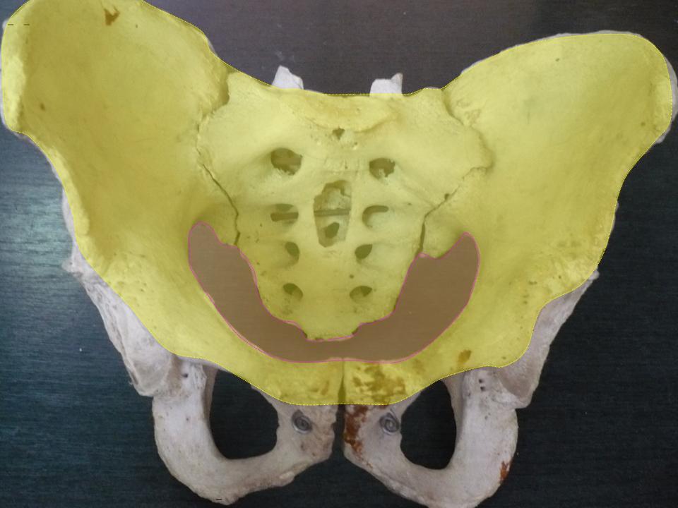
The sacrum (plural: ''sacra'' or ''sacrums''), in human anatomy, is a large, triangular bone at the base of the spine that forms by the fusing of the sacral vertebrae (S1S5) between ages 18 and 30. The sacrum situates at the upper, back part of the pelvic cavity, between the two wings of the pelvis. It forms joints with four other bones. The two projections at the sides of the sacrum are called the alae (wings), and articulate with the ilium at the L-shaped sacroiliac joints. The upper part of the sacrum connects with the last lumbar vertebra (L5), and its lower part with the coccyx (tailbone) via the sacral and coccygeal cornua. The sacrum has three different surfaces which are shaped to accommodate surrounding pelvic structures. Overall it is concave (curved upon itself). The base of the sacrum, the broadest and uppermost part, is tilted forward as the sacral promontory internally. The central part is curved outward toward the posterior, allowing greater room for the p ... [...More Info...] [...Related Items...] OR: [Wikipedia] [Google] [Baidu] |
Nerve
A nerve is an enclosed, cable-like bundle of nerve fibers (called axons) in the peripheral nervous system. A nerve transmits electrical impulses. It is the basic unit of the peripheral nervous system. A nerve provides a common pathway for the electrochemical nerve impulses called action potentials that are transmitted along each of the axons to peripheral organs or, in the case of sensory nerves, from the periphery back to the central nervous system. Each axon, within the nerve, is an extension of an individual neuron, along with other supportive cells such as some Schwann cells that coat the axons in myelin. Within a nerve, each axon is surrounded by a layer of connective tissue called the endoneurium. The axons are bundled together into groups called fascicles, and each fascicle is wrapped in a layer of connective tissue called the perineurium. Finally, the entire nerve is wrapped in a layer of connective tissue called the epineurium. Nerve cells (often called neurons) ... [...More Info...] [...Related Items...] OR: [Wikipedia] [Google] [Baidu] |
Parietal Fascia
A fascia (; plural fasciae or fascias; adjective fascial; from Latin: "band") is a band or sheet of connective tissue, primarily collagen, beneath the skin that attaches to, stabilizes, encloses, and separates muscles and other internal organs. Fascia is classified by layer, as superficial fascia, deep fascia, and ''visceral'' or ''parietal'' fascia, or by its function and anatomical location. Like ligaments, aponeuroses, and tendons, fascia is made up of fibrous connective tissue containing closely packed bundles of collagen fibers oriented in a wavy pattern parallel to the direction of pull. Fascia is consequently flexible and able to resist great unidirectional tension forces until the wavy pattern of fibers has been straightened out by the pulling force. These collagen fibers are produced by fibroblasts located within the fascia. Fasciae are similar to ligaments and tendons as they have collagen as their major component. They differ in their location and function: ... [...More Info...] [...Related Items...] OR: [Wikipedia] [Google] [Baidu] |
Pelvic Cavity
The pelvic cavity is a body cavity that is bounded by the bones of the pelvis. Its oblique roof is the pelvic inlet (the superior opening of the pelvis). Its lower boundary is the pelvic floor. The pelvic cavity primarily contains the reproductive organs, urinary bladder, distal ureters, proximal urethra, terminal sigmoid colon, rectum, and anal canal. In females, the uterus, Fallopian tubes, ovaries and upper vagina occupy the area between the other viscera. The rectum is located at the back of the pelvis, in the curve of the sacrum and coccyx; the bladder is in front, behind the pubic symphysis. The pelvic cavity also contains major arteries, veins, muscles, and nerves. These structures coexist in a crowded space, and disorders of one pelvic component may impact upon another; for example, constipation may overload the rectum and compress the urinary bladder, or childbirth might damage the pudendal nerves and later lead to anal weakness. Structure The pelvis has a ... [...More Info...] [...Related Items...] OR: [Wikipedia] [Google] [Baidu] |
Levator Ani
The levator ani is a broad, thin muscle group, situated on either side of the pelvis. It is formed from three muscle components: the pubococcygeus, the iliococcygeus, and the puborectalis. It is attached to the inner surface of each side of the lesser pelvis, and these unite to form the greater part of the pelvic floor. The coccygeus muscle completes the pelvic floor, which is also called the ''pelvic diaphragm''. It supports the viscera in the pelvic cavity, and surrounds the various structures that pass through it. The levator ani is the main pelvic floor muscle and painfully contracts during vaginismus. It also contracts rhythmically during orgasm. Structure The levator ani is made up of 3 parts: * Iliococcygeus muscle * Pubococcygeus muscle * Puborectalis muscle The iliococcygeus arises from the inner side of the ischium (the lower and back part of the hip bone) and from the posterior part of the tendinous arch of the obturator fascia, and is attached to the coccyx ... [...More Info...] [...Related Items...] OR: [Wikipedia] [Google] [Baidu] |
Surgery
Surgery ''cheirourgikē'' (composed of χείρ, "hand", and ἔργον, "work"), via la, chirurgiae, meaning "hand work". is a medical specialty that uses operative manual and instrumental techniques on a person to investigate or treat a pathological condition such as a disease or injury, to help improve bodily function, appearance, or to repair unwanted ruptured areas. The act of performing surgery may be called a surgical procedure, operation, or simply "surgery". In this context, the verb "operate" means to perform surgery. The adjective surgical means pertaining to surgery; e.g. surgical instruments or surgical nurse. The person or subject on which the surgery is performed can be a person or an animal. A surgeon is a person who practices surgery and a surgeon's assistant is a person who practices surgical assistance. A surgical team is made up of the surgeon, the surgeon's assistant, an anaesthetist, a circulating nurse and a surgical technologist. Surgery usually ... [...More Info...] [...Related Items...] OR: [Wikipedia] [Google] [Baidu] |
Total Mesorectal Excision
Total mesorectal excision (TME) is a standard surgical technique for treatment of rectal cancer, first described in 1982 by Professor Bill Heald at the UK's Basingstoke District Hospital. It is a precise dissection of the mesorectal envelope comprising rectum containing the tumour together with all the surrounding fatty tissue and the sheet of tissue that contains lymph nodes and blood vessels. Dissection is along the avascular alveolar plane between the presacral and mesorectal fascia, described as holy plane (Heald's "holy plane"). Dissection along this plane facilitates a straightforward dissection and preserves the sacral vessels and hypogastric nerves and is a sphincter-sparing resection and decreases permanent stoma rates. It is possible to rejoin the two ends of the colon; however, most patients require a temporary ileostomy pouch to bypass the colon, allowing it to heal with less risk of infection, perforation or leakage. TME has become the "gold standard" treatment for ... [...More Info...] [...Related Items...] OR: [Wikipedia] [Google] [Baidu] |
Hypogastric Nerve
The hypogastric nerve is the nerve that transitions between the superior hypogastric plexus and the inferior hypogastric plexus. The hypogastric nerve enters the sympathetic chain at T12- L3. Structure The hypogastric nerve begins where the superior hypogastric plexus splits into a right and left plexus. Each of these divisions is considered a hypogastric nerve. The hypogastric nerve continues inferiorly on its corresponding side of the body, where it descends into the pelvis to form the inferior hypogastric plexus. Contents of the right and left hypogastric nerves include preganglionic and postganglionic sympathetic fibers from vertebral levels of T10 to L2, passing through the spinal nerve roots of T12- L3. The hypogastric nerve provides sympathetic innervation.Le, Bhushhan, Hoffman. First Aid for the USMLE Step1. p.613. 2019. It is responsible for the emission of semen into the posterior urethra. Emission is the first phase of male ejaculation (followed by the second pha ... [...More Info...] [...Related Items...] OR: [Wikipedia] [Google] [Baidu] |
Rectal Cancer
Colorectal cancer (CRC), also known as bowel cancer, colon cancer, or rectal cancer, is the development of cancer from the colon or rectum (parts of the large intestine). Signs and symptoms may include blood in the stool, a change in bowel movements, weight loss, and fatigue. Most colorectal cancers are due to old age and lifestyle factors, with only a small number of cases due to underlying genetic disorders. Risk factors include diet, obesity, smoking, and lack of physical activity. Dietary factors that increase the risk include red meat, processed meat, and alcohol. Another risk factor is inflammatory bowel disease, which includes Crohn's disease and ulcerative colitis. Some of the inherited genetic disorders that can cause colorectal cancer include familial adenomatous polyposis and hereditary non-polyposis colon cancer; however, these represent less than 5% of cases. It typically starts as a benign tumor, often in the form of a polyp, which over time becomes cancerou ... [...More Info...] [...Related Items...] OR: [Wikipedia] [Google] [Baidu] |
Rectus Abdominis Muscle
The rectus abdominis muscle, ( la, straight abdominal) also known as the "abdominal muscle" or simply the "abs", is a paired straight muscle. It is a paired muscle, separated by a midline band of connective tissue called the linea alba. It extends from the pubic symphysis, pubic crest and pubic tubercle inferiorly, to the xiphoid process and costal cartilages of ribs V to VII superiorly. The proximal attachments are the pubic crest and the pubic symphysis. It attaches distally at the costal cartilages of ribs 5-7 and the xiphoid process of the sternum. The rectus abdominis muscle is contained in the rectus sheath, which consists of the aponeuroses of the lateral abdominal muscles. The outer, most lateral line, defining the rectus is the linea semilunaris. Bands of connective tissue traverse the rectus abdominis, separating it into distinct muscle bellies. In the abdomens of people with low body fat, these muscle bellies can be viewed externally. They can appear in s ... [...More Info...] [...Related Items...] OR: [Wikipedia] [Google] [Baidu] |


.jpg)
