|
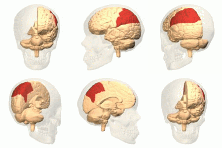
Premotor Cortex
The premotor cortex is an area of the motor cortex lying within the frontal lobe of the brain just anterior to the primary motor cortex. It occupies part of Brodmann's area 6. It has been studied mainly in primates, including monkeys and humans. The functions of the premotor cortex are diverse and not fully understood. It projects directly to the spinal cord and therefore may play a role in the direct control of behavior, with a relative emphasis on the trunk muscles of the body. It may also play a role in planning movement, in the spatial guidance of movement, in the sensory guidance of movement, in understanding the actions of others, and in using abstract rules to perform specific tasks. Different subregions of the premotor cortex have different properties and presumably emphasize different functions. Nerve signals generated in the premotor cortex cause much more complex patterns of movement than the discrete patterns generated in the primary motor cortex. Structure The premo ... [...More Info...] [...Related Items...] OR: [Wikipedia] [Google] [Baidu] |
Cortex Praemotorius
Cortex or cortical may refer to: Biology * Cortex (anatomy), the outermost layer of an organ ** Cerebral cortex, the outer layer of the vertebrate cerebrum, part of which is the ''forebrain'' *** Motor cortex, the regions of the cerebral cortex involved in voluntary motor functions *** Prefrontal cortex, the anterior part of the frontal lobes of the brain *** Visual cortex, regions of the cerebral cortex involved in visual functions ** Cerebellar cortex, the outer layer of the vertebrate cerebellum ** Renal cortex, the outer portion of the kidney ** Adrenal cortex, a portion of the adrenal gland * Cell cortex, the region of a cell directly underneath the membrane * Cortex (hair), the middle layer of a strand of hair * Cortex (botany), the outer portion of the stem or root of a plant Entertainment * Cortex (film), ''Cortex'' (film), a 2008 French film directed by Nicolas Boukhrief * Cortex (podcast), a 2015 podcast * ''Cortex Command'', a 2012 video game * Doctor Neo Cortex, a fic ... [...More Info...] [...Related Items...] OR: [Wikipedia] [Google] [Baidu] |
Cécile Vogt-Mugnier
Cécile Vogt-Mugnier (27 March 1875 – 4 May 1962) was a French neurologist from Haute-Savoie. She and her husband Oskar Vogt are known for their extensive cytology, cytoarchetectonic studies on the brain. Professional life Education and career Vogt-Mugnier obtained her medical Doctorate degree, doctorate in Paris in 1900 and studied under Pierre Marie at the Bicêtre Hospital. At the time, women only made up 6% of those receiving medical doctorates, even though it had been thirty years since women were first admitted to medical studies.Helga Satzinger �Femininity and Science: The Brain Researcher Cécile Vogt (1875-1962). Translation of: Weiblichkeit und Wissenschaft. In: Bleker, Johanna (ed.): Der Eintritt der Frauen in die Gelehrtenrepublik. Husum, 1998, 75-93. Vogt-Mugnier and her husband's findings on myelinogenesis led to her dissertation work on the fiber systems in the cat cerebral cortex (''Étude sur la myelination of hémishères cérébraux'') and the beginning of t ... [...More Info...] [...Related Items...] OR: [Wikipedia] [Google] [Baidu] |
Wilder Penfield
Wilder Graves Penfield (January 26, 1891April 5, 1976) was an American Canadians, American-Physicians in Canada, Canadian neurosurgeon. He expanded brain surgery's methods and techniques, including mapping the functions of various regions of the brain such as the cortical homunculus. His scientific contributions on neural stimulation expand across a variety of topics including hallucinations, illusions, and ''déjà vu''. Penfield devoted much of his thinking to mental processes, including contemplation of whether there was any scientific basis for the existence of the human soul. Biography Early life and education Born in Spokane, Washington, on January 26, 1891, Penfield spent most of his early life in Hudson, Wisconsin. He studied at Princeton University, where he was a member of Cap and Gown Club and played on the Princeton Tigers football, football team. After graduation in 1913, he was hired briefly as the team coach. In 1915 he obtained a Rhodes Scholarship to Merton C ... [...More Info...] [...Related Items...] OR: [Wikipedia] [Google] [Baidu] |
Mirror Neurons
A mirror neuron is a neuron that fires both when an animal acts and when the animal observes the same action performed by another. Thus, the neuron "mirrors" the behavior of the other, as though the observer were itself acting. Such neurons have been directly observed in human and primate species, and in birds. In humans, brain activity consistent with that of mirror neurons has been found in the premotor cortex, the supplementary motor area, the primary somatosensory cortex, and the inferior parietal cortex. The function of the mirror system in humans is a subject of much speculation. Birds have been shown to have imitative resonance behaviors and neurological evidence suggests the presence of some form of mirroring system. To date, no widely accepted neural or computational models have been put forward to describe how mirror neuron activity supports cognitive functions. The subject of mirror neurons continues to generate intense debate. In 2014, Philosophical Transactions o ... [...More Info...] [...Related Items...] OR: [Wikipedia] [Google] [Baidu] |
Michael Graziano
Michael Steven Anthony Graziano (born May 22, 1967) is an American scientist and novelist who is currently a professor of Psychology and Neuroscience at Princeton University. His scientific research focuses on the brain basis of awareness. He has proposed the "attention schema" theory, an explanation of how, and for what adaptive advantage, brains attribute the property of awareness to themselves. His previous work focused on how the |
Thalamus
The thalamus (from Greek θάλαμος, "chamber") is a large mass of gray matter located in the dorsal part of the diencephalon (a division of the forebrain). Nerve fibers project out of the thalamus to the cerebral cortex in all directions, allowing hub-like exchanges of information. It has several functions, such as the relaying of sensory signals, including motor signals to the cerebral cortex and the regulation of consciousness, sleep, and alertness. Anatomically, it is a paramedian symmetrical structure of two halves (left and right), within the vertebrate brain, situated between the cerebral cortex and the midbrain. It forms during embryonic development as the main product of the diencephalon, as first recognized by the Swiss embryologist and anatomist Wilhelm His Sr. in 1893. Anatomy The thalamus is a paired structure of gray matter located in the forebrain which is superior to the midbrain, near the center of the brain, with nerve fibers projecting out to the ... [...More Info...] [...Related Items...] OR: [Wikipedia] [Google] [Baidu] |
Striatum
The striatum, or corpus striatum (also called the striate nucleus), is a nucleus (a cluster of neurons) in the subcortical basal ganglia of the forebrain. The striatum is a critical component of the motor and reward systems; receives glutamatergic and dopaminergic inputs from different sources; and serves as the primary input to the rest of the basal ganglia. Functionally, the striatum coordinates multiple aspects of cognition, including both motor and action planning, decision-making, motivation, reinforcement, and reward perception. The striatum is made up of the caudate nucleus and the lentiform nucleus. The lentiform nucleus is made up of the larger putamen, and the smaller globus pallidus. Strictly speaking the globus pallidus is part of the striatum. It is common practice, however, to implicitly exclude the globus pallidus when referring to striatal structures. In primates, the striatum is divided into a ventral striatum, and a dorsal striatum, subdivisions that are ... [...More Info...] [...Related Items...] OR: [Wikipedia] [Google] [Baidu] |
Prefrontal Cortex
In mammalian brain anatomy, the prefrontal cortex (PFC) covers the front part of the frontal lobe of the cerebral cortex. The PFC contains the Brodmann areas BA8, BA9, BA10, BA11, BA12, BA13, BA14, BA24, BA25, BA32, BA44, BA45, BA46, and BA47. The basic activity of this brain region is considered to be orchestration of thoughts and actions in accordance with internal goals. Many authors have indicated an integral link between a person's will to live, personality, and the functions of the prefrontal cortex. This brain region has been implicated in executive functions, such as planning, decision making, short-term memory, personality expression, moderating social behavior and controlling certain aspects of speech and language. Executive function relates to abilities to differentiate among conflicting thoughts, determine good and bad, better and best, same and different, future consequences of current activities, working toward a defined goal, prediction of outcomes, e ... [...More Info...] [...Related Items...] OR: [Wikipedia] [Google] [Baidu] |
Parietal Lobe
The parietal lobe is one of the four major lobes of the cerebral cortex in the brain of mammals. The parietal lobe is positioned above the temporal lobe and behind the frontal lobe and central sulcus. The parietal lobe integrates sensory information among various modalities, including spatial sense and navigation (proprioception), the main sensory receptive area for the sense of touch in the somatosensory cortex which is just posterior to the central sulcus in the postcentral gyrus, and the dorsal stream of the visual system. The major sensory inputs from the skin (touch, temperature, and pain receptors), relay through the thalamus to the parietal lobe. Several areas of the parietal lobe are important in language processing. The somatosensory cortex can be illustrated as a distorted figure – the cortical homunculus (Latin: "little man") in which the body parts are rendered according to how much of the somatosensory cortex is devoted to them. The superior parietal lobule and in ... [...More Info...] [...Related Items...] OR: [Wikipedia] [Google] [Baidu] |
Oskar Vogt
Oskar Vogt (6 April 1870, in Husum – 30 July 1959, in Freiburg im Breisgau) was a German physician and neurologist. He and his wife Cécile Vogt-Mugnier are known for their extensive cytoarchetectonic studies on the brain. Personal life He was born in Husum, Schleswig-Holstein, Germany. Vogt studied medicine at Kiel and Jena, obtaining his doctorate from Jena in 1894. The Vogts met in 1897 in Paris, and eventually married in 1899. The Vogts were close to the Krupp family. Friedrich Alfred Krupp financially supported them, and in 1898, Oskar and Cécile founded a private research institute called the ''Neurologische Zentralstation'' (Neurological Center) in Berlin, which was formally associated with the Physiological Institute of the Charité as the Neurobiological Laboratory of the Berlin University in 1902. This institute served as the basis for the 1914 formation of the ''Kaiser Institut für Hirnforschung'' (Kaiser Wilhelm Institute for Brain Research), of which Oskar was a ... [...More Info...] [...Related Items...] OR: [Wikipedia] [Google] [Baidu] |
Cytoarchitecture
Cytoarchitecture (Greek '' κύτος''= "cell" + '' ἀρχιτεκτονική''= "architecture"), also known as cytoarchitectonics, is the study of the cellular composition of the central nervous system's tissues under the microscope. Cytoarchitectonics is one of the ways to parse the brain, by obtaining sections of the brain using a microtome and staining them with chemical agents which reveal where different neurons are located. The study of the parcellation of ''nerve fibers'' (primarily axons) into layers forms the subject of myeloarchitectonics ( History of the cerebral cytoarchitecture Defining cerebral cytoarchitecture began with the advent of —the science of slicing a ...[...More Info...] [...Related Items...] OR: [Wikipedia] [Google] [Baidu] |
Brodmann Area 6
Brodmann area 6 (BA6) is part of the frontal cortex in the human brain. Situated just anterior to the primary motor cortex ( BA4), it is composed of the premotor cortex and, medially, the supplementary motor area (SMA). This large area of the frontal cortex is believed to play a role in planning complex, coordinated movements. Brodmann area 6 is also called agranular frontal area 6 in humans because it lacks an internal granular cortical layer (layer IV). It is a subdivision of the cytoarchitecturally defined precentral region of cerebral cortex. In the human brain, it is located on the portions of the precentral gyrus that are not occupied by Brodmann area 4; furthermore, BA6 extends onto the caudal portions of the superior frontal and middle frontal gyri. It extends from the cingulate sulcus on the medial aspect of the hemisphere to the lateral sulcus on the lateral aspect. It is bounded rostrally by the granular frontal region and caudally by the gigantopyramidal area 4 (Brod ... [...More Info...] [...Related Items...] OR: [Wikipedia] [Google] [Baidu] |


