|
Preload (cardiology)
In cardiac physiology, preload is the amount of sarcomere stretch experienced by cardiac muscle cells, called cardiomyocytes, at the end of ventricular filling during diastole. Preload is directly related to ventricular filling. As the relaxed ventricle fills during diastole, the walls are stretched and the length of sarcomeres increases. Sarcomere length can be approximated by the volume of the ventricle because each shape has a conserved surface-area-to-volume ratio. This is useful clinically because measuring the sarcomere length is destructive to heart tissue. It requires cutting out a piece of cardiac muscle to look at the sarcomeres under a microscope. It is currently not possible to directly measure preload in the beating heart of a living animal. Preload is estimated from end-diastolic ventricular pressure and is measured in millimeters of mercury (mmHg). Estimating preload Though not exactly equivalent to the strict definition of ''preload,'' end-diastolic volume is ... [...More Info...] [...Related Items...] OR: [Wikipedia] [Google] [Baidu] |
Heart Diasystole
The heart is a muscular organ in most animals. This organ pumps blood through the blood vessels of the circulatory system. The pumped blood carries oxygen and nutrients to the body, while carrying metabolic waste such as carbon dioxide to the lungs. In humans, the heart is approximately the size of a closed fist and is located between the lungs, in the middle compartment of the chest. In humans, other mammals, and birds, the heart is divided into four chambers: upper left and right atria and lower left and right ventricles. Commonly the right atrium and ventricle are referred together as the right heart and their left counterparts as the left heart. Fish, in contrast, have two chambers, an atrium and a ventricle, while most reptiles have three chambers. In a healthy heart blood flows one way through the heart due to heart valves, which prevent backflow. The heart is enclosed in a protective sac, the pericardium, which also contains a small amount of fluid. The wall of ... [...More Info...] [...Related Items...] OR: [Wikipedia] [Google] [Baidu] |
Boyle's Law
Boyle's law, also referred to as the Boyle–Mariotte law, or Mariotte's law (especially in France), is an experimental gas law that describes the relationship between pressure and volume of a confined gas. Boyle's law has been stated as: The absolute pressure exerted by a given mass of an ideal gas is inversely proportional to the volume it occupies if the temperature and amount of gas remain unchanged within a closed system.Levine, Ira. N. (1978), p. 12 gives the original definition. Mathematically, Boyle's law can be stated as: or where is the pressure of the gas, is the volume of the gas, and is a constant. Boyle's Law states that when the temperature of a given mass of confined gas is constant, the product of its pressure and volume is also constant. When comparing the same substance under two different sets of conditions, the law can be expressed as: :P_1 V_1 = P_2 V_2. showing that as volume increases, the pressure of a gas decreases proportionally, and ... [...More Info...] [...Related Items...] OR: [Wikipedia] [Google] [Baidu] |
Passive Leg Raising Test
Passive leg raise, also known as shock position, is a treatment for shock or a test to evaluate the need for further fluid resuscitation in a critically ill person. It is the position of a person who is lying flat on their back with the legs elevated approximately . The purpose of the position is to elevate the legs above the heart in a manner that will help blood flow to the heart. This test involves raising the legs of a person's (without their active participation), which causes gravity to pull blood from the legs, thus increasing circulatory volume available to the heart ( cardiac preload) by around 150-300 milliliters, depending on the amount of venous reservoir. The real-time effects of this maneuver on hemodynamic parameters such as blood pressure and heart rate are used to guide the decision whether or not more fluid will be beneficial. The assessment is easier when invasive monitoring is present (such as an arterial catheter). The maneuver might be reinforced in a ... [...More Info...] [...Related Items...] OR: [Wikipedia] [Google] [Baidu] |
Cardiac Output
In cardiac physiology, cardiac output (CO), also known as heart output and often denoted by the symbols Q, \dot Q, or \dot Q_ , edited by Catherine E. Williamson, Phillip Bennett is the volumetric flow rate of the heart's pumping output: that is, the volume of blood being pumped by both ventricles of the heart, per unit time (usually measured per minute). Cardiac output (CO) is the product of the heart rate (HR), i.e. the number of heartbeats per minute (bpm), and the stroke volume (SV), which is the volume of blood pumped from the left ventricle per beat; thus giving the formula: :CO = HR \times SV Values for cardiac output are usually denoted as L/min. For a healthy individual weighing 70 kg, the cardiac output at rest averages about 5 L/min; assuming a heart rate of 70 beats/min, the stroke volume would be approximately 70 mL. Because cardiac output is related to the quantity of blood delivered to various parts of the body, it is an important component of how eff ... [...More Info...] [...Related Items...] OR: [Wikipedia] [Google] [Baidu] |
Afterload
Afterload is the pressure that the heart must work against to eject blood during systole (ventricular contraction). Afterload is proportional to the average arterial pressure. As aortic and pulmonary pressures increase, the afterload increases on the left and right ventricles respectively. Afterload changes to adapt to the continually changing demands on an animal's cardiovascular system. Afterload is proportional to mean systolic blood pressure and is measured in millimeters of mercury (mm Hg). Hemodynamics Afterload is a determinant of cardiac output. Cardiac output is the product of stroke volume and heart rate. Afterload is a determinant of stroke volume (in addition to preload, and strength of myocardial contraction). Following Laplace's law, the tension upon the muscle fibers in the heart wall is the pressure within the ventricle multiplied by the volume within the ventricle divided by the wall thickness (this ratio is the other factor in setting the afterload). T ... [...More Info...] [...Related Items...] OR: [Wikipedia] [Google] [Baidu] |
Arteriovenous Fistula
An arteriovenous fistula is an abnormal connection or passageway between an artery and a vein. It may be congenital, surgically created for hemodialysis treatments, or acquired due to pathologic process, such as trauma or erosion of an arterial aneurysm. Clinical features Pathological Hereditary hemorrhagic telangiectasia is a condition where there is direct connection between arterioles and venules without intervening capillary beds, at the mucocutaneous region and internal bodily organs. Those who are affected by this conditions usually do not experience any symptoms. Difficulty in breathing is the most common symptom for those who experience symptoms. Just like berry aneurysm, a cerebral arteriovenous malformation can rupture causing subarachnoid hemorrhage. Causes The cause of this condition include * Congenital (developmental defect) * Rupture of arterial aneurysm into an adjacent vein * Penetrating injuries * Inflammatory necrosis of adjacent vessels * Complication of ... [...More Info...] [...Related Items...] OR: [Wikipedia] [Google] [Baidu] |
Polycythemia
Polycythemia (also known as polycythaemia) is a laboratory finding in which the hematocrit (the volume percentage of red blood cells in the blood) and/or hemoglobin concentration are increased in the blood. Polycythemia is sometimes called erythrocytosis, and there is significant overlap in the two findings, but the terms are not the same: polycythemia describes any increase in hematocrit and/or hemoglobin, while erythrocytosis describes an increase specifically in the number of red blood cells in the blood. Polycythemia has many causes. It can describe an increase in the number of red blood cells ("absolute polycythemia") or to a decrease in the volume of plasma ("relative polycythemia"). Absolute polycythemia can be due to genetic mutations in the bone marrow ("primary polycythemia"), physiologic adaptations to one's environment, medications, and/or other health conditions. Laboratory studies such as serum erythropoeitin levels and genetic testing might be helpful to clarify ... [...More Info...] [...Related Items...] OR: [Wikipedia] [Google] [Baidu] |
End Diastolic Volume
In cardiovascular physiology, end-diastolic volume (EDV) is the volume of blood in the right or left ventricle at end of filling in diastole which is ammount of blood present in ventricle at the end of diastole systole. Because greater EDVs cause greater distention of the ventricle, ''EDV'' is often used synonymously with '' preload'', which refers to the length of the sarcomeres in cardiac muscle prior to contraction (systole). An increase in EDV increases the preload on the heart and, through the Frank-Starling mechanism of the heart, increases the amount of blood ejected from the ventricle during systole ( stroke volume). __TOC__ Sample values The right ventricular end-diastolic volume (RVEDV) ranges between 100 and 160 mL. The right ventricular end-diastolic volume index (RVEDVI) is calculated by RVEDV/ BSA and ranges between 60 and 100 mL/m2. See also * End-systolic volume End-systolic volume (ESV) is the volume of blood in a ventricle at the end of contraction, or systo ... [...More Info...] [...Related Items...] OR: [Wikipedia] [Google] [Baidu] |
Dilated Cardiomyopathy
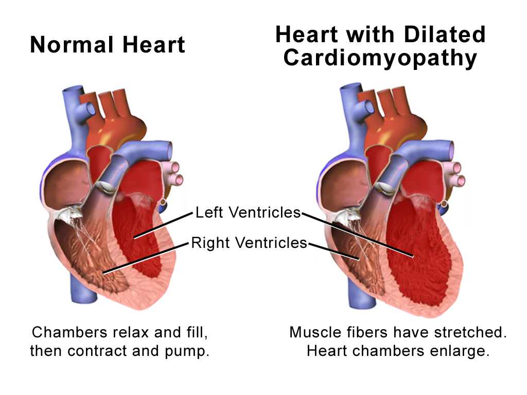
Dilated cardiomyopathy (DCM) is a condition in which the heart becomes enlarged and cannot pump blood effectively. Symptoms vary from none to feeling tired, leg swelling, and shortness of breath. It may also result in chest pain or fainting. Complications can include heart failure, heart valve disease, or an irregular heartbeat. Causes include genetics, alcohol, cocaine, certain toxins, complications of pregnancy, and certain infections. Coronary artery disease and high blood pressure may play a role, but are not the primary cause. In many cases the cause remains unclear. It is a type of cardiomyopathy, a group of diseases that primarily affects the heart muscle. The diagnosis may be supported by an electrocardiogram, chest X-ray, or echocardiogram. In those with heart failure, treatment may include medications in the ACE inhibitor, beta blocker, and diuretic families. A low salt diet may also be helpful. In those with certain types of irregular heartbeat, blood thinners ... [...More Info...] [...Related Items...] OR: [Wikipedia] [Google] [Baidu] |
Mitral Valve Stenosis
Mitral stenosis is a valvular heart disease characterized by the narrowing of the opening of the mitral valve of the heart. It is almost always caused by rheumatic valvular heart disease. Normally, the mitral valve is about 5 cm2 during diastole. Any decrease in area below 2 cm2 causes mitral stenosis. Early diagnosis of mitral stenosis in pregnancy is very important as the heart cannot tolerate increased cardiac output demand as in the case of exercise and pregnancy. Atrial fibrillation is a common complication of resulting left atrial enlargement, which can lead to systemic thromboembolic complications like stroke. Signs and symptoms Signs and symptoms of mitral stenosis include the following: * Heart failure symptoms, such as dyspnea on exertion, orthopnea and paroxysmal nocturnal dyspnea (PND) * Palpitations * Chest pain * Hemoptysis * Thromboembolism in later stages when the left atrial volume is increased (i.e., dilation). The latter leads to increase risk of ... [...More Info...] [...Related Items...] OR: [Wikipedia] [Google] [Baidu] |


