|
Pre-Bötzinger Complex
The preBötzinger complex, sometimes written pre-Bötzinger complex (preBötC), is a functionally and anatomically specialized site in the ventral-lateral region of the lower medulla oblongata (i.e., lower brainstem). The preBötC is part of the ventral respiratory group of respiratory related interneurons. Its foremost function is to generate the inexorable rhythm for inspiratory breathing movements in mammals. In addition, the preBötC is widely and paucisynaptically connected to higher brain centers that regulate arousal and excitability more generally such that respiratory brain function is intimately connected with many other rhythmic and cognitive functions of the brain and central nervous system. Further, the preBötC receives mechanical sensory information from the airways that encode lung volume as well as pH, oxygen, and carbon dioxide content of circulating blood and the cerebrospinal fluid. The preBötC spans approximately 250‒500 µm in the anterior-posterior axis (dep ... [...More Info...] [...Related Items...] OR: [Wikipedia] [Google] [Baidu] |
Medulla Oblongata
The medulla oblongata or simply medulla is a long stem-like structure which makes up the lower part of the brainstem. It is anterior and partially inferior to the cerebellum. It is a cone-shaped neuronal mass responsible for autonomic (involuntary) functions, ranging from vomiting to sneezing. The medulla contains the cardiac, respiratory, vomiting and vasomotor centers, and therefore deals with the autonomic functions of breathing, heart rate and blood pressure as well as the sleep–wake cycle. During embryonic development, the medulla oblongata develops from the myelencephalon. The myelencephalon is a secondary vesicle which forms during the maturation of the rhombencephalon, also referred to as the hindbrain. The bulb is an archaic term for the medulla oblongata. In modern clinical usage, the word bulbar (as in bulbar palsy) is retained for terms that relate to the medulla oblongata, particularly in reference to medical conditions. The word bulbar can refer to the nerves ... [...More Info...] [...Related Items...] OR: [Wikipedia] [Google] [Baidu] |
CGS-21680
CGS-21680 is a specific adenosine A2A subtype receptor agonist. It is usually presented as an organic hydrochloride salt with a molecular weight of 536.0 g/''M''. It is soluble up to 3.4 mg/mL in DMSO and 20 mg/mL in 45% (w/v) ''aq'' 2-hydroxypropyl-β-cyclodextrin. The chemical is currently used by researchers interested in studying neuronal transmission with a high-affinity, subtype specific analogue for adenosine. This includes research in respiration where it is believed that A2A receptors are involved in rhythm generation in the pre-Bötzinger complex. The drug is not currently approved for use in a therapeutic capacity. See also * Adenosine receptor The adenosine receptors (or P1 receptors) are a class of purinergic G protein-coupled receptors with adenosine as the endogenous ligand. There are four known types of adenosine receptors in humans: A1, A2A, A2B and A3; each is encoded by a di ... References * * Nucleosides Purines Carboxamides Adenosin ... [...More Info...] [...Related Items...] OR: [Wikipedia] [Google] [Baidu] |
Neuromodulation
Neuromodulation is the physiological process by which a given neuron uses one or more chemicals to regulate diverse populations of neurons. Neuromodulators typically bind to metabotropic, G-protein coupled receptors (GPCRs) to initiate a second messenger signaling cascade that induces a broad, long-lasting signal. This modulation can last for hundreds of milliseconds to several minutes. Some of the effects of neuromodulators include: alter intrinsic firing activity, increase or decrease voltage-dependent currents, alter synaptic efficacy, increase bursting activity and reconfiguration of synaptic connectivity. Major neuromodulators in the central nervous system include: dopamine, serotonin, acetylcholine, histamine, norepinephrine, nitric oxide, and several neuropeptides. Cannabinoids can also be powerful CNS neuromodulators. Neuromodulators can be packaged into vesicles and released by neurons, secreted as hormones and delivered through the circulatory system. A neuromodulator ... [...More Info...] [...Related Items...] OR: [Wikipedia] [Google] [Baidu] |
Oxygenation (medicine)
Oxygen saturation is the fraction of oxygen-saturated hemoglobin relative to total hemoglobin (unsaturated + saturated) in the blood. The human body requires and regulates a very precise and specific balance of oxygen in the blood. Normal arterial blood oxygen saturation levels in humans are 97–100 percent. If the level is below 90 percent, it is considered low and called hypoxemia. Arterial blood oxygen levels below 80 percent may compromise organ function, such as the brain and heart, and should be promptly addressed. Continued low oxygen levels may lead to respiratory or cardiac arrest. Oxygen therapy may be used to assist in raising blood oxygen levels. Oxygenation occurs when oxygen molecules () enter the tissues of the body. For example, blood is oxygenated in the lungs, where oxygen molecules travel from the air and into the blood. Oxygenation is commonly used to refer to medical oxygen saturation. Definition In medicine, oxygen saturation, commonly referred to as "sa ... [...More Info...] [...Related Items...] OR: [Wikipedia] [Google] [Baidu] |
Hyperpolarization (biology)
Hyperpolarization is a change in a cell's membrane potential that makes it more negative. It is the opposite of a depolarization. It inhibits action potentials by increasing the stimulus required to move the membrane potential to the action potential threshold. Hyperpolarization is often caused by efflux of K+ (a cation) through K+ channels, or influx of Cl– (an anion) through Cl– channels. On the other hand, influx of cations, e.g. Na+ through Na+ channels or Ca2+ through Ca2+ channels, inhibits hyperpolarization. If a cell has Na+ or Ca2+ currents at rest, then inhibition of those currents will also result in a hyperpolarization. This voltage-gated ion channel response is how the hyperpolarization state is achieved. In neurons, the cell enters a state of hyperpolarization immediately following the generation of an action potential. While hyperpolarized, the neuron is in a refractory period that lasts roughly 2 milliseconds, during which the neuron is unabl ... [...More Info...] [...Related Items...] OR: [Wikipedia] [Google] [Baidu] |
Hypoglossal
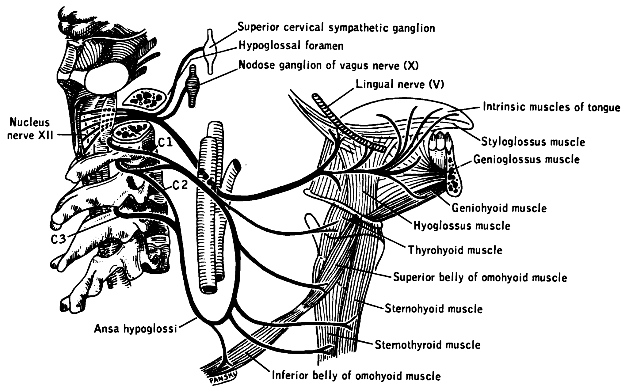
The hypoglossal nerve, also known as the twelfth cranial nerve, cranial nerve XII, or simply CN XII, is a cranial nerve that innervates all the extrinsic and intrinsic muscles of the tongue except for the palatoglossus, which is innervated by the vagus nerve. CN XII is a nerve with a solely motor function. The nerve arises from the hypoglossal nucleus in the medulla as a number of small rootlets, passes through the hypoglossal canal and down through the neck, and eventually passes up again over the tongue muscles it supplies into the tongue. The nerve is involved in controlling tongue movements required for speech and swallowing, including sticking out the tongue and moving it from side to side. Damage to the nerve or the neural pathways which control it can affect the ability of the tongue to move and its appearance, with the most common sources of damage being injury from trauma or surgery, and motor neuron disease. The first recorded description of the nerve is by Herophil ... [...More Info...] [...Related Items...] OR: [Wikipedia] [Google] [Baidu] |
Phrenic
The phrenic nerve is a mixed motor/sensory nerve which originates from the C3-C5 spinal nerves in the neck. The nerve is important for breathing because it provides exclusive motor control of the diaphragm, the primary muscle of respiration. In humans, the right and left phrenic nerves are primarily supplied by the C4 spinal nerve, but there is also contribution from the C3 and C5 spinal nerves. From its origin in the neck, the nerve travels downward into the chest to pass between the heart and lungs towards the diaphragm. In addition to motor fibers, the phrenic nerve contains sensory fibers, which receive input from the central tendon of the diaphragm and the mediastinal pleura, as well as some sympathetic nerve fibers. Although the nerve receives contributions from nerves roots of the cervical plexus and the brachial plexus, it is usually considered separate from either plexus. The name of the nerve comes from Ancient Greek ''phren'' 'diaphragm'. Structure The phrenic ne ... [...More Info...] [...Related Items...] OR: [Wikipedia] [Google] [Baidu] |
Synaptic Inhibition
An inhibitory postsynaptic potential (IPSP) is a kind of synaptic potential that makes a postsynaptic neuron less likely to generate an action potential.Purves et al. Neuroscience. 4th ed. Sunderland (MA): Sinauer Associates, Incorporated; 2008. IPSP were first investigated in motorneurons by David P. C. Lloyd, John Eccles and Rodolfo Llinás in the 1950s and 1960s. The opposite of an inhibitory postsynaptic potential is an excitatory postsynaptic potential (EPSP), which is a synaptic potential that makes a postsynaptic neuron ''more'' likely to generate an action potential. IPSPs can take place at all chemical synapses, which use the secretion of neurotransmitters to create cell to cell signalling. Inhibitory presynaptic neurons release neurotransmitters that then bind to the postsynaptic receptors; this induces a change in the permeability of the postsynaptic neuronal membrane to particular ions. An electric current that changes the postsynaptic membrane potential to create a ... [...More Info...] [...Related Items...] OR: [Wikipedia] [Google] [Baidu] |
Synaptic Augmentation
Augmentation is one of four components of short-term synaptic plasticity that increases the probability of releasing synaptic vesicles during and after repetitive stimulation such that :A(t) = t)/ (0)- 1, when all the other components of enhancement and depression are zero, where A is augmentation at time t and 0 refers to the baseline response to a single stimulus. The increase in the number of synaptic vesicles that release their transmitter leads to enhancement of the post synaptic response. Augmentation can be differentiated from the other components of enhancement by its kinetics of decay and by pharmacology. Augmentation selectively decays with a time constant of about 7 seconds and its magnitude is enhanced in the presence of barium. All four components are thought to be associated with or triggered by increases in internal calcium ions Calcium ions (Ca2+) contribute to the physiology and biochemistry of organisms' cells. They play an important role in signal tran ... [...More Info...] [...Related Items...] OR: [Wikipedia] [Google] [Baidu] |
Pons
The pons (from Latin , "bridge") is part of the brainstem that in humans and other bipeds lies inferior to the midbrain, superior to the medulla oblongata and anterior to the cerebellum. The pons is also called the pons Varolii ("bridge of Varolius"), after the Italian anatomist and surgeon Costanzo Varolio (1543–75). This region of the brainstem includes neural pathways and tracts that conduct signals from the brain down to the cerebellum and medulla, and tracts that carry the sensory signals up into the thalamus.Saladin Kenneth S.(2007) Anatomy & physiology the unity of form and function. Dubuque, IA: McGraw-Hill Structure The pons is in the brainstem situated between the midbrain and the medulla oblongata, and in front of the cerebellum. A separating groove between the pons and the medulla is the inferior pontine sulcus. The superior pontine sulcus separates the pons from the midbrain. The pons can be broadly divided into two parts: the basilar part of the pons (ventral ... [...More Info...] [...Related Items...] OR: [Wikipedia] [Google] [Baidu] |
Glutamatergic
Glutamatergic means "related to glutamate". A glutamatergic agent (or drug) is a chemical that directly modulates the excitatory amino acid (glutamate/ aspartate) system in the body or brain. Examples include excitatory amino acid receptor agonists, excitatory amino acid receptor antagonists, and excitatory amino acid reuptake inhibitors. See also * Adenosinergic * Adrenergic * Cannabinoidergic * Cholinergic * Dopaminergic * GABAergic * GHBergic * Glycinergic * Histaminergic * Melatonergic * Monoaminergic * Opioidergic * Serotonergic * Sigmaergic Sigma receptors (σ-receptors) are protein cell surface receptors that bind ligands such as 4-PPBP (4-phenyl-1-(4-phenylbutyl) piperidine), SA 4503 (cutamesine), ditolylguanidine, dimethyltryptamine, and siramesine. There are two subtypes, ... References Neurochemistry Neurotransmitters {{nervous-system-drug-stub ... [...More Info...] [...Related Items...] OR: [Wikipedia] [Google] [Baidu] |
Tidal Volume
Tidal volume (symbol VT or TV) is the volume of air moved into or out of the lungs during a normal breath. In a healthy, young human adult, tidal volume is approximately 500 ml per inspiration or 7 ml/kg of body mass. Mechanical ventilation Tidal volume plays a significant role during mechanical ventilation to ensure adequate ventilation without causing trauma to the lungs. Tidal volume is measured in milliliters and ventilation volumes are estimated based on a patient's ideal body mass. Measurement of tidal volume can be affected (usually overestimated) by leaks in the breathing circuit or the introduction of additional gas, for example during the introduction of nebulized drugs. Ventilator-induced lung injury such as Acute lung injury (ALI) /Acute Respiratory Distress Syndrome (ARDS) can be caused by ventilation with very large tidal volumes in normal lungs, as well as ventilation with moderate or small volumes in previously injured lungs, and research shows that t ... [...More Info...] [...Related Items...] OR: [Wikipedia] [Google] [Baidu] |


