|
Posterior Sternoclavicular Ligament
The posterior sternoclavicular ligament is a band of fibers, covering the posterior surface of the sternoclavicular joint. It is attached above to the upper and back part of the sternal end of the clavicle, and, passing obliquely downward and medialward, is fixed below to the back of the upper part of the manubrium sterni. It is in relation, in front, with the articular disk and synovial membrane The synovial membrane (also known as the synovial stratum, synovium or stratum synoviale) is a specialized connective tissue that lines the inner surface of capsules of synovial joints and tendon sheath. It makes direct contact with the fibrous ...s; behind, with the Sternohyoideus and Sternothyreoideus. References Ligaments of the upper limb {{ligament-stub ... [...More Info...] [...Related Items...] OR: [Wikipedia] [Google] [Baidu] |
Human Sternum
The sternum or breastbone is a long flat bone located in the central part of the chest. It connects to the ribs via cartilage and forms the front of the rib cage, thus helping to protect the heart, lungs, and major blood vessels from injury. Shaped roughly like a necktie, it is one of the largest and longest flat bones of the body. Its three regions are the manubrium, the body, and the xiphoid process. The word "sternum" originates from the Ancient Greek στέρνον (stérnon), meaning "chest". Structure The sternum is a narrow, flat bone, forming the middle portion of the front of the chest. The top of the sternum supports the clavicles (collarbones) and its edges join with the costal cartilages of the first two pairs of ribs. The inner surface of the sternum is also the attachment of the sternopericardial ligaments. Its top is also connected to the sternocleidomastoid muscle. The sternum consists of three main parts, listed from the top: * Manubrium * Body (gladiolus) * ... [...More Info...] [...Related Items...] OR: [Wikipedia] [Google] [Baidu] |
Manubrium
The sternum or breastbone is a long flat bone located in the central part of the chest. It connects to the ribs via cartilage and forms the front of the rib cage, thus helping to protect the heart, lungs, and major blood vessels from injury. Shaped roughly like a necktie, it is one of the largest and longest flat bones of the body. Its three regions are the manubrium, the body, and the xiphoid process. The word "sternum" originates from the Ancient Greek στέρνον (stérnon), meaning "chest". Structure The sternum is a narrow, flat bone, forming the middle portion of the front of the chest. The top of the sternum supports the clavicles (collarbones) and its edges join with the costal cartilages of the first two pairs of ribs. The inner surface of the sternum is also the attachment of the sternopericardial ligaments. Its top is also connected to the sternocleidomastoid muscle. The sternum consists of three main parts, listed from the top: * Manubrium * Body (gladi ... [...More Info...] [...Related Items...] OR: [Wikipedia] [Google] [Baidu] |
Clavicle
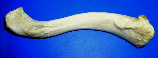
The clavicle, or collarbone, is a slender, S-shaped long bone approximately 6 inches (15 cm) long that serves as a strut between the shoulder blade and the sternum (breastbone). There are two clavicles, one on the left and one on the right. The clavicle is the only long bone in the body that lies horizontally. Together with the shoulder blade, it makes up the shoulder girdle. It is a palpable bone and, in people who have less fat in this region, the location of the bone is clearly visible. It receives its name from the Latin ''clavicula'' ("little key"), because the bone rotates along its axis like a key when the shoulder is abducted. The clavicle is the most commonly fractured bone. It can easily be fractured by impacts to the shoulder from the force of falling on outstretched arms or by a direct hit. Structure The collarbone is a thin doubly curved long bone that connects the arm to the trunk of the body. Located directly above the first rib, it acts as a strut t ... [...More Info...] [...Related Items...] OR: [Wikipedia] [Google] [Baidu] |
Manubrium Sterni
The sternum or breastbone is a long flat bone located in the central part of the chest. It connects to the ribs via cartilage and forms the front of the rib cage, thus helping to protect the heart, lungs, and major blood vessels from injury. Shaped roughly like a necktie, it is one of the largest and longest flat bones of the body. Its three regions are the manubrium, the body, and the xiphoid process. The word "sternum" originates from the Ancient Greek στέρνον (stérnon), meaning "chest". Structure The sternum is a narrow, flat bone, forming the middle portion of the front of the chest. The top of the sternum supports the clavicles (collarbones) and its edges join with the costal cartilages of the first two pairs of ribs. The inner surface of the sternum is also the attachment of the sternopericardial ligaments. Its top is also connected to the sternocleidomastoid muscle. The sternum consists of three main parts, listed from the top: * Manubrium * Body (gladi ... [...More Info...] [...Related Items...] OR: [Wikipedia] [Google] [Baidu] |
Articular Disk
The articular disk (or disc) is a thin, oval plate of fibrocartilage present in several joints which separates synovial cavities. This separation of the cavity space allows for separate movements to occur in each space. The presence of an articular disk also permits a more even distribution of forces between the articulating surfaces of bones, increases the stability of the joint, and aids in directing the flow of synovial fluid to areas of the articular cartilage that experience the most friction. The term " meniscus" has a very similar meaning. Additional images File:Gray325.png, Sternoclavicular articulation. Anterior view. File:Gray300.png , Diagrammatic section of a diarthrodial joint, with an articular disk. See also * Triangular fibrocartilage ("articular disk of the distal radioulnar articulation") * Articular disk of the temporomandibular joint * Articular disk of sternoclavicular articulation The articular disc of the sternoclavicular joint is flat and nearly circu ... [...More Info...] [...Related Items...] OR: [Wikipedia] [Google] [Baidu] |
Synovial Membrane
The synovial membrane (also known as the synovial stratum, synovium or stratum synoviale) is a specialized connective tissue that lines the inner surface of capsules of synovial joints and tendon sheath. It makes direct contact with the fibrous membrane on the outside surface and with the synovial fluid lubricant on the inside surface. In contact with the synovial fluid at the tissue surface are many rounded macrophage-like synovial cells (type A) and also type B cells, which are also known as fibroblast-like synoviocytes (FLS). Type A cells maintain the synovial fluid by removing wear-and-tear debris. As for the FLS, they produce hyaluronan, as well as other extracellular components in the synovial fluid. Structure The synovial membrane is variable but often has two layers: * The outer layer, or subintima, can be of almost any type of connective tissue – fibrous (dense collagenous type), adipose (fatty; e.g. in intra-articular fat pads) or areolar (loose collagenous ty ... [...More Info...] [...Related Items...] OR: [Wikipedia] [Google] [Baidu] |
Sternohyoideus
The sternohyoid muscle is a thin, narrow muscle attaching the hyoid bone to the sternum. It is one of the paired strap muscles of the infrahyoid muscles. It is supplied by the ansa cervicalis. It depresses the hyoid bone. Structure The sternohyoid muscle is one of the paired strap muscles of the infrahyoid muscles. It arises from the posterior border of the medial end of the clavicle, the posterior sternoclavicular ligament, and the upper and posterior part of the manubrium of the sternum. Passing upward and medially, it is inserted by short tendinous fibers into the lower border of the body of the hyoid bone. It runs lateral to the trachea. Nerve supply The sternohyoid muscle is supplied by a branch of the ansa cervicalis. Variations The sternohyoid muscle may be doubled, have accessory slips (Cleidohyoideus) or be completely absent in some people. It sometimes presents a transverse tendinous inscription immediately above its origin. Function The sternohyoid muscle p ... [...More Info...] [...Related Items...] OR: [Wikipedia] [Google] [Baidu] |
Sternothyreoideus
The sternothyroid muscle, or sternothyroideus, is an infrahyoid muscle in the neck. It acts to depress the hyoid bone. It is below the sternohyoid muscle. It is shorter and wider than the sternohyoid. Structure The sternothyroid arises from the posterior surface of the manubrium of the sternum, below the origin of the sternohyoid. It also arises from the edge of the cartilage of the first rib. It is inserted into the oblique line on the lamina of the thyroid cartilage. It is in close contact with its fellow at the lower part of the neck, but diverges somewhat as it ascends. It is occasionally traversed by a transverse or oblique tendinous inscription. Innervation The sternothyroid muscle is innervated by the ansa cervicalis. Variations Doubling; absence; accessory slips to the thyrohyoid, inferior pharyngeal constrictor, or to the carotid sheath. Function The sternothyroid muscle depresses the hyoid bone, along with the other infrahyoid muscle. Clinical significance Th ... [...More Info...] [...Related Items...] OR: [Wikipedia] [Google] [Baidu] |


