|
Posterior Auricular Artery
The posterior auricular artery is a small artery that arises from the external carotid artery, above the digastric muscle and stylohyoid muscle, opposite the apex of the styloid process. It ascends posteriorly beneath the parotid gland, along the styloid process of the temporal bone, between the cartilage of the ear and the mastoid process of the temporal bone along the lateral side of the head. The posterior auricular artery gives off the stylomastoid artery, small branches to the auricle, and supplies blood to the scalp posterior to the auricle. A person may be able to "hear" their own heart rate via this artery, under certain conditions. See also * Anterior auricular branches of superficial temporal artery The anterior auricular branches of the superficial temporal artery are distributed to the anterior portion of the auricula, the lobule, and part of the external meatus, anastomosing with the posterior auricular. They supply the external acousti ... * Posterior auric ... [...More Info...] [...Related Items...] OR: [Wikipedia] [Google] [Baidu] |
External Carotid Artery
The external carotid artery is a major artery of the head and neck. It arises from the common carotid artery when it splits into the external and internal carotid artery. External carotid artery supplies blood to the face and neck. Structure The external carotid artery begins at the upper border of thyroid cartilage, and curves, passing forward and upward, and then inclining backward to the space behind the neck of the mandible, where it divides into the superficial temporal and maxillary artery within the parotid gland. It rapidly diminishes in size as it travels up the neck, owing to the number and large size of its branches. At its origin, this artery is closer to the skin and more medial than the internal carotid, and is situated within the carotid triangle. Development In children, the external carotid artery is somewhat smaller than the internal carotid; but in the adult, the two vessels are of nearly equal size. Relations At the origin, external carotid artery is mo ... [...More Info...] [...Related Items...] OR: [Wikipedia] [Google] [Baidu] |
Posterior Auricular Vein
The posterior auricular vein is a vein of the head. It begins from a plexus with the occipital vein and the superficial temporal vein, descends behind the auricle, and drains into the external jugular vein. Structure The posterior auricular vein begins upon the side of the head, in a plexus which communicates with the tributaries of the occipital vein and the superficial temporal vein. It descends behind the auricle. It joins the posterior division of the retromandibular vein. It drains into the external jugular vein. It receive the stylomastoid vein, and some tributaries from the cranial surface of the auricle. Variation The posterior auricular vein may drain into the internal jugular vein or a posterior jugular vein if there are variations in the external jugular vein The external jugular vein receives the greater part of the blood from the exterior of the cranium and the deep parts of the face, being formed by the junction of the posterior division of the retromandib ... [...More Info...] [...Related Items...] OR: [Wikipedia] [Google] [Baidu] |
Scalp
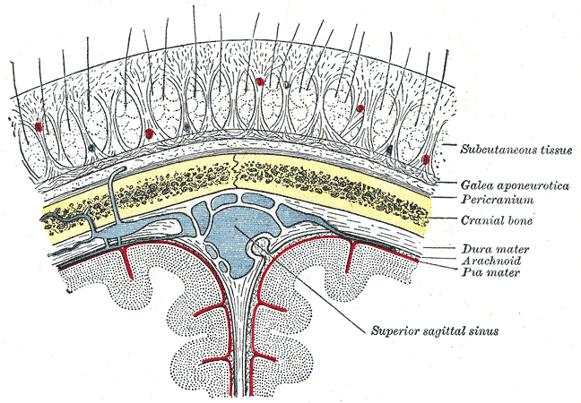
The scalp is the anatomical area bordered by the human face at the front, and by the neck at the sides and back. Structure The scalp is usually described as having five layers, which can conveniently be remembered as a mnemonic: * S: The skin on the head from which head hair grows. It contains numerous sebaceous glands and hair follicles. * C: Connective tissue. A dense subcutaneous layer of fat and fibrous tissue that lies beneath the skin, containing the nerves and vessels of the scalp. * A: The aponeurosis called epicranial aponeurosis (or galea aponeurotica) is the next layer. It is a tough layer of dense fibrous tissue which runs from the frontalis muscle anteriorly to the occipitalis posteriorly. * L: The loose areolar connective tissue layer provides an easy plane of separation between the upper three layers and the pericranium. In scalping the scalp is torn off through this layer. It also provides a plane of access in craniofacial surgery and neurosurgery. This layer i ... [...More Info...] [...Related Items...] OR: [Wikipedia] [Google] [Baidu] |
Pinna (anatomy)
The auricle or auricula is the visible part of the ear that is outside the head. It is also called the pinna (Latin for "wing" or "fin", plural pinnae), a term that is used more in zoology. Structure The diagram shows the shape and location of most of these components: * ''antihelix'' forms a 'Y' shape where the upper parts are: ** ''Superior crus'' (to the left of the ''fossa triangularis'' in the diagram) ** ''Inferior crus'' (to the right of the ''fossa triangularis'' in the diagram) * ''Antitragus'' is below the ''tragus'' * ''Aperture'' is the entrance to the ear canal * ''Auricular sulcus'' is the depression behind the ear next to the head * ''Concha'' is the hollow next to the ear canal * Conchal angle is the angle that the back of the ''concha'' makes with the side of the head * ''Crus'' of the helix is just above the ''tragus'' * ''Cymba conchae'' is the narrowest end of the ''concha'' * External auditory meatus is the ear canal * ''Fossa triangularis'' is the depres ... [...More Info...] [...Related Items...] OR: [Wikipedia] [Google] [Baidu] |
Artery
An artery (plural arteries) () is a blood vessel in humans and most animals that takes blood away from the heart to one or more parts of the body (tissues, lungs, brain etc.). Most arteries carry oxygenated blood; the two exceptions are the pulmonary and the umbilical arteries, which carry deoxygenated blood to the organs that oxygenate it (lungs and placenta, respectively). The effective arterial blood volume is that extracellular fluid which fills the arterial system. The arteries are part of the circulatory system, that is responsible for the delivery of oxygen and nutrients to all cells, as well as the removal of carbon dioxide and waste products, the maintenance of optimum blood pH, and the circulation of proteins and cells of the immune system. Arteries contrast with veins, which carry blood back towards the heart. Structure The anatomy of arteries can be separated into gross anatomy, at the macroscopic level, and microanatomy, which must be studied with a microscop ... [...More Info...] [...Related Items...] OR: [Wikipedia] [Google] [Baidu] |
Digastric Muscle
The digastric muscle (also digastricus) (named ''digastric'' as it has two 'bellies') is a small muscle located under the jaw. The term "digastric muscle" refers to this specific muscle. However, other muscles that have two separate muscle bellies include the suspensory muscle of duodenum, omohyoid, occipitofrontalis. It lies below the body of the mandible, and extends, in a curved form, from the mastoid notch to the mandibular symphysis. It belongs to the suprahyoid muscles group. A broad aponeurotic layer is given off from the tendon of the digastric muscle on either side, to be attached to the body and greater cornu of the hyoid bone; this is termed the suprahyoid aponeurosis. Structure The digastricus (digastric muscle) consists of two muscular bellies united by an intermediate rounded tendon. The two bellies of the digastric muscle have different embryological origins, and are supplied by different cranial nerves. Each person has a right and left digastric muscle. ... [...More Info...] [...Related Items...] OR: [Wikipedia] [Google] [Baidu] |
Stylohyoid Muscle
The stylohyoid muscle is a slender muscle, lying anterior and superior of the posterior belly of the digastric muscle. It is one of the suprahyoid muscles. It shares this muscle's innervation by the facial nerve, and functions to draw the hyoid bone backwards and elevate the tongue. Its origin is the styloid process of the temporal bone. It inserts on the body of the hyoid. Structure The stylohyoid muscle originates from the posterior and lateral surface of the styloid process of the temporal bone, near the base. Passing inferior and anterior, it inserts into the body of the hyoid bone, at its junction with the greater cornu, and just superior to the omohyoid muscle. It belongs to the group of suprahyoid muscles. It is perforated, near its insertion, by the intermediate tendon of the digastric muscle. The stylohyoid muscle has vascular supply from the lingual artery, a branch of the external carotid artery. Nerve supply A branch of the facial nerve The facial n ... [...More Info...] [...Related Items...] OR: [Wikipedia] [Google] [Baidu] |
Temporal Styloid Process
The temporal styloid process is a slender bony process of the temporal bone extending downward and forward from the undersurface of the temporal bone just below the ear. The styloid process gives attachments to several muscles, and ligaments. Structure The styloid process is a slender and pointed bony process of the temporal bone projecting anteroinferiorly from the inferior surface of the temporal bone just below the ear. Its length normally ranges from just under 3 cm to just over 4 cm. It is usually nearly straight, but may be curved in some individuals. Its ''proximal'' (''tympanohyal'') ''part'' is ensheathed by the tympanic part of the temporal bone ''(vaginal process), whereas'' its ''distal (stylohyal)'' ''part'' gives attachment to several structures. Attachments The styloid process gives attachments to several muscles, and ligaments. It serves as an anchor point for several muscles associated with the tongue and larynx. * stylohyoid ligament * stylomandibu ... [...More Info...] [...Related Items...] OR: [Wikipedia] [Google] [Baidu] |
Parotid Gland
The parotid gland is a major salivary gland in many animals. In humans, the two parotid glands are present on either side of the mouth and in front of both ears. They are the largest of the salivary glands. Each parotid is wrapped around the mandibular ramus, and secretes serous saliva through the parotid duct into the mouth, to facilitate mastication and swallowing and to begin the digestion of starches. There are also two other types of salivary glands; they are submandibular and sublingual glands. Sometimes accessory parotid glands are found close to the main parotid glands. Etymology The word ''parotid'' literally means "beside the ear". From Greek παρωτίς (stem παρωτιδ-) : (gland) behind the ear < παρά - pará : in front, and οὖς - ous (stem ὠτ-, ōt-) : ear. Structure The parotid glands are a pair of mainly |
Temporal Bone
The temporal bones are situated at the sides and base of the skull, and lateral to the temporal lobes of the cerebral cortex. The temporal bones are overlaid by the sides of the head known as the temples, and house the structures of the ears. The lower seven cranial nerves and the major vessels to and from the brain traverse the temporal bone. Structure The temporal bone consists of four parts— the squamous, mastoid, petrous and tympanic parts. The squamous part is the largest and most superiorly positioned relative to the rest of the bone. The zygomatic process is a long, arched process projecting from the lower region of the squamous part and it articulates with the zygomatic bone. Posteroinferior to the squamous is the mastoid part. Fused with the squamous and mastoid parts and between the sphenoid and occipital bones lies the petrous part, which is shaped like a pyramid. The tympanic part is relatively small and lies inferior to the squamous part, anterior to the mast ... [...More Info...] [...Related Items...] OR: [Wikipedia] [Google] [Baidu] |
Mastoid Process
The mastoid part of the temporal bone is the posterior (back) part of the temporal bone, one of the bones of the skull. Its rough surface gives attachment to various muscles (via tendons) and it has openings for blood vessels. From its borders, the mastoid part articulates with two other bones. Etymology The word "mastoid" is derived from the Greek word for "breast", a reference to the shape of this bone. Surfaces Outer surface Its outer surface is rough and gives attachment to the occipitalis and posterior auricular muscles. It is perforated by numerous foramina (holes); for example, the mastoid foramen is situated near the posterior border and transmits a vein to the transverse sinus and a small branch of the occipital artery to the dura mater. The position and size of this foramen are very variable; it is not always present; sometimes it is situated in the occipital bone, or in the suture between the temporal and the occipital. Mastoid process The mastoid process is ... [...More Info...] [...Related Items...] OR: [Wikipedia] [Google] [Baidu] |
Stylomastoid Artery
The stylomastoid artery enters the stylomastoid foramen and supplies the tympanic cavity, the tympanic antrum and mastoid cells, and the semicircular canals. It is a branch of the posterior auricular artery, and thus part of the external carotid arterial system. In the young subject a branch from this vessel forms, with the anterior tympanic artery from the internal maxillary, a vascular circle, which surrounds the tympanic membrane, and from which delicate vessels ramify on that membrane. It anastomoses with the superficial petrosal branch of the middle meningeal artery by a twig which enters the hiatus canalis facialis The hiatus for the greater petrosal nerve is a small hole in the petrous part of the temporal bone which connects the facial canal to the middle cranial fossa. The greater petrosal nerve travels through it to branch from the facial nerve and reac .... References External links ArcLab Arteries of the head and neck {{circulatory-stub ... [...More Info...] [...Related Items...] OR: [Wikipedia] [Google] [Baidu] |


