|
Point-of-care Ultrasound
Emergency ultrasound employing point-of-care ultrasound (POCUS) is the application of ultrasound at the point of care to make immediate patient-care decisions. It is performed by the health care professional caring for the injured or ill persons. This point-of-care use of ultrasound is often to evaluate an emergency medical condition, in settings such as an emergency department, critical care unit, ambulance, or combat zone.Beck-Razi N, Fischer D, Michaelson M et al. The utility of focused assessment with sonography for trauma as a triage tool in multiple-casualty incidents during the second Lebanon war. J Ultrasound Med 2007;26:1149–1156.Stawicki SP, Howard JM, Pryor JP, et al. Portable ultrasonography in mass casualty incidents: The CAVEAT examination. World J Orthopedics 2010;1(1):10–19. Setting Emergency ultrasound is used to quickly diagnose a limited set of injuries or pathologic conditions, specifically those where conventional diagnostic methods would either take too l ... [...More Info...] [...Related Items...] OR: [Wikipedia] [Google] [Baidu] |
Medical Ultrasonography
Medical ultrasound includes diagnostic techniques (mainly imaging techniques) using ultrasound, as well as therapeutic applications of ultrasound. In diagnosis, it is used to create an image of internal body structures such as tendons, muscles, joints, blood vessels, and internal organs, to measure some characteristics (e.g. distances and velocities) or to generate an informative audible sound. Its aim is usually to find a source of disease or to exclude pathology. The usage of ultrasound to produce visual images for medicine is called medical ultrasonography or simply sonography. The practice of examining pregnant women using ultrasound is called obstetric ultrasonography, and was an early development of clinical ultrasonography. Ultrasound is composed of sound waves with frequencies which are significantly higher than the range of human hearing (>20,000 Hz). Ultrasonic images, also known as sonograms, are created by sending pulses of ultrasound into tissue usin ... [...More Info...] [...Related Items...] OR: [Wikipedia] [Google] [Baidu] |
Abdominal Aortic Aneurysm
Abdominal aortic aneurysm (AAA) is a localized enlargement of the abdominal aorta such that the diameter is greater than 3 cm or more than 50% larger than normal. They usually cause no symptoms, except during rupture. Occasionally, abdominal, back, or leg pain may occur. Large aneurysms can sometimes be felt by pushing on the abdomen. Rupture may result in pain in the abdomen or back, low blood pressure, or loss of consciousness, and often results in death. AAAs occur most commonly in those over 50 years old, in men, and among those with a family history. Additional risk factors include smoking, high blood pressure, and other heart or blood vessel diseases. Genetic conditions with an increased risk include Marfan syndrome and Ehlers–Danlos syndrome. AAAs are the most common form of aortic aneurysm. About 85% occur below the kidneys with the rest either at the level of or above the kidneys. In the United States, screening with abdominal ultrasound is recommended for mal ... [...More Info...] [...Related Items...] OR: [Wikipedia] [Google] [Baidu] |
American Journal Of Emergency Medicine
The ''American Journal of Emergency Medicine'', the oldest (1983) independent peer-reviewed medical journal, covering emergency medicine, is a monthly journal covering all aspects of emergency medicine care. The editor-in-chief is J. Douglas White and it is published by Elsevier Elsevier () is a Dutch academic publishing company specializing in scientific, technical, and medical content. Its products include journals such as '' The Lancet'', '' Cell'', the ScienceDirect collection of electronic journals, '' Trends'', .... External links * Publications established in 1983 Elsevier academic journals Emergency medicine journals 9 times per year journals English-language journals {{med-journal-stub ... [...More Info...] [...Related Items...] OR: [Wikipedia] [Google] [Baidu] |
Thoracentesis
Thoracentesis , also known as thoracocentesis (from Greek ''thōrax'' 'chest, thorax'—GEN ''thōrakos''—and ''kentēsis'' 'pricking, puncture'), pleural tap, needle thoracostomy, or needle decompression (often used term), is an invasive medical procedure to remove fluid or air from the pleural space for diagnostic or therapeutic purposes. A cannula, or hollow needle, is carefully introduced into the thorax, generally after administration of local anesthesia. The procedure was first performed by Morrill Wyman in 1850 and then described by Henry Ingersoll Bowditch in 1852. The recommended location varies depending upon the source. Some sources recommend the midaxillary line, in the eighth, ninth, or tenth intercostal space. Whenever possible, the procedure should be performed under ultrasound guidance, which has shown to reduce complications. Tension pneumothorax is a medical emergency that requires emergent needle decompression before a chest tube is placed. Indication ... [...More Info...] [...Related Items...] OR: [Wikipedia] [Google] [Baidu] |
Arterial Line
An arterial line (also art-line or a-line) is a thin catheter inserted into an artery. Use Arterial lines are most commonly used in intensive care medicine and anesthesia to monitor blood pressure directly and in real-time (rather than by intermittent and indirect measurement) and to obtain samples for arterial blood gas analysis. Arterial lines are generally not used to administer medication, since many injectable drugs may lead to serious tissue damage and even require amputation of the limb if administered into an artery rather than a vein. An arterial line is usually inserted into the radial artery in the wrist, but can also be inserted into the brachial artery at the elbow, into the femoral artery in the groin, into the dorsalis pedis artery in the foot, or into the ulnar artery in the wrist. A golden rule is that there has to be collateral circulation to the area affected by the chosen artery, so that peripheral circulation is maintained by another artery even if circula ... [...More Info...] [...Related Items...] OR: [Wikipedia] [Google] [Baidu] |
Central Venous Catheter
A central venous catheter (CVC), also known as a central line(c-line), central venous line, or central venous access catheter, is a catheter placed into a large vein. It is a form of venous access. Placement of larger catheters in more centrally located veins is often needed in critically ill patients, or in those requiring prolonged intravenous therapies, for more reliable vascular access. These catheters are commonly placed in veins in the neck (internal jugular vein), chest ( subclavian vein or axillary vein), groin (femoral vein), or through veins in the arms (also known as a PICC line, or peripherally inserted central catheters). Central lines are used to administer medication or fluids that are unable to be taken by mouth or would harm a smaller peripheral vein, obtain blood tests (specifically the "central venous oxygen saturation"), administer fluid or blood products for large volume resuscitation, and measure central venous pressure. The catheters used are commonly ... [...More Info...] [...Related Items...] OR: [Wikipedia] [Google] [Baidu] |
Intravenous Therapy
Intravenous therapy (abbreviated as IV therapy) is a medical technique that administers fluids, medications and nutrients directly into a person's vein. The intravenous route of administration is commonly used for rehydration or to provide nutrients for those who cannot, or will not—due to reduced mental states or otherwise—consume food or water by mouth. It may also be used to administer medications or other medical therapy such as blood products or electrolytes to correct electrolyte imbalances. Attempts at providing intravenous therapy have been recorded as early as the 1400s, but the practice did not become widespread until the 1900s after the development of techniques for safe, effective use. The intravenous route is the fastest way to deliver medications and fluid replacement throughout the body as they are introduced directly into the circulatory system and thus quickly distributed. For this reason, the intravenous route of administration is also used for the cons ... [...More Info...] [...Related Items...] OR: [Wikipedia] [Google] [Baidu] |
Pericardiocentesis
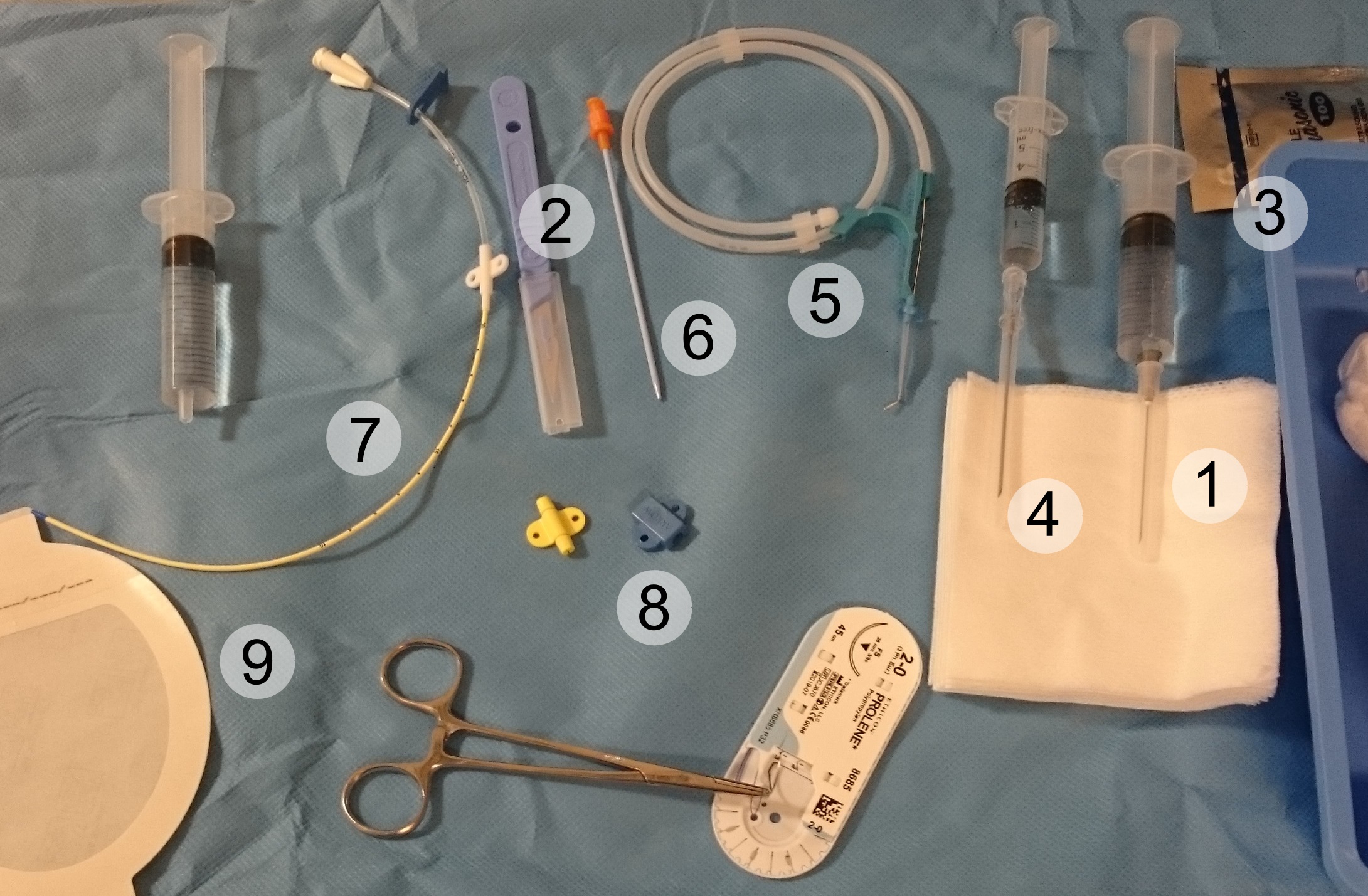
Pericardiocentesis (PCC), also called pericardial tap, is a medical procedure where fluid is aspirated from the pericardium (the sac enveloping the heart). Anatomy and Physiology The pericardium is a fibrous sac surrounding the heart composed of two layers: an inner visceral pericardium and an outer parietal pericardium. The area between these two layers is known as the pericardial space and normally contains 15 to 50 mL of serous fluid. This fluid protects the heart by serving as a shock absorber and provides lubrication to the heart during contraction. The elastic nature of the pericardium allows it to accommodate a small amount of extra fluid, roughly 80 to 120 mL, in the acute setting. However, once a critical volume is reached, even small amounts of extra fluid can rapidly increase pressure within the pericardium. This pressure can significantly hinder the ability of the heart to contract, leading to cardiac tamponade. If accumulation of fluid is slow and occurs over w ... [...More Info...] [...Related Items...] OR: [Wikipedia] [Google] [Baidu] |
Acad Emerg Med
''Academic Emergency Medicine'' is a monthly peer reviewed medical journal published by Wiley on behalf of the Society for Academic Emergency Medicine. The editor in chief is Jeffrey A. Kline, MD. Coverage includes basic science, clinical research, education information, and clinical practice related to emergency medicine. Abstracting and indexing This journal is indexed by the following services: NLM ID 9418450 * Current Contents/ Clinical Medicine * Journal Citation Reports/Science Edition * Research Alert (Thomson Reuters) * Science Citation Index * Abstracts in Anthropology * Embase * MEDLINE/Index Medicus According to the ''Journal Citation Reports'', the journal has a 2020 impact factor The impact factor (IF) or journal impact factor (JIF) of an academic journal is a scientometric index calculated by Clarivate that reflects the yearly mean number of citations of articles published in the last two years in a given journal, as ... of 3.451. External links * ... [...More Info...] [...Related Items...] OR: [Wikipedia] [Google] [Baidu] |
Hospital Emergency Codes
Hospital emergency codes are coded messages often announced over a public address system of a hospital to alert staff to various classes of on-site emergencies. The use of codes is intended to convey essential information quickly and with minimal misunderstanding to staff while preventing stress and panic among visitors to the hospital. Such codes are sometimes posted on placards throughout the hospital or are printed on employee identification badges for ready reference. Hospital emergency codes have varied widely by location, even between hospitals in the same community. Confusion over these codes has led to the proposal for and sometimes adoption of standardized codes. In many American, Canadian, New Zealand and Australian hospitals, for example "code blue" indicates a patient has entered cardiac arrest, while "code red" indicates that a fire has broken out somewhere in the hospital facility. In order for a code call to be useful in activating the response of specific hospital ... [...More Info...] [...Related Items...] OR: [Wikipedia] [Google] [Baidu] |
Pulmonary Embolus
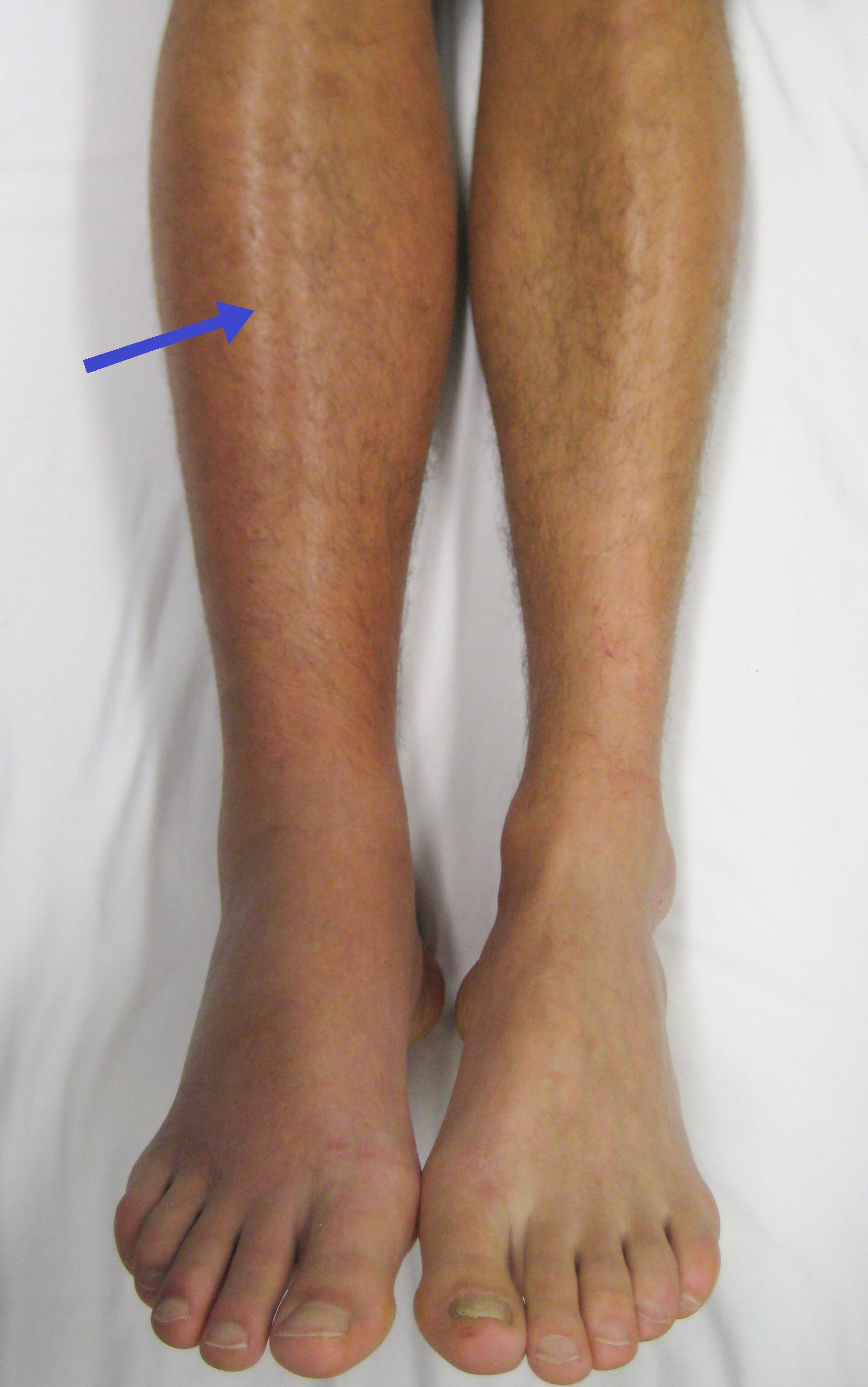
Pulmonary embolism (PE) is a blockage of an artery in the lungs by a substance that has moved from elsewhere in the body through the bloodstream (embolism). Symptoms of a PE may include shortness of breath, chest pain particularly upon breathing in, and coughing up blood. Symptoms of a blood clot in the leg may also be present, such as a red, warm, swollen, and painful leg. Signs of a PE include low blood oxygen levels, rapid breathing, rapid heart rate, and sometimes a mild fever. Severe cases can lead to passing out, abnormally low blood pressure, obstructive shock, and sudden death. PE usually results from a blood clot in the leg that travels to the lung. The risk of blood clots is increased by advanced age, cancer, prolonged bed rest and immobilization, smoking, stroke, long-haul travel over 4 hours, certain genetic conditions, estrogen-based medication, pregnancy, obesity, trauma or bone fracture, and after some types of surgery. A small proportion of cases ... [...More Info...] [...Related Items...] OR: [Wikipedia] [Google] [Baidu] |
Pericardial Effusion
A pericardial effusion is an abnormal accumulation of fluid in the pericardial cavity. The pericardium is a two-part membrane surrounding the heart: the outer fibrous connective membrane and an inner two-layered serous membrane. The two layers of the serous membrane enclose the pericardial cavity (the potential space) between them.Phelan, D., Collier, P., Grimm, R. Pericardial Disease'. Cleveland Clinic. July 2015. Retrieved Nov 2020. This pericardial space contains a small amount of pericardial fluid. The fluid is normally 15-50 mL in volume. The pericardium, specifically the pericardial fluid provides lubrication, maintains the anatomic position of the heart in the chest, and also serves as a barrier to protect the heart from infection and inflammation in adjacent tissues and organs.Vogiatzidis, Konstantinos et al.Physiology of pericardial fluid production and drainage" ''Frontiers in physiology'' vol. 6 62. 18 Mar. 2015, doi:10.3389/fphys.2015.00062 By definition, a pericardial e ... [...More Info...] [...Related Items...] OR: [Wikipedia] [Google] [Baidu] |


