|
Pneumoretroperitoneum
Pneumoretroperitoneum is the presence of air in the retroperitoneum. It is always a pathological condition and can be caused by a perforation of a retroperitoneal hollow organ such as the duodenum, colon or rectum. Pneumoretroperitoneum can best be identified by CT scan.{{cn, date=May 2022 See also * Pneumoperitoneum Pneumoperitoneum is pneumatosis (abnormal presence of air or other gas) in the peritoneal cavity, a potential space within the abdominal cavity. The most common cause is a perforated abdominal organ, generally from a perforated peptic ulcer, althou ... References Peritoneum disorders ... [...More Info...] [...Related Items...] OR: [Wikipedia] [Google] [Baidu] |
Pneumoperitoneum
Pneumoperitoneum is pneumatosis (abnormal presence of air or other gas) in the peritoneal cavity, a potential space within the abdominal cavity. The most common cause is a perforated abdominal organ, generally from a perforated peptic ulcer, although any part of the bowel may perforate from a benign ulcer, tumor or abdominal trauma. A perforated appendix seldom causes a pneumoperitoneum. Spontaneous pneumoperitoneum is a rare case that is not caused by an abdominal organ rupture. This is also called an idiopathic spontaneous pneumoperitoneum when the cause is not known. In the mid-twentieth century, an "artificial" pneumoperitoneum was sometimes intentionally administered as a treatment for a hiatal hernia. This was achieved by insufflating the abdomen with carbon dioxide. The practice is currently used by surgical teams in order to aid in performing laparoscopic surgery. Causes * Perforated duodenal ulcer – The most common cause of rupture in the abdomen. Especially of the ... [...More Info...] [...Related Items...] OR: [Wikipedia] [Google] [Baidu] |
Retroperitoneum
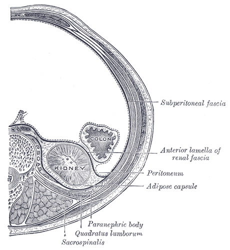
The retroperitoneal space (retroperitoneum) is the anatomical space (sometimes a potential space) behind (''retro'') the peritoneum. It has no specific delineating anatomical structures. Organs are retroperitoneal if they have peritoneum on their anterior side only. Structures that are not suspended by mesentery in the abdominal cavity and that lie between the parietal peritoneum and abdominal wall are classified as retroperitoneal. This is different from organs that are not retroperitoneal, which have peritoneum on their posterior side and are suspended by mesentery in the abdominal cavity. The retroperitoneum can be further subdivided into the following: *Perirenal (or perinephric) space *Anterior pararenal (or paranephric) space *Posterior pararenal (or paranephric) space Retroperitoneal structures Structures that lie behind the peritoneum are termed "retroperitoneal". Organs that were once suspended within the abdominal cavity by mesentery but migrated posterior to the p ... [...More Info...] [...Related Items...] OR: [Wikipedia] [Google] [Baidu] |
Pathological
Pathology is the study of the causal, causes and effects of disease or injury. The word ''pathology'' also refers to the study of disease in general, incorporating a wide range of biology research fields and medical practices. However, when used in the context of modern medical treatment, the term is often used in a narrower fashion to refer to processes and tests that fall within the contemporary medical field of "general pathology", an area which includes a number of distinct but inter-related medical specialties that diagnose disease, mostly through analysis of tissue (biology), tissue, human cell, cell, and body fluid samples. Idiomatically, "a pathology" may also refer to the predicted or actual progression of particular diseases (as in the statement "the many different forms of cancer have diverse pathologies", in which case a more proper choice of word would be "Pathophysiology, pathophysiologies"), and the affix ''pathy'' is sometimes used to indicate a state of disease ... [...More Info...] [...Related Items...] OR: [Wikipedia] [Google] [Baidu] |
Perforation
A perforation is a small hole in a thin material or web. There is usually more than one perforation in an organized fashion, where all of the holes collectively are called a ''perforation''. The process of creating perforations is called perforating, which involves puncturing the workpiece with a tool. Perforations are usually used to allow easy separation of two sections of the material, such as allowing paper to be torn easily along the line. Packaging with perforations in paperboard or plastic film is easy for consumers to open. Other purposes include filtrating fluids, sound deadening, allowing light or fluids to pass through, and to create an aesthetic design. Various applications include plastic films to allow the packages to breathe, medical films, micro perforated plate and sound and vapor barriers. Processes Pins and needles Rotary pinned perforation rollers are precision tools that can be used to perforate a wide variety of materials. The pins or needles can be ... [...More Info...] [...Related Items...] OR: [Wikipedia] [Google] [Baidu] |
Hollow Organ
In biology, an organ is a collection of tissues joined in a structural unit to serve a common function. In the hierarchy of life, an organ lies between tissue and an organ system. Tissues are formed from same type cells to act together in a function. Tissues of different types combine to form an organ which has a specific function. The intestinal wall for example is formed by epithelial tissue and smooth muscle tissue. Two or more organs working together in the execution of a specific body function form an organ system, also called a biological system or body system. An organ's tissues can be broadly categorized as parenchyma, the functional tissue, and stroma, the structural tissue with supportive, connective, or ancillary functions. For example, the gland's tissue that makes the hormones is the parenchyma, whereas the stroma includes the nerves that innervate the parenchyma, the blood vessels that oxygenate and nourish it and carry away its metabolic wastes, and the co ... [...More Info...] [...Related Items...] OR: [Wikipedia] [Google] [Baidu] |
Duodenum
The duodenum is the first section of the small intestine in most higher vertebrates, including mammals, reptiles, and birds. In fish, the divisions of the small intestine are not as clear, and the terms anterior intestine or proximal intestine may be used instead of duodenum. In mammals the duodenum may be the principal site for iron absorption. The duodenum precedes the jejunum and ileum and is the shortest part of the small intestine. In humans, the duodenum is a hollow jointed tube about 25–38 cm (10–15 inches) long connecting the stomach to the middle part of the small intestine. It begins with the duodenal bulb and ends at the suspensory muscle of duodenum. Duodenum can be divided into four parts: the first (superior), the second (descending), the third (horizontal) and the fourth (ascending) parts. Structure The duodenum is a C-shaped structure lying adjacent to the stomach. It is divided anatomically into four sections. The first part of the duodenum lies ... [...More Info...] [...Related Items...] OR: [Wikipedia] [Google] [Baidu] |
Large Intestine
The large intestine, also known as the large bowel, is the last part of the gastrointestinal tract and of the digestive system in tetrapods. Water is absorbed here and the remaining waste material is stored in the rectum as feces before being removed by defecation. The colon is the longest portion of the large intestine, and the terms are often used interchangeably but most sources define the large intestine as the combination of the cecum, colon, rectum, and anal canal. Some other sources exclude the anal canal. In humans, the large intestine begins in the right iliac region of the pelvis, just at or below the waist, where it is joined to the end of the small intestine at the cecum, via the ileocecal valve. It then continues as the colon ascending the abdomen, across the width of the abdominal cavity as the transverse colon, and then descending to the rectum and its endpoint at the anal canal. Overall, in humans, the large intestine is about long, which is about one-fi ... [...More Info...] [...Related Items...] OR: [Wikipedia] [Google] [Baidu] |
Rectum
The rectum is the final straight portion of the large intestine in humans and some other mammals, and the Gastrointestinal tract, gut in others. The adult human rectum is about long, and begins at the rectosigmoid junction (the end of the sigmoid colon) at the level of the third sacral vertebra or the sacral promontory depending upon what definition is used. Its diameter is similar to that of the sigmoid colon at its commencement, but it is dilated near its termination, forming the rectal ampulla. It terminates at the level of the anorectal ring (the level of the puborectalis sling) or the dentate line, again depending upon which definition is used. In humans, the rectum is followed by the anal canal which is about long, before the gastrointestinal tract terminates at the anal verge. The word rectum comes from the Latin ''Wikt:rectum, rectum Wikt:intestinum, intestinum'', meaning ''straight intestine''. Structure The rectum is a part of the lower gastrointestinal tract ... [...More Info...] [...Related Items...] OR: [Wikipedia] [Google] [Baidu] |
CT Scan
A computed tomography scan (CT scan; formerly called computed axial tomography scan or CAT scan) is a medical imaging technique used to obtain detailed internal images of the body. The personnel that perform CT scans are called radiographers or radiology technologists. CT scanners use a rotating X-ray tube and a row of detectors placed in a gantry (medical), gantry to measure X-ray Attenuation#Radiography, attenuations by different tissues inside the body. The multiple X-ray measurements taken from different angles are then processed on a computer using tomographic reconstruction algorithms to produce Tomography, tomographic (cross-sectional) images (virtual "slices") of a body. CT scans can be used in patients with metallic implants or pacemakers, for whom magnetic resonance imaging (MRI) is Contraindication, contraindicated. Since its development in the 1970s, CT scanning has proven to be a versatile imaging technique. While CT is most prominently used in medical diagnosis, ... [...More Info...] [...Related Items...] OR: [Wikipedia] [Google] [Baidu] |

.jpg)


