|
Pneumococcal Pneumonia
Pneumococcal pneumonia is a type of bacterial pneumonia that is caused by Streptococcus pneumoniae (pneumococcus). It is the most common bacterial pneumonia found in adults, the most common type of community-acquired pneumonia, and one of the common types of pneumococcal infection. The estimated number of Americans with pneumococcal pneumonia is 900,000 annually, with almost 400,000 cases hospitalized and fatalities accounting for 5-7% of these cases. Symptoms The symptoms of pneumococcal pneumonia can occur suddenly, presenting as a severe chill, followed by a severe fever, cough, shortness of breath, rapid breathing, and chest pains. Other symptoms like nausea, vomiting, headache, fatigue, and muscle aches could also accompany initial symptoms. The coughing can occasionally produce rusty or blood-streaked sputum. In 25% of cases, a parapneumonic effusion may occur. Chest X-rays will typically show lobar consolidation or patchy infiltrates. Treatment In most cases, once pne ... [...More Info...] [...Related Items...] OR: [Wikipedia] [Google] [Baidu] |
Bacterial Pneumonia
Bacterial pneumonia is a type of pneumonia caused by bacterial infection. Types Gram-positive '' Streptococcus pneumoniae'' () is the most common bacterial cause of pneumonia in all age groups except newborn infants. ''Streptococcus pneumoniae'' is a Gram-positive bacterium that often lives in the throat of people who do not have pneumonia. Other important Gram-positive causes of pneumonia are ''Staphylococcus aureus'' () and ''Bacillus anthracis''. Gram-negative Gram-negative bacteria are seen less frequently: '' Haemophilus influenzae'' (), '' Klebsiella pneumoniae'' (), '' Escherichia coli'' (), '' Pseudomonas aeruginosa'' (), '' Bordetella pertussis'', and '' Moraxella catarrhalis'' are the most common. These bacteria often live in the gut and enter the lungs when contents of the gut (such as vomit or faeces) are inhaled. Pneumonia caused by '' Yersinia pestis'' is usually called pneumonic plague. Atypical Atypical bacteria causing pneumonia are '' Coxiella burne ... [...More Info...] [...Related Items...] OR: [Wikipedia] [Google] [Baidu] |
Fragment Crystallizable Region
The fragment crystallizable region (Fc region) is the tail region of an antibody that interacts with cell surface receptors called Fc receptors and some proteins of the complement system. This property allows antibodies to activate the immune system. In IgG, IgA and IgD antibody isotypes, the Fc region is composed of two identical protein fragments, derived from the second and third constant domains of the antibody's two heavy chains; IgM and IgE Fc regions contain three heavy chain constant domains (CH domains 2–4) in each polypeptide chain. The Fc regions of IgGs bear a highly conserved N-glycosylation site. Glycosylation of the Fc fragment is essential for Fc receptor-mediated activity. The N-glycans attached to this site are predominantly core- fucosylated diantennary structures of the complex type. In addition, small amounts of these N-glycans also bear bisecting GlcNAc and α-2,6 linked sialic acid residues. The other part of an antibody, called the Fab regio ... [...More Info...] [...Related Items...] OR: [Wikipedia] [Google] [Baidu] |
Neuraminidase
Exo-α-sialidase (EC 3.2.1.18, sialidase, neuraminidase; systematic name acetylneuraminyl hydrolase) is a glycoside hydrolase that cleaves the glycosidic linkages of neuraminic acids: : Hydrolysis of α-(2→3)-, α-(2→6)-, α-(2→8)- glycosidic linkages of terminal sialic acid residues in oligosaccharides, glycoproteins, glycolipids, colominic acid and synthetic substrates Neuraminidase enzymes are a large family, found in a range of organisms. The best-known neuraminidase is the viral neuraminidase, a drug target for the prevention of the spread of influenza infection. The viral neuraminidases are frequently used as antigenic determinants found on the surface of the influenza virus. Some variants of the influenza neuraminidase confer more virulence to the virus than others. Other homologues are found in mammalian cells, which have a range of functions. At least four mammalian sialidase homologues have been described in the human genome (see NEU1, NEU2, NEU3, NEU4) ... [...More Info...] [...Related Items...] OR: [Wikipedia] [Google] [Baidu] |
Mucociliary Escalator
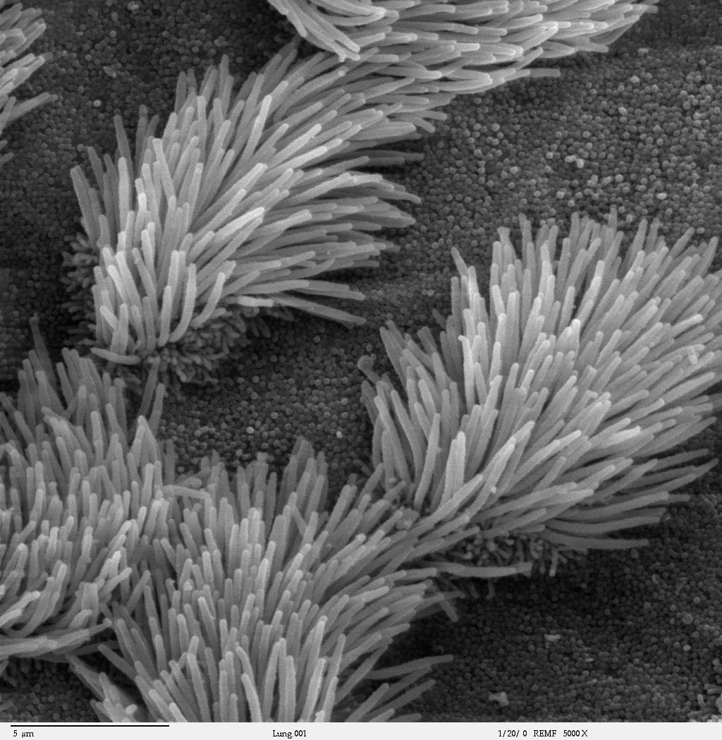
Mucociliary clearance (MCC), mucociliary transport, or the mucociliary escalator, describes the self-clearing mechanism of the airways in the respiratory system. It is one of the two protective processes for the lungs in removing inhaled particles including pathogens before they can reach the delicate tissue of the lungs. The other clearance mechanism is provided by the cough reflex. Mucociliary clearance has a major role in pulmonary hygiene. MCC effectiveness relies on the correct properties of the airway surface liquid produced, both of the periciliary sol layer and the overlying mucus gel layer, and of the number and quality of the cilia present in the lining of the airways. An important factor is the rate of mucin secretion. The ion channels CFTR and ENaC work together to maintain the necessary hydration of the airway surface liquid. Any disturbance in the closely regulated functioning of the cilia can cause a disease. Disturbances in the structural formation of the ... [...More Info...] [...Related Items...] OR: [Wikipedia] [Google] [Baidu] |
Immunoglobulin M
Immunoglobulin M (IgM) is one of several isotypes of antibody (also known as immunoglobulin) that are produced by vertebrates. IgM is the largest antibody, and it is the first antibody to appear in the response to initial exposure to an antigen. In humans and other mammals that have been studied, plasmablasts residing in the spleen are the main source for specific IgM production. History In 1937, an antibody was observed in horses hyper-immunized with pneumococcus polysaccharide that was much larger in size than the typical rabbit γ-globulin, with a molecular weight of 990,000 daltons. In accordance with its larger size, the new antibody was originally referred to as γ-macroglobulin, and subsequently termed as IgM—M for “macro”. The V domains of normal immunoglobulin are highly heterogeneous, reflecting their role in protecting against the great variety of infectious microbes, and this heterogeneity impeded detailed structural analysis of IgM. Two sources of homoge ... [...More Info...] [...Related Items...] OR: [Wikipedia] [Google] [Baidu] |
Polymeric Immunoglobulin Receptor
Polymeric immunoglobulin receptor (pIgR) is a Protein, transmembrane protein that in humans is encoded by the ''PIGR'' gene. It is an Fc receptor which facilitates the transcytosis of the soluble polymeric isoforms of immunoglobulin A and immunoglobulin M (pIg) and immune complexes. pIgRs are mainly located on the epithelial lining of mucosal surfaces of the gastrointestinal tract. The composition of the receptor is complex, including 6 immunoglobulin-like domains, a transmembrane region, and an intracellular domain. pIgR expression is under the strong regulation of cytokines, hormones, and pathogenic stimuli. Structure pIgR is produced among others by Intestinal epithelium, intestinal epithelial cells (IECs) and bronchial epithelial cells. pIgR belongs to the family of type I transmembrane proteins. The extracellular portion of the protein contains 6 domains: 5 evolutionary conserved immunoglobulin-like domains, and 1 non-homologous domain, which is involved in Proteolysis, prot ... [...More Info...] [...Related Items...] OR: [Wikipedia] [Google] [Baidu] |
Phosphorylcholine
:''Phosphorylcholine refers to the functional group derived from phosphocholine. Also not to be confused with phosphatidylcholine.'' Phosphorylcholine (abbreviated ChoP) is the hydrophilic polar head group of some phospholipids, which is composed of a negatively charged phosphate bonded to a small, positively charged choline group. Phosphorylcholine is part of the platelet-activating factor; the phospholipid phosphatidylcholine and sphingomyelin, the only phospholipid of the membrane that is not built with a glycerol backbone. Treatment of cell membranes, like those of RBCs, by certain enzymes, like some phospholipase A2 renders the phosphorylcholine moiety exposed to the external aqueous phase, and thus accessible for recognition by the immune system. Antibodies against phosphorylcholine are naturally occurring autoantibodies that are created by CD5+/ B-1 B cells and are referred to as non-pathogenic autoantibodies. Thrombus-resistant stents In interventional cardiology, phosp ... [...More Info...] [...Related Items...] OR: [Wikipedia] [Google] [Baidu] |
Oligosaccharide
An oligosaccharide (/ˌɑlɪgoʊˈsækəˌɹaɪd/; from the Greek ὀλίγος ''olígos'', "a few", and σάκχαρ ''sácchar'', "sugar") is a saccharide polymer containing a small number (typically two to ten) of monosaccharides (simple sugars). Oligosaccharides can have many functions including cell recognition and cell adhesion. They are normally present as glycans: oligosaccharide chains are linked to lipids or to compatible amino acid side chains in proteins, by ''N''- or ''O''-glycosidic bonds. ''N''-Linked oligosaccharides are always pentasaccharides attached to asparagine via a beta linkage to the amine nitrogen of the side chain.. Alternately, ''O''-linked oligosaccharides are generally attached to threonine or serine on the alcohol group of the side chain. Not all natural oligosaccharides occur as components of glycoproteins or glycolipids. Some, such as the raffinose series, occur as storage or transport carbohydrates in plants. Others, such as maltodextrin ... [...More Info...] [...Related Items...] OR: [Wikipedia] [Google] [Baidu] |
Glycoprotein
Glycoproteins are proteins which contain oligosaccharide chains covalently attached to amino acid side-chains. The carbohydrate is attached to the protein in a cotranslational or posttranslational modification. This process is known as glycosylation. Secreted extracellular proteins are often glycosylated. In proteins that have segments extending extracellularly, the extracellular segments are also often glycosylated. Glycoproteins are also often important integral membrane proteins, where they play a role in cell–cell interactions. It is important to distinguish endoplasmic reticulum-based glycosylation of the secretory system from reversible cytosolic-nuclear glycosylation. Glycoproteins of the cytosol and nucleus can be modified through the reversible addition of a single GlcNAc residue that is considered reciprocal to phosphorylation and the functions of these are likely to be an additional regulatory mechanism that controls phosphorylation-based signalling. In co ... [...More Info...] [...Related Items...] OR: [Wikipedia] [Google] [Baidu] |
Glycolipid
Glycolipids are lipids with a carbohydrate attached by a glycosidic (covalent) bond. Their role is to maintain the stability of the cell membrane and to facilitate cellular recognition, which is crucial to the immune response and in the connections that allow cells to connect to one another to form tissues. Glycolipids are found on the surface of all eukaryotic cell membranes, where they extend from the phospholipid bilayer into the extracellular environment. Structure The essential feature of a glycolipid is the presence of a monosaccharide or oligosaccharide bound to a lipid moiety. The most common lipids in cellular membranes are glycerolipids and sphingolipids, which have glycerol or a sphingosine backbones, respectively. Fatty acids are connected to this backbone, so that the lipid as a whole has a polar head and a non-polar tail. The lipid bilayer of the cell membrane consists of two layers of lipids, with the inner and outer surfaces of the membrane made up of the ... [...More Info...] [...Related Items...] OR: [Wikipedia] [Google] [Baidu] |
N-Acetylneuraminic Acid
''N''-Acetylneuraminic acid (Neu5Ac or NANA) is the predominant sialic acid found in human cells, and many mammalian cells. Other forms, such as N-Glycolylneuraminic acid, may also occur in cells. This residue is negatively charged at physiological pH and is found in complex glycans on mucins and glycoproteins found at the cell membrane. Neu5Ac residues are also found in glycolipids, known as gangliosides, a crucial component of neuronal membranes found in the brain. Along with involvement in preventing infections (mucus associated with mucous membranes—mouth, nose, GI, respiratory tract), Neu5Ac acts as a receptor for influenza viruses, allowing attachment to mucous cells via hemagglutinin (an early step in acquiring influenzavirus infection). In the biology of bacterial pathogens Neu5Ac is also important in the biology of a number of pathogenic and symbiotic bacteria as it can used either as a nutrient, providing both carbon and nitrogen to the bacteria, or in some pathogen ... [...More Info...] [...Related Items...] OR: [Wikipedia] [Google] [Baidu] |
Neuraminidase
Exo-α-sialidase (EC 3.2.1.18, sialidase, neuraminidase; systematic name acetylneuraminyl hydrolase) is a glycoside hydrolase that cleaves the glycosidic linkages of neuraminic acids: : Hydrolysis of α-(2→3)-, α-(2→6)-, α-(2→8)- glycosidic linkages of terminal sialic acid residues in oligosaccharides, glycoproteins, glycolipids, colominic acid and synthetic substrates Neuraminidase enzymes are a large family, found in a range of organisms. The best-known neuraminidase is the viral neuraminidase, a drug target for the prevention of the spread of influenza infection. The viral neuraminidases are frequently used as antigenic determinants found on the surface of the influenza virus. Some variants of the influenza neuraminidase confer more virulence to the virus than others. Other homologues are found in mammalian cells, which have a range of functions. At least four mammalian sialidase homologues have been described in the human genome (see NEU1, NEU2, NEU3, NEU4) ... [...More Info...] [...Related Items...] OR: [Wikipedia] [Google] [Baidu] |


