|
Pioneer Axon
Pioneer axon is the classification given to axons that are the first to grow in a particular region. They originate from pioneer neurons, and have the main function of laying down the initial growing path that subsequent growing axons, dubbed follower axons, from other neurons will eventually follow. Several theories relating to the structure and function of pioneer axons are currently being explored. The first theory is that pioneer axons are specialized structures, and that they play a crucial role in guiding follower axons. The second is that pioneer axons are no different from follower axons, and that they play no role in guiding follower axons. Anatomically, there are no differences between pioneer and follower axons, although there are morphological differences. The mechanisms of pioneer axons and their role in axon guidance is currently being explored. In addition, many studies are being conducted in model organisms, such grasshoppers, zebrafish, and fruit flies to study t ... [...More Info...] [...Related Items...] OR: [Wikipedia] [Google] [Baidu] |
Axons
An axon (from Greek ἄξων ''áxōn'', axis), or nerve fiber (or nerve fibre: see American and British English spelling differences#-re, -er, spelling differences), is a long, slender projection of a nerve cell, or neuron, in vertebrates, that typically conducts electrical impulses known as action potentials away from the Soma (biology), nerve cell body. The function of the axon is to transmit information to different neurons, muscles, and glands. In certain sensory neurons (pseudounipolar neurons), such as those for touch and warmth, the axons are called afferent nerve fibers and the electrical impulse travels along these from the peripheral nervous system, periphery to the cell body and from the cell body to the spinal cord along another branch of the same axon. Axon dysfunction can be the cause of many inherited and acquired neurological disorders that affect both the Peripheral nervous system, peripheral and Central nervous system, central neurons. Nerve fibers are Axon#Cl ... [...More Info...] [...Related Items...] OR: [Wikipedia] [Google] [Baidu] |
Notch Signaling Pathway
The Notch signaling pathway is a highly Conserved sequence, conserved cell signaling system present in most animals. Mammals possess four different Notch proteins, notch receptors, referred to as NOTCH1, NOTCH2, Notch 3, NOTCH3, and NOTCH4. The notch receptor is a single-pass Cell surface receptor, transmembrane receptor protein. It is a hetero-oligomer composed of a large extracellular portion, which associates in a calcium-dependent, non-covalent interaction with a smaller piece of the notch protein composed of a short extracellular region, a single transmembrane-pass, and a small intracellular region. Notch signaling promotes proliferative signaling during neurogenesis, and its activity is inhibited by NUMB (gene), Numb to promote neural differentiation. It plays a major role in the regulation of embryonic development. Notch signaling is dysregulated in many cancers, and faulty notch signaling is implicated in many diseases, including T-cell acute lymphoblastic leukemia (Pre ... [...More Info...] [...Related Items...] OR: [Wikipedia] [Google] [Baidu] |
Posterior Commissure
The posterior commissure (also known as the epithalamic commissure) is a rounded band of white fibers crossing the middle line on the dorsal aspect of the rostral end of the cerebral aqueduct. It is important in the bilateral pupillary light reflex. Its fibers acquire their medullary sheaths early, but their connections have not been definitively determined. Most of them have their origin in a nucleus, the ''nucleus of the posterior commissure'' (nucleus of Darkschewitsch), which lies in the periaqueductal grey at rostral end of the cerebral aqueduct, in front of the oculomotor nucleus. Some are thought to be derived from the posterior part of the thalamus and from the superior colliculus, whereas others are believed to be continued downward into the medial longitudinal fasciculus. For the pupillary light reflex, the olivary pretectal nucleus In neuroanatomy, the pretectal area, or pretectum, is a midbrain structure composed of seven nuclei and comprises part of the subcortical ... [...More Info...] [...Related Items...] OR: [Wikipedia] [Google] [Baidu] |
Axonal Pathfinding And Fasciculation Behaviour In The Embryonic Ventral Nerve Cord Of Drosophila
An axon (from Greek ἄξων ''áxōn'', axis), or nerve fiber (or nerve fibre: see spelling differences), is a long, slender projection of a nerve cell, or neuron, in vertebrates, that typically conducts electrical impulses known as action potentials away from the nerve cell body. The function of the axon is to transmit information to different neurons, muscles, and glands. In certain sensory neurons (pseudounipolar neurons), such as those for touch and warmth, the axons are called afferent nerve fibers and the electrical impulse travels along these from the periphery to the cell body and from the cell body to the spinal cord along another branch of the same axon. Axon dysfunction can be the cause of many inherited and acquired neurological disorders that affect both the peripheral and central neurons. Nerve fibers are classed into three typesgroup A nerve fibers, group B nerve fibers, and group C nerve fibers. Groups A and B are myelinated, and group C are unmyelinated. The ... [...More Info...] [...Related Items...] OR: [Wikipedia] [Google] [Baidu] |
Peripheral Nervous System
The peripheral nervous system (PNS) is one of two components that make up the nervous system of bilateral animals, with the other part being the central nervous system (CNS). The PNS consists of nerves and ganglia, which lie outside the brain and the spinal cord. The main function of the PNS is to connect the CNS to the limbs and organs, essentially serving as a relay between the brain and spinal cord and the rest of the body. Unlike the CNS, the PNS is not protected by the vertebral column and skull, or by the blood–brain barrier, which leaves it exposed to toxins. The peripheral nervous system can be divided into the somatic nervous system and the autonomic nervous system. In the somatic nervous system, the cranial nerves are part of the PNS with the exception of the optic nerve (cranial nerve II), along with the retina. The second cranial nerve is not a true peripheral nerve but a tract of the diencephalon. Cranial nerve ganglia, as with all ganglia, are part of the P ... [...More Info...] [...Related Items...] OR: [Wikipedia] [Google] [Baidu] |
Central Nervous System
The central nervous system (CNS) is the part of the nervous system consisting primarily of the brain and spinal cord. The CNS is so named because the brain integrates the received information and coordinates and influences the activity of all parts of the bodies of bilaterally symmetric and triploblastic animals—that is, all multicellular animals except sponges and diploblasts. It is a structure composed of nervous tissue positioned along the rostral (nose end) to caudal (tail end) axis of the body and may have an enlarged section at the rostral end which is a brain. Only arthropods, cephalopods and vertebrates have a true brain (precursor structures exist in onychophorans, gastropods and lancelets). The rest of this article exclusively discusses the vertebrate central nervous system, which is radically distinct from all other animals. Overview In vertebrates, the brain and spinal cord are both enclosed in the meninges. The meninges provide a barrier to chemicals dissolv ... [...More Info...] [...Related Items...] OR: [Wikipedia] [Google] [Baidu] |
Lamellipodium
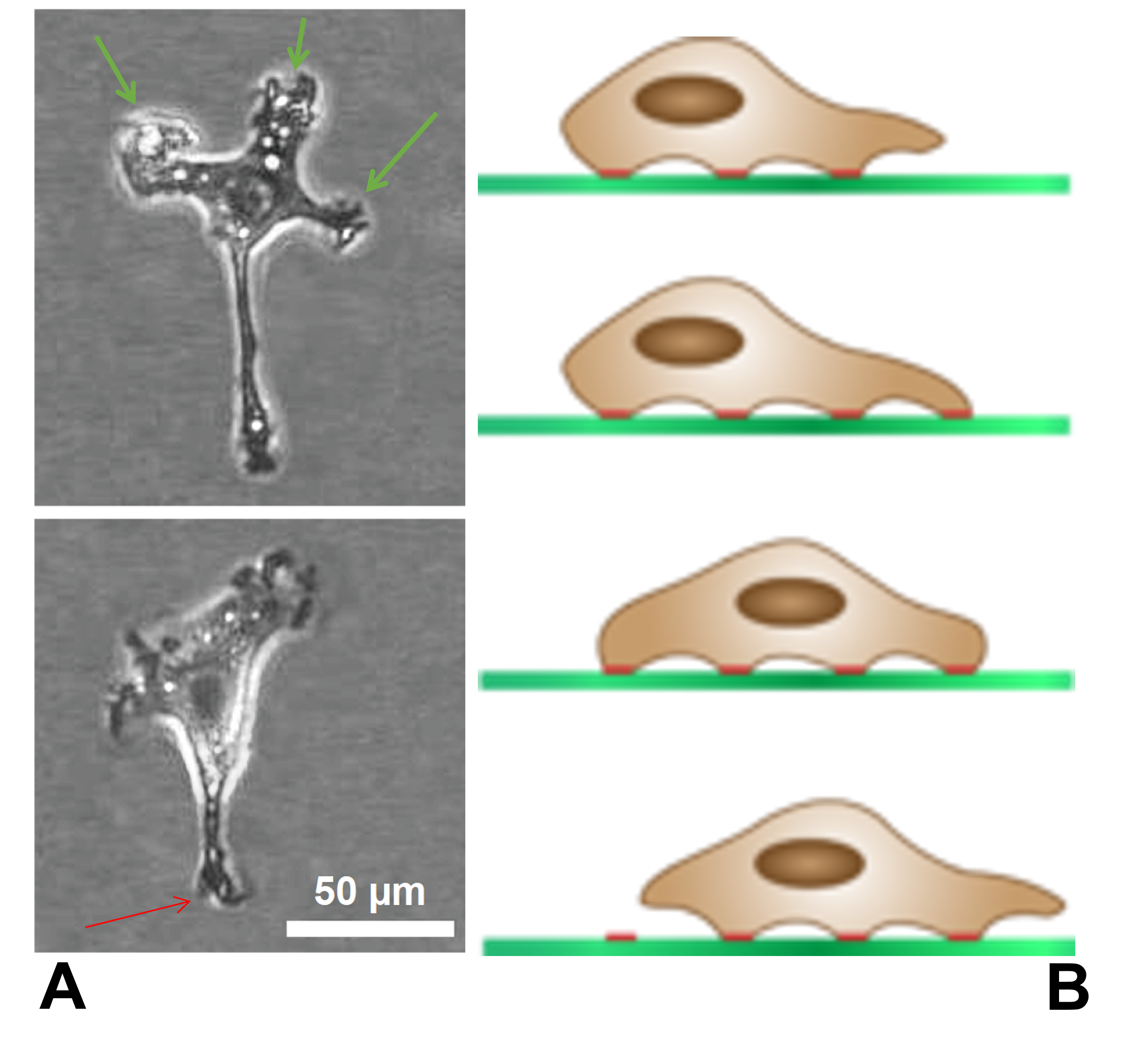
The lamellipodium (plural lamellipodia) (from Latin ''lamella'', related to ', "thin sheet", and the Greek radical ''pod-'', "foot") is a cytoskeletal protein actin projection on the leading edge of the cell. It contains a quasi-two-dimensional actin mesh; the whole structure propels the cell across a substrate. Within the lamellipodia are ribs of actin called microspikes, which, when they spread beyond the lamellipodium frontier, are called filopodia. The lamellipodium is born of actin nucleation in the plasma membrane of the cell and is the primary area of actin incorporation or microfilament formation of the cell. Description Lamellipodia are found primarily in all mobile cells, such as the keratinocytes of fish and frogs, which are involved in the quick repair of wounds. The lamellipodia of these keratinocytes allow them to move at speeds of 10–20 μm / min over epithelial surfaces. When separated from the main part of a cell, a lamellipodium can still cra ... [...More Info...] [...Related Items...] OR: [Wikipedia] [Google] [Baidu] |
Growth Cones
Growth may refer to: Biology * Auxology, the study of all aspects of human physical growth * Bacterial growth * Cell growth * Growth hormone, a peptide hormone that stimulates growth * Human development (biology) * Plant growth * Secondary growth, growth that thickens woody plants Economics * Economic growth, the increase in the inflation-adjusted market value of the goods and services * Growth investing, a style of investment strategy focused on capital appreciation Mathematics * Exponential growth, also called geometric growth * Hyperbolic growth * Linear growth, refers to two distinct but related notions * Logistic growth, characterized as an S curve Social science * Developmental psychology * Erikson's stages of psychosocial development * Human development (humanity) * Personal development * Population growth Other uses * ''Growth'' (film), a 2010 American horror film * Izaugsme (''Growth''), a Latvian political party * ''Grown'' (album), by 2PM See also * Grow (disambi ... [...More Info...] [...Related Items...] OR: [Wikipedia] [Google] [Baidu] |
Ventral Nerve Cord
The ventral nerve cord is a major structure of the invertebrate central nervous system. It is the functional equivalent of the vertebrate spinal cord. The ventral nerve cord coordinates neural signaling from the brain to the body and vice versa, integrating sensory input and locomotor output. Decapitated insects can still walk, groom, and mate, illustrating that the circuitry of the ventral nerve cord is sufficient to perform complex motor programs without brain input. Structure The ventral nerve cord runs down the Anatomical terms of location, ventral ("belly", as opposed to back) plane of the organism. It contains ascending and Descending neuron, descending neurons that relay information to and from the brain, Motor neuron, motor neurons that project into the body and synapse onto muscles, Sensory neuron, sensory neurons that receive information from the body and environment, and Interneuron, interneurons that coordinate circuitry of all of these neurons. Ventral nerve cord ... [...More Info...] [...Related Items...] OR: [Wikipedia] [Google] [Baidu] |
Floor Plate
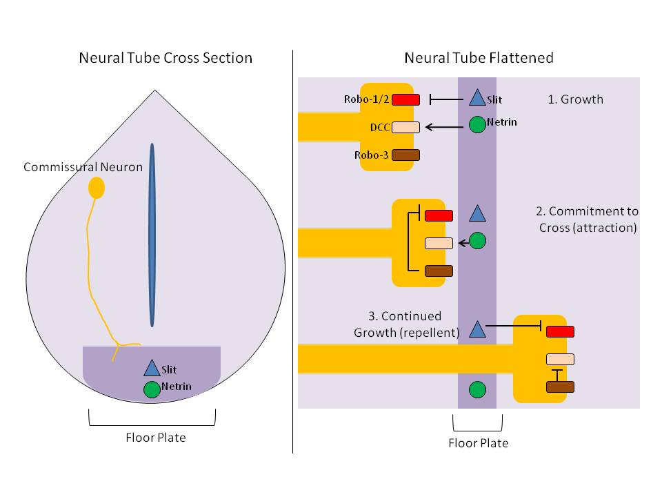
The floor plate is a structure integral to the developing nervous system of vertebrate organisms. Located on the ventral midline of the embryonic neural tube, the floor plate is a specialized glial structure that spans the anteroposterior axis from the midbrain to the tail regions. It has been shown that the floor plate is conserved among vertebrates, such as zebrafish and mice, with homologous structures in invertebrates such as the fruit fly ''Drosophila'' and the nematode ''C. elegans''. Functionally, the structure serves as an organizer to ventralize tissues in the embryo as well as to guide neuronal positioning and differentiation along the dorsoventral axis of the neural tube. Induction Induction of the floor plate during embryogenesis of vertebrate embryos has been studied extensively in chick and zebrafish and occurs as a result of a complex signaling network among tissues, the details of which have yet to be fully refined. Currently there are several competing lines o ... [...More Info...] [...Related Items...] OR: [Wikipedia] [Google] [Baidu] |
Hindbrain
The hindbrain or rhombencephalon or lower brain is a developmental categorization of portions of the central nervous system in vertebrates. It includes the medulla, pons, and cerebellum. Together they support vital bodily processes. Metencephalon Rhombomeres Rh3-Rh1 form the metencephalon. The metencephalon is composed of the pons and the cerebellum; it contains: * a portion of the fourth (IV) ventricle, * the trigeminal nerve (CN V), * abducens nerve (CN VI), * facial nerve (CN VII), * and a portion of the vestibulocochlear nerve (CN VIII). Myelencephalon Rhombomeres Rh8-Rh4 form the myelencephalon. The myelencephalon forms the medulla oblongata in the adult brain; it contains: * a portion of the fourth ventricle, * the glossopharyngeal nerve (CN IX), * vagus nerve (CN X), * accessory nerve (CN XI), * hypoglossal nerve (CN XII), * and a portion of the vestibulocochlear nerve (CN VIII). Evolution The hindbrain is homologous to a part of the arthropod brain known as the sub- ... [...More Info...] [...Related Items...] OR: [Wikipedia] [Google] [Baidu] |
Midbrain
The midbrain or mesencephalon is the forward-most portion of the brainstem and is associated with vision, hearing, motor control, sleep and wakefulness, arousal (alertness), and temperature regulation. The name comes from the Greek ''mesos'', "middle", and ''enkephalos'', "brain". Structure The principal regions of the midbrain are the tectum, the cerebral aqueduct, tegmentum, and the cerebral peduncles. Rostrally the midbrain adjoins the diencephalon (thalamus, hypothalamus, etc.), while caudally it adjoins the hindbrain (pons, medulla and cerebellum). In the rostral direction, the midbrain noticeably splays laterally. Sectioning of the midbrain is usually performed axially, at one of two levels – that of the superior colliculi, or that of the inferior colliculi. One common technique for remembering the structures of the midbrain involves visualizing these cross-sections (especially at the level of the superior colliculi) as the upside-down face of a be ... [...More Info...] [...Related Items...] OR: [Wikipedia] [Google] [Baidu] |


