|
Picornain 3C
Picornain 3C () is a protease found in picornaviruses, which cleaves peptide bonds of non-terminal sequences. Picornain 3C’s endopeptidase activity is primarily responsible for the catalytic process of selectively cleaving Gln-Gly bonds in the polyprotein of poliovirus and with substitution of Glu for Gln, and Ser or Thr for Gly in other picornaviruses. Picornain 3C are cysteine proteases related by amino acid sequence to trypsin-like serine proteases. Picornain 3C is encoded by enteroviruses, rhinoviruses, aphtoviruses and cardioviruses. These genera of picoviruses cause a wide range of infections in humans and mammals. Picornavirus belongs to the family ''Picornaviridae''. Picornavirus virions are nonenveloped and the +ssRNA nonsegmented genome is encapsulated in an icosahedral protein structure made from four capsid proteins encoded by the virus. Picornavirus viral replication typically takes place in the cytoplasm of the cell. Picornavirus +ssRNA genome then gets translated ... [...More Info...] [...Related Items...] OR: [Wikipedia] [Google] [Baidu] |
Ribbon Diagram
Ribbon diagrams, also known as Richardson diagrams, are three-dimensional space, 3D schematic representations of protein structure and are one of the most common methods of protein depiction used today. The ribbon shows the overall path and organization of the protein backbone in 3D, and serves as a visual framework on which to hang details of the full atomic structure, such as the balls for the oxygen atoms bound to the active site of myoglobin in the adjacent image. Ribbon diagrams are generated by interpolating a smooth curve through the polypeptide backbone. Alpha helix, α-helices are shown as coiled ribbons or thick tubes, Beta strand, β-strands as arrows, and non-repetitive coils or loops as lines or thin tubes. The direction of the Peptide, polypeptide chain is shown locally by the arrows, and may be indicated overall by a colour ramp along the length of the ribbon. Ribbon diagrams are simple yet powerful, expressing the visual basics of a molecular structure (twist, fold ... [...More Info...] [...Related Items...] OR: [Wikipedia] [Google] [Baidu] |
RNA Polymerase
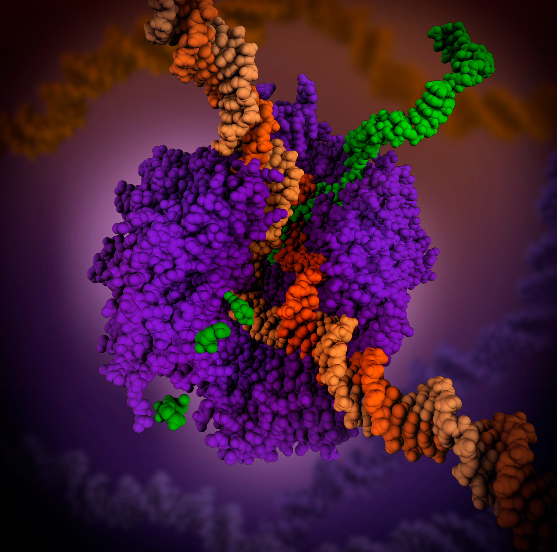
In molecular biology, RNA polymerase (abbreviated RNAP or RNApol), or more specifically DNA-directed/dependent RNA polymerase (DdRP), is an enzyme that synthesizes RNA from a DNA template. Using the enzyme helicase, RNAP locally opens the double-stranded DNA so that one strand of the exposed nucleotides can be used as a template for the synthesis of RNA, a process called transcription. A transcription factor and its associated transcription mediator complex must be attached to a DNA binding site called a promoter region before RNAP can initiate the DNA unwinding at that position. RNAP not only initiates RNA transcription, it also guides the nucleotides into position, facilitates attachment and elongation, has intrinsic proofreading and replacement capabilities, and termination recognition capability. In eukaryotes, RNAP can build chains as long as 2.4 million nucleotides. RNAP produces RNA that, functionally, is either for protein coding, i.e. messenger RNA (mRNA); or n ... [...More Info...] [...Related Items...] OR: [Wikipedia] [Google] [Baidu] |
Hepatitis A
Hepatitis A is an infectious disease of the liver caused by ''Hepatovirus A'' (HAV); it is a type of viral hepatitis. Many cases have few or no symptoms, especially in the young. The time between infection and symptoms, in those who develop them, is 2–6 weeks. When symptoms occur, they typically last 8 weeks and may include nausea, vomiting, diarrhea, jaundice, fever, and abdominal pain. Around 10–15% of people experience a recurrence of symptoms during the 6 months after the initial infection. Acute liver failure may rarely occur, with this being more common in the elderly. It is usually spread by eating food or drinking water contaminated with infected feces. Undercooked or raw shellfish are relatively common sources. It may also be spread through close contact with an infectious person. While children often do not have symptoms when infected, they are still able to infect others. After a single infection, a person is immune for the rest of his or her life. Diagnosis requir ... [...More Info...] [...Related Items...] OR: [Wikipedia] [Google] [Baidu] |
Foot And Mouth Disease Virus
''Foot-and-mouth disease virus'' (FMDV) is the pathogen that causes foot-and-mouth disease. It is a picornavirus, the prototypical member of the genus ''Aphthovirus''. The disease, which causes vesicles (blisters) in the mouth and feet of cattle, pigs, sheep, goats, and other cloven-hoofed animals is highly infectious and a major plague of animal farming. Structure and genome The virus particle (25-30 nm) has an icosahedral capsid made of protein, without envelope, containing a positive-sense (mRNA sense) single-stranded ribonucleic acid (RNA) genome. Replication When the virus comes in contact with the membrane of a host cell, it binds to a receptor site and triggers a folding-in of the membrane. Once the virus is inside the host cell, the capsid dissolves, and the RNA gets replicated, and translated into viral proteins by the cell's ribosomes using a cap-independent mechanism driven by the internal ribosome entry site element. The synthesis of viral proteins i ... [...More Info...] [...Related Items...] OR: [Wikipedia] [Google] [Baidu] |
Rhinovirus
The rhinovirus (from the grc, ῥίς, rhis "nose", , romanized: "of the nose", and the la, vīrus) is the most common viral infectious agent in humans and is the predominant cause of the common cold. Rhinovirus infection proliferates in temperatures of 33–35 °C (91–95 °F), the temperatures found in the nose. Rhinoviruses belong to the genus ''Enterovirus'' in the family ''Picornaviridae''. The three species of rhinovirus (A, B, and C) include around 160 recognized types (called serotypes) of human rhinovirus that differ according to their surface antigens. They are lytic in nature and are among the smallest viruses, with diameters of about 30 nanometers. By comparison, other viruses, such as smallpox and vaccinia, are around ten times larger at about 300 nanometers, while flu viruses are around 80–120 nm. History In 1953, when a cluster of nurses developed a mild respiratory illness, Winston Price, from the Johns Hopkins University, took nasal pas ... [...More Info...] [...Related Items...] OR: [Wikipedia] [Google] [Baidu] |
CREB
CREB-TF (CREB, cAMP response element-binding protein) is a cellular transcription factor. It binds to certain DNA sequences called cAMP response elements (CRE), thereby increasing or decreasing the transcription of the genes. CREB was first described in 1987 as a cAMP-responsive transcription factor regulating the somatostatin gene. Genes whose transcription is regulated by CREB include: '' c-fos'', BDNF, tyrosine hydroxylase, numerous neuropeptides (such as somatostatin, enkephalin, VGF, corticotropin-releasing hormone), and genes involved in the mammalian circadian clock (PER1, PER2). CREB is closely related in structure and function to CREM (cAMP response element modulator) and ATF-1 (activating transcription factor-1) proteins. CREB proteins are expressed in many animals, including humans. CREB has a well-documented role in neuronal plasticity and long-term memory formation in the brain and has been shown to be integral in the formation of spatial memory. CREB downreg ... [...More Info...] [...Related Items...] OR: [Wikipedia] [Google] [Baidu] |
Coxsackievirus
Coxsackieviruses are a few related enteroviruses that belong to the ''Picornaviridae'' family of viral envelope, nonenveloped, linear, positive-sense single-stranded RNA viruses, as well as its genus ''Enterovirus'', which also includes poliovirus and echovirus. Enteroviruses are among the most common and important human pathogens, and ordinarily its members are transmitted by the fecal–oral route. Coxsackieviruses share many characteristics with poliovirus. With control of poliovirus infections in much of the world, more attention has been focused on understanding the nonpolio enteroviruses such as coxsackievirus. Coxsackieviruses are among the leading causes of aseptic meningitis (the other usual suspects being echovirus and mumps virus). The entry of coxsackievirus into cells, especially endothelial cells, is mediated by coxsackievirus and adenovirus receptor. Groups Coxsackieviruses are divided into Coxsackie A virus, group A and Coxsackie B virus, group B viruses based ... [...More Info...] [...Related Items...] OR: [Wikipedia] [Google] [Baidu] |
EIF4E
Eukaryotic translation initiation factor 4E, also known as eIF4E, is a protein that in humans is encoded by the ''EIF4E'' gene. Structure and function Most eukaryotic cellular mRNAs are blocked at their 5'-ends with the 7-methyl-guanosine five-prime cap structure, m7GpppX (where X is any nucleotide). This structure is involved in several cellular processes including enhanced translational efficiency, splicing, mRNA stability, and RNA nuclear export. eIF4E is a eukaryotic translation initiation factor involved in directing ribosomes to the cap structure of mRNAs. It is a 24-kD polypeptide that exists as both a free form and as part of the eIF4F pre-initiation complex. Almost all cellular mRNA require eIF4E in order to be translated into protein. The eIF4E polypeptide is the rate-limiting component of the eukaryotic translation apparatus and is involved in the mRNA-ribosome binding step of eukaryotic protein synthesis. The other subunits of eIF4F are a 47-kD polypeptide, terme ... [...More Info...] [...Related Items...] OR: [Wikipedia] [Google] [Baidu] |
Internal Ribosome Entry Site
An internal ribosome entry site, abbreviated IRES, is an RNA element that allows for translation initiation in a cap-independent manner, as part of the greater process of protein synthesis. In eukaryotic translation, initiation typically occurs at the 5' end of mRNA molecules, since 5' cap recognition is required for the assembly of the initiation complex. The location for IRES elements is often in the 5'UTR, but can also occur elsewhere in mRNAs. History IRES sequences were first discovered in 1988 in the poliovirus (PV) and encephalomyocarditis virus (EMCV) RNA genomes in the labs of Nahum Sonenberg and Eckard Wimmer, respectively. They are described as distinct regions of RNA molecules that are able to recruit the eukaryotic ribosome to the mRNA. This process is also known as cap-independent translation. It has been shown that IRES elements have a distinct secondary or even tertiary structure, but similar structural features at the levels of either primary or secondary structur ... [...More Info...] [...Related Items...] OR: [Wikipedia] [Google] [Baidu] |
Protease
A protease (also called a peptidase, proteinase, or proteolytic enzyme) is an enzyme that catalyzes (increases reaction rate or "speeds up") proteolysis, breaking down proteins into smaller polypeptides or single amino acids, and spurring the formation of new protein products. They do this by cleaving the peptide bonds within proteins by hydrolysis, a reaction where water breaks bonds. Proteases are involved in many biological functions, including digestion of ingested proteins, protein catabolism (breakdown of old proteins), and cell signaling. In the absence of functional accelerants, proteolysis would be very slow, taking hundreds of years. Proteases can be found in all forms of life and viruses. They have independently evolved multiple times, and different classes of protease can perform the same reaction by completely different catalytic mechanisms. Hierarchy of proteases Based on catalytic residue Proteases can be classified into seven broad groups: * Serine protease ... [...More Info...] [...Related Items...] OR: [Wikipedia] [Google] [Baidu] |
Cardiovirus
Cardiovirus are a group of viruses within order ''Picornavirales'', family ''Picornaviridae''. Vertebrates serve as natural hosts for these viruses. Taxonomy There are currently six species in the genus: * ''Cardiovirus A'' * '' Cardiovirus B'' * '' Cardiovirus C'' * '' Cardiovirus D'' * '' Cardiovirus E'' * '' Cardiovirus F'' ''Cardiovirus A'' is composed of only one serotype, encephalomyocarditis virus (EMCV). ''Cardiovirus B'' consists of four viruses that are most probably serologically distinct. These are Theiler's Murine encephalomyelitis virus (TMEV), Vilyuisk human encephalomyelitis virus (VHEV), a Theiler-like rat virus (TRV) (which has yet to be named) and Saffold virus (SAF-V). Of these 4, only VHEV and SAF-V are thought to cause infection in humans. Thus far, ''Cardiovirus C'' has only been observed in the brown rat. Structure Cardioviruses are single-stranded RNA, non-enveloped viruses with icosahedral or spherical geometries, and a T=pseudo3 icosahedral capsid ... [...More Info...] [...Related Items...] OR: [Wikipedia] [Google] [Baidu] |


