|
Peritoneal Cavity
The peritoneal cavity is a potential space between the parietal peritoneum (the peritoneum that surrounds the abdominal wall) and visceral peritoneum (the peritoneum that surrounds the internal organs). The parietal and visceral peritonea are layers of the peritoneum named depending on their function/location. It is one of the spaces derived from the coelomic cavity of the embryo, the others being the pleural cavities around the lungs and the pericardial cavity around the heart. It is the largest serosal sac, and the largest fluid-filled cavity, in the body and secretes approximately 50 ml of fluid per day. This fluid acts as a lubricant and has anti-inflammatory properties. The peritoneal cavity is divided into two compartments – one above, and one below the transverse colon. Compartments The peritoneal cavity is divided by the transverse colon (and its mesocolon) into an upper supracolic compartment, and a lower infracolic compartment. The liver, spleen, stomach, and lesser ... [...More Info...] [...Related Items...] OR: [Wikipedia] [Google] [Baidu] |
Intraembryonic Coelom
In the development of the human embryo the intraembryonic coelom (or somatic coelom) is a portion of the conceptus forming in the mesoderm during the third week of development. During the third week of development, the lateral plate mesoderm splits into a dorsal somatic mesoderm (somatopleure) and a ventral splanchnic mesoderm (splanchnopleure). The resulting cavity between the somatopleure and splanchnopleure is called the intraembryonic coelom. This space will give rise to the thoracic and abdominal cavities. The coelomic spaces in the lateral mesoderm and cardiogenic area are isolated. The isolated coelom begins to organize into a horseshoe shape. The spaces soon join together and form a single horseshoe-shaped cavity: the intraembryonic coelom. It then separates the mesoderm into two layers. It briefly has a connection with the extraembryonic coelom. See also * Cavitation (embryology) Cavitation is a process in early embryonic development that follows cleavage. Cavitation ... [...More Info...] [...Related Items...] OR: [Wikipedia] [Google] [Baidu] |
Small Intestine
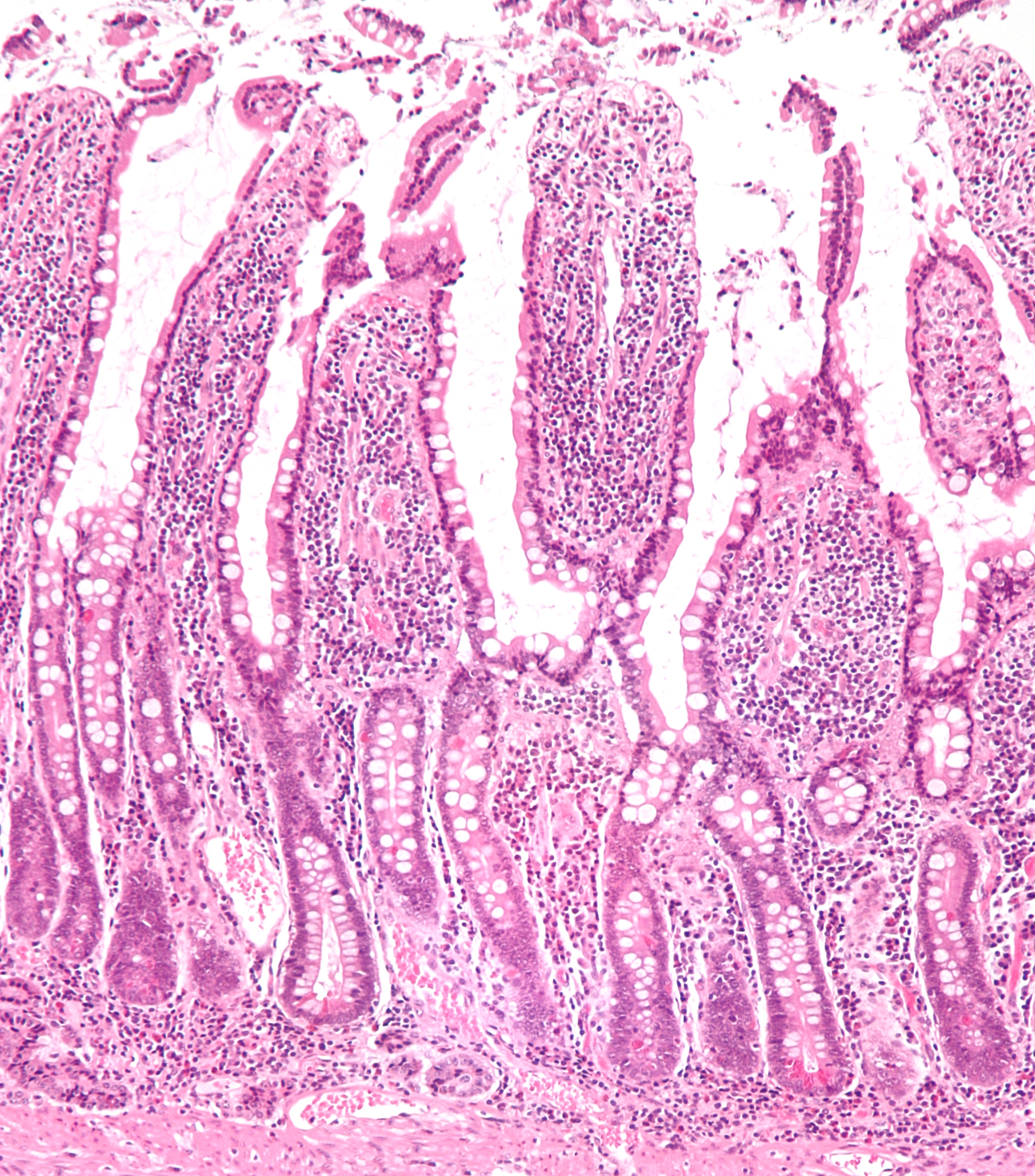
The small intestine or small bowel is an organ in the gastrointestinal tract where most of the absorption of nutrients from food takes place. It lies between the stomach and large intestine, and receives bile and pancreatic juice through the pancreatic duct to aid in digestion. The small intestine is about long and folds many times to fit in the abdomen. Although it is longer than the large intestine, it is called the small intestine because it is narrower in diameter. The small intestine has three distinct regions – the duodenum, jejunum, and ileum. The duodenum, the shortest, is where preparation for absorption through small finger-like protrusions called villi begins. The jejunum is specialized for the absorption through its lining by enterocytes: small nutrient particles which have been previously digested by enzymes in the duodenum. The main function of the ileum is to absorb vitamin B12, bile salts, and whatever products of digestion that were not absorbed by the ... [...More Info...] [...Related Items...] OR: [Wikipedia] [Google] [Baidu] |
Lesser Sac
The lesser sac, also known as the omental bursa, is a part of the peritoneal cavity that is formed by the lesser and greater omentum. Usually found in mammals, it is connected with the greater sac via the omental foramen or ''Foramen of Winslow''. In mammals, it is common for the lesser sac to contain considerable amounts of fat. Anatomic margins ;Anterior margin: listed from the top-to-bottom margin: Quadrate lobe of the liver, lesser omentum, stomach, gastrocolic ligament ;Lateral margin: listed from the most anterior to the most posterior margin: Gastrosplenic ligament, spleen, phrenicosplenic ligament ;Posterior margin: Left kidney and adrenal gland, pancreas ;Inferior margin: Greater omentum ;Superior margin: Liver If any of the marginal structures rupture their contents could leak into the lesser sac. If the stomach were to rupture on its anterior side though the leak would collect in the greater sac. The lesser sac is formed during embryogenesis from an infolding of ... [...More Info...] [...Related Items...] OR: [Wikipedia] [Google] [Baidu] |
Peritoneal Dialysis
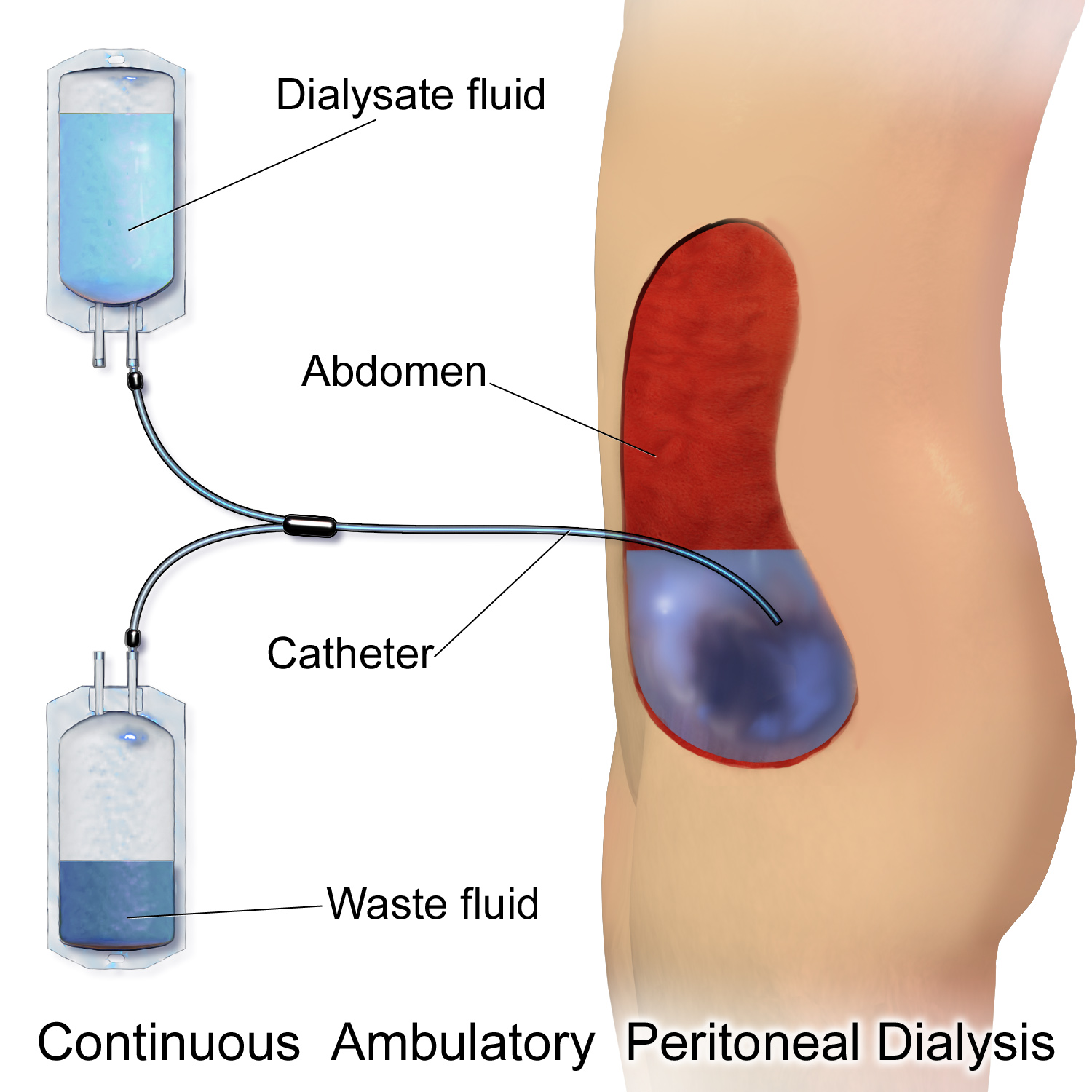
Peritoneal dialysis (PD) is a type of dialysis which uses the peritoneum in a person's abdomen as the membrane through which fluid and dissolved substances are exchanged with the blood. It is used to remove excess fluid, correct electrolyte problems, and remove toxins in those with kidney failure. Peritoneal dialysis has better outcomes than hemodialysis during the first couple of years. Other benefits include greater flexibility and better tolerability in those with significant heart disease. Complications may include infections within the abdomen, hernias, high blood sugar, bleeding in the abdomen, and blockage of the catheter. Use is not possible in those with significant prior abdominal surgery or inflammatory bowel disease. It requires some degree of technical skill to be done properly. In peritoneal dialysis, a specific solution is introduced through a permanent tube in the lower abdomen and then removed. This may either occur at regular intervals throughout the day, kn ... [...More Info...] [...Related Items...] OR: [Wikipedia] [Google] [Baidu] |
Peritoneocentesis
Paracentesis (from Greek κεντάω, "to pierce") is a form of body fluid sampling procedure, generally referring to peritoneocentesis (also called laparocentesis or abdominal paracentesis) in which the peritoneal cavity is punctured by a needle to sample peritoneal fluid. The procedure is used to remove fluid from the peritoneal cavity, particularly if this cannot be achieved with medication. The most common indication is ascites that has developed in people with cirrhosis. Indications It is used for a number of reasons: * to relieve abdominal pressure from ascites * to diagnose spontaneous bacterial peritonitis and other infections (e.g. abdominal TB) * to diagnose metastatic cancer * to diagnose blood in peritoneal space in trauma Paracentesis for ascites The procedure is often performed in a doctor's office or an outpatient clinic. In an expert's hands, it is usually very safe, although there is a small risk of infection, excessive bleeding or perforating a loop of bowel ... [...More Info...] [...Related Items...] OR: [Wikipedia] [Google] [Baidu] |
Body Fluid Sampling
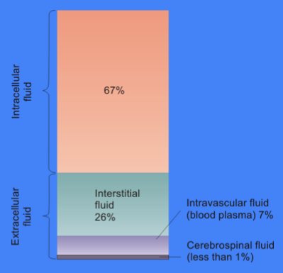
Body fluids, bodily fluids, or biofluids, sometimes body liquids, are liquids within the human body. In lean healthy adult men, the total body water is about 60% (60–67%) of the total body weight; it is usually slightly lower in women (52-55%). The exact percentage of fluid relative to body weight is inversely proportional to the percentage of body fat. A lean 70 kg (160 pound) man, for example, has about 42 (42–47) liters of water in his body. The total body of water is divided into fluid compartments, between the intracellular fluid (ICF) compartment (also called space, or volume) and the extracellular fluid (ECF) compartment (space, volume) in a ''two-to-one ratio'': 28 (28–32) liters are inside cells and 14 (14–15) liters are outside cells. The ECF compartment is divided into the interstitial fluid volume – the fluid outside both the cells and the blood vessels – and the intravascular volume (also called the vascular volume and blood plasma volume) – the fluid in ... [...More Info...] [...Related Items...] OR: [Wikipedia] [Google] [Baidu] |
Shunt (medical)
In medicine, a shunt is a hole or a small passage that moves, or allows movement of, fluid from one part of the body to another. The term may describe either congenital or acquired shunts; acquired shunts (sometimes referred to as iatrogenic shunts) may be either biological or mechanical. __TOC__ Types * Cardiac shunts may be described as right-to-left, left-to-right or bidirectional, or as systemic-to-pulmonary or pulmonary-to-systemic. * Cerebral shunt: In cases of hydrocephalus and other conditions that cause chronic increased intracranial pressure, a one-way valve is used to drain excess cerebrospinal fluid from the brain and carry it to other parts of the body. This valve usually sits outside the skull but beneath the skin, somewhere behind the ear. Cerebral shunts that drain fluid to the peritoneal cavity (located in the upper abdomen) are called ''ventriculoperitoneal'' (''VP'') shunts. * Lumbar-peritoneal shunt (a.k.a. ''lumboperitoneal'', ''LP''): In cases of chronic ... [...More Info...] [...Related Items...] OR: [Wikipedia] [Google] [Baidu] |
Hydrocephalus
Hydrocephalus is a condition in which an accumulation of cerebrospinal fluid (CSF) occurs within the brain. This typically causes increased intracranial pressure, pressure inside the skull. Older people may have headaches, double vision, poor balance, urinary incontinence, personality changes, or mental impairment. In babies, it may be seen as a rapid increase in head size. Other symptoms may include vomiting, sleepiness, seizures, and Parinaud's syndrome, downward pointing of the eyes. Hydrocephalus can occur due to birth defects or be acquired later in life. Associated birth defects include neural tube defects and those that result in aqueductal stenosis. Other causes include meningitis, brain tumors, traumatic brain injury, intraventricular hemorrhage, and subarachnoid hemorrhage. The four types of hydrocephalus are communicating, noncommunicating, ''ex vacuo'', and normal pressure hydrocephalus, normal pressure. Diagnosis is typically made by physical examination and medic ... [...More Info...] [...Related Items...] OR: [Wikipedia] [Google] [Baidu] |
Cerebrospinal Fluid
Cerebrospinal fluid (CSF) is a clear, colorless body fluid found within the tissue that surrounds the brain and spinal cord of all vertebrates. CSF is produced by specialised ependymal cells in the choroid plexus of the ventricles of the brain, and absorbed in the arachnoid granulations. There is about 125 mL of CSF at any one time, and about 500 mL is generated every day. CSF acts as a shock absorber, cushion or buffer, providing basic mechanical and immunological protection to the brain inside the skull. CSF also serves a vital function in the cerebral autoregulation of cerebral blood flow. CSF occupies the subarachnoid space (between the arachnoid mater and the pia mater) and the ventricular system around and inside the brain and spinal cord. It fills the ventricles of the brain, cisterns, and sulci, as well as the central canal of the spinal cord. There is also a connection from the subarachnoid space to the bony labyrinth of the inner ear via the perilymphat ... [...More Info...] [...Related Items...] OR: [Wikipedia] [Google] [Baidu] |
Ascites
Ascites is the abnormal build-up of fluid in the abdomen. Technically, it is more than 25 ml of fluid in the peritoneal cavity, although volumes greater than one liter may occur. Symptoms may include increased abdominal size, increased weight, abdominal discomfort, and shortness of breath. Complications can include spontaneous bacterial peritonitis. In the developed world, the most common cause is liver cirrhosis. Other causes include cancer, heart failure, tuberculosis, pancreatitis, and blockage of the hepatic vein. In cirrhosis, the underlying mechanism involves high blood pressure in the portal system and dysfunction of blood vessels. Diagnosis is typically based on an examination together with ultrasound or a CT scan. Testing the fluid can help in determining the underlying cause. Treatment often involves a low-salt diet, medication such as diuretics, and draining the fluid. A transjugular intrahepatic portosystemic shunt (TIPS) may be placed but is associated with co ... [...More Info...] [...Related Items...] OR: [Wikipedia] [Google] [Baidu] |
Intraperitoneal Injection
Intraperitoneal injection or IP injection is the injection of a substance into the peritoneum (body cavity). It is more often applied to animals than to humans. In general, it is preferred when large amounts of blood replacement fluids are needed or when low blood pressure or other problems prevent the use of a suitable blood vessel for intravenous injection. In humans, the method is widely used to administer chemotherapy drugs to treat some cancers, particularly ovarian cancer. Although controversial, intraperitoneal use in ovarian cancer has been recommended as a standard of care. Fluids are injected intraperitoneally in infants, also used for peritoneal dialysis. Background Intraperitoneal injections are a way to administer therapeutics and drugs through a peritoneal route (body cavity). They are one of the few ways drugs can be administered through injection, and have uses in research involving animals, drug administration to treat ovarian cancers, and much more. Understanding ... [...More Info...] [...Related Items...] OR: [Wikipedia] [Google] [Baidu] |
Paracolic Gutter
The paracolic gutters (paracolic sulci, paracolic recesses) are peritoneal recesses – spaces between the colon and the abdominal wall. Structure There are two paracolic gutters: * The right lateral paracolic gutter. * The left medial paracolic gutter. The right and left paracolic gutters are peritoneal recesses on the posterior abdominal wall lying alongside the ascending and descending colon. The main paracolic gutter lies lateral to the colon on each side. A less obvious medial paracolic gutter may be formed, especially on the right side, if the colon possesses a short mesentery for part of its length. The right (lateral) paracolic gutter runs from the superiolateral aspect of the hepatic flexure of the colon, down the lateral aspect of the ascending colon, and around the cecum. It is continuous with the peritoneum as it descends into the pelvis over the pelvic brim. Superiorly, it is continuous with the peritoneum which lines the hepatorenal pouch and, through the epiploic fo ... [...More Info...] [...Related Items...] OR: [Wikipedia] [Google] [Baidu] |


