|
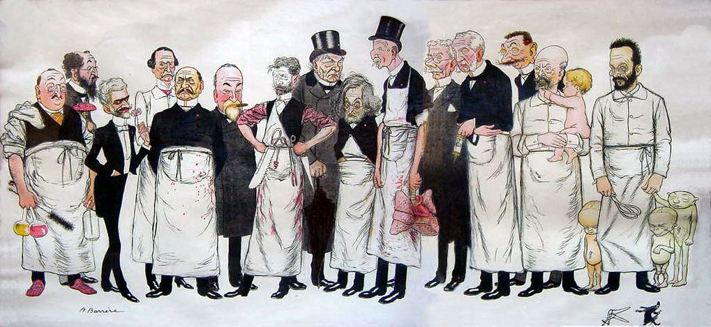
Paul Georges Dieulafoy
Paul Georges Dieulafoy (18 November 1839 – 16 August 1911) was a French physician and surgeon. He is best known for his study of acute appendicitis and his description of Dieulafoy's lesion, a rare cause of gastric bleeding. Life, studies, and career Dieulafoy was born in Toulouse. He studied medicine in Paris and earned his doctorate in 1869. In 1863, during his third year of medical school, Dieulafoy went to Paris to attend the clinical department of Professor Armand Trousseau. The two men remained close until the former's death in 1867, with Dieulafoy being referred to as Trousseau's spiritual son. Dieulafoy later led an ambulance service at the Holy Trinity Church of Paris during the Franco-Prussian war, became Chief of Medicine at the famed Hôtel-Dieu de Paris, taught pathology in the University of Paris, and was elected president of the French Academy of Medicine in 1910 after being a member since 1890. Dieulafoy married his cousin Claire Bessaignet in 1872, however ... [...More Info...] [...Related Items...] OR: [Wikipedia] [Google] [Baidu] |
Hydatid Disease
Hydatid may refer to: * Echinococcosis * ''Echinococcus granulosus'', known as the hydatid tapeworm * Hydatid of Morgagni The hydatid of Morgagni can refer to one of two closely related bodily structures: * Appendix of testis (in the male) * Paraovarian cyst Paraovarian cysts or paratubal cysts are epithelium-lined fluid-filled cysts in the adnexa adjacent to the ... * Hydatidiform mole or hydatid mole {{disambig ... [...More Info...] [...Related Items...] OR: [Wikipedia] [Google] [Baidu] |
Pleural Cavity
The pleural cavity, pleural space, or interpleural space is the potential space between the pleurae of the pleural sac that surrounds each lung. A small amount of serous pleural fluid is maintained in the pleural cavity to enable lubrication between the membranes, and also to create a pressure gradient. The serous membrane that covers the surface of the lung is the visceral pleura and is separated from the outer membrane the parietal pleura by just the film of pleural fluid in the pleural cavity. The visceral pleura follows the fissures of the lung and the root of the lung structures. The parietal pleura is attached to the mediastinum, the upper surface of the diaphragm, and to the inside of the ribcage. Structure In humans, the left and right lungs are completely separated by the mediastinum, and there is no communication between their pleural cavities. Therefore, in cases of a unilateral pneumothorax, the contralateral lung will remain functioning normally unless there is ... [...More Info...] [...Related Items...] OR: [Wikipedia] [Google] [Baidu] |
Medical Sign
Signs and symptoms are the observed or detectable signs, and experienced symptoms of an illness, injury, or condition. A sign for example may be a higher or lower temperature than normal, raised or lowered blood pressure or an abnormality showing on a medical scan. A symptom is something out of the ordinary that is experienced by an individual such as feeling feverish, a headache or other pain or pains in the body. Signs and symptoms Signs A medical sign is an objective observable indication of a disease, injury, or abnormal physiological state that may be detected during a physical examination, examining the patient history, or diagnostic procedure. These signs are visible or otherwise detectable such as a rash or bruise. Medical signs, along with symptoms, assist in formulating diagnostic hypothesis. Examples of signs include elevated blood pressure, nail clubbing of the fingernails or toenails, staggering gait, and arcus senilis and arcus juvenilis of the eyes. Indicati ... [...More Info...] [...Related Items...] OR: [Wikipedia] [Google] [Baidu] |
McBurney's Point
McBurney's point is the name given to the point over the right side of the abdomen that is one-third of the distance from the anterior superior iliac spine to the umbilicus (navel). This is near the most common location of the appendix. Location McBurney's point is located one third of the distance from the right anterior superior iliac spine to the umbilicus (navel). This point roughly corresponds to the most common location of the base of the appendix, where it is attached to the cecum. Clinical significance Appendicitis Deep tenderness at McBurney's point, known as McBurney's sign, is a sign of acute appendicitis. The clinical sign of referred pain in the epigastrium when pressure is applied is also known as Aaron's sign. Specific localization of tenderness to McBurney's point indicates that inflammation is no longer limited to the lumen of the bowel (which localizes pain poorly), and is irritating the lining of the peritoneum at the place where the peritoneum comes ... [...More Info...] [...Related Items...] OR: [Wikipedia] [Google] [Baidu] |
Hyperesthesia
Hyperesthesia is a condition that involves an abnormal increase in sensitivity to stimuli of the sense. Stimuli of the senses can include sound that one hears, foods that one tastes, textures that one feels, and so forth. Increased touch sensitivity is referred to as "tactile hyperesthesia", and increased sound sensitivity is called "auditory hyperesthesia". In the context of pain hyperaesthesia can refer to an increase in sensitivity where there is both allodynia and hyperalgesia. In psychology, Jeanne Siaud-Facchin uses the term by defining it as an "exacerbation des sens" that characterizes gifted individuals: for them, the sensory information reaches the brain much faster than the average, and the information is processed in a significantly shorter time. Other animals Feline hyperesthesia syndrome is an uncommon but recognized condition in cats, particularly Siamese, Burmese, Himalayan, and Abyssinian cats. It can affect cats of all ages, though it is most prevalent durin ... [...More Info...] [...Related Items...] OR: [Wikipedia] [Google] [Baidu] |
Esophagus
The esophagus (American English) or oesophagus (British English; both ), non-technically known also as the food pipe or gullet, is an organ in vertebrates through which food passes, aided by peristaltic contractions, from the pharynx to the stomach. The esophagus is a fibromuscular tube, about long in adults, that travels behind the trachea and heart, passes through the diaphragm, and empties into the uppermost region of the stomach. During swallowing, the epiglottis tilts backwards to prevent food from going down the larynx and lungs. The word ''oesophagus'' is from Ancient Greek οἰσοφάγος (oisophágos), from οἴσω (oísō), future form of φέρω (phérō, “I carry”) + ἔφαγον (éphagon, “I ate”). The wall of the esophagus from the lumen outwards consists of mucosa, submucosa (connective tissue), layers of muscle fibers between layers of fibrous tissue, and an outer layer of connective tissue. The mucosa is a stratified squamous epithel ... [...More Info...] [...Related Items...] OR: [Wikipedia] [Google] [Baidu] |
Mucosa
A mucous membrane or mucosa is a membrane that lines various cavities in the body of an organism and covers the surface of internal organs. It consists of one or more layers of epithelial cells overlying a layer of loose connective tissue. It is mostly of endodermal origin and is continuous with the skin at body openings such as the eyes, eyelids, ears, inside the nose, inside the mouth, lips, the genital areas, the urethral opening and the anus. Some mucous membranes secrete mucus, a thick protective fluid. The function of the membrane is to stop pathogens and dirt from entering the body and to prevent bodily tissues from becoming dehydrated. Structure The mucosa is composed of one or more layers of epithelial cells that secrete mucus, and an underlying lamina propria of loose connective tissue. The type of cells and type of mucus secreted vary from organ to organ and each can differ along a given tract. Mucous membranes line the digestive, respiratory and reproductive trac ... [...More Info...] [...Related Items...] OR: [Wikipedia] [Google] [Baidu] |
Arteriole
An arteriole is a small-diameter blood vessel in the microcirculation that extends and branches out from an artery and leads to capillaries. Arterioles have muscular walls (usually only one to two layers of smooth muscle cells) and are the primary site of vascular resistance. The greatest change in blood pressure and velocity of blood flow occurs at the transition of arterioles to capillaries.This function is extremely important because it prevents the thin, one-layer capillaries from exploding upon pressure. The arterioles achieve this decrease in pressure, as they are the site with the highest resistance (a large contributor to total peripheral resistance) which translates to a large decrease in the pressure. Structure Microanatomy In a healthy vascular system the endothelium lines all blood-contacting surfaces, including arteries, arterioles, veins, venules, capillaries, and heart chambers. This healthy condition is promoted by the ample production of nitric oxide by the end ... [...More Info...] [...Related Items...] OR: [Wikipedia] [Google] [Baidu] |
Allergic Bronchopulmonary Aspergillosis
Allergic bronchopulmonary aspergillosis (ABPA) is a condition characterised by an exaggerated response of the immune system (a hypersensitivity response) to the fungus ''Aspergillus'' (most commonly ''Aspergillus fumigatus''). It occurs most often in people with asthma or cystic fibrosis. ''Aspergillus'' spores are ubiquitous in soil and are commonly found in the sputum of healthy individuals. ''A. fumigatus'' is responsible for a spectrum of lung diseases known as aspergilloses. ABPA causes airway inflammation, leading to bronchiectasis—a condition marked by abnormal dilation of the airways. Left untreated, the immune system and fungal spores can damage sensitive lung tissues and lead to scarring. The exact criteria for the diagnosis of ABPA are not agreed upon. Chest X-rays and CT scans, raised blood levels of IgE and eosinophils, immunological tests for ''Aspergillus'' together with sputum staining and sputum cultures can be useful. Treatment consists of corticosteroids and ... [...More Info...] [...Related Items...] OR: [Wikipedia] [Google] [Baidu] |
Georges-Fernand Widal
Georges-Fernand-Isidor Widal (March 9, 1862 in Dellys, Algeria – January 14, 1929 in Paris ) was a French people, French physician. From 1886 to 1888 he devoted himself to public demonstrations of the researches of the faculty of pathological anatomy, and during the 2 years following was in charge of a course in bacteriology in the laboratory of Professor Victor André Cornil (1837–1908). In 1895 he was appointed visiting physician to the hospitals of Paris, and in 1904 became an professor, instructor in the faculty of medicine. In 1905 he became a physician to the Hôpital Cochin, and was in charge of the medical clinics at the same institution. During the WWI, he developed a vaccine against typhoid fever which reduced the spread of this disease in French army and more generally allied troops. Widal was the author of a remarkable series of essays on infectious diseases, erysipelas, diseases of the heart (anatomy), heart, liver, nervous system, etc., besides being a prolific c ... [...More Info...] [...Related Items...] OR: [Wikipedia] [Google] [Baidu] |
André Chantemesse
André Chantemesse (23 October 1851 – 25 February 1919) was a French bacteriologist born in Le Puy-en-Velay, Haute-Loire. From 1880 to 1885 he served as ''interne des hôpitaux'' in Paris, earning his doctorate in 1884 with a dissertation on adult tuberculous meningitis titled ''Étude sur la méningite tuberculeuse de l'adulte : les formes anormales en particulier''. In 1885 he traveled to Berlin to study bacteriology at the laboratory of Robert Koch (1843–1910). After his return to Paris, he became associated with the work of Louis Pasteur. In 1886, he began extensive research of typhoid fever. In collaboration with Georges-Fernand Widal (1862–1929), he studied the aetiology of the disease, and in 1888 developed an experimental antityphoid inoculation. Also with Widal, he isolated the bacillus that was the cause of dysentery, however the two scientists were unable to establish the aetiological link to the disease. [...More Info...] [...Related Items...] OR: [Wikipedia] [Google] [Baidu] |


