|
Partial Volume (imaging)
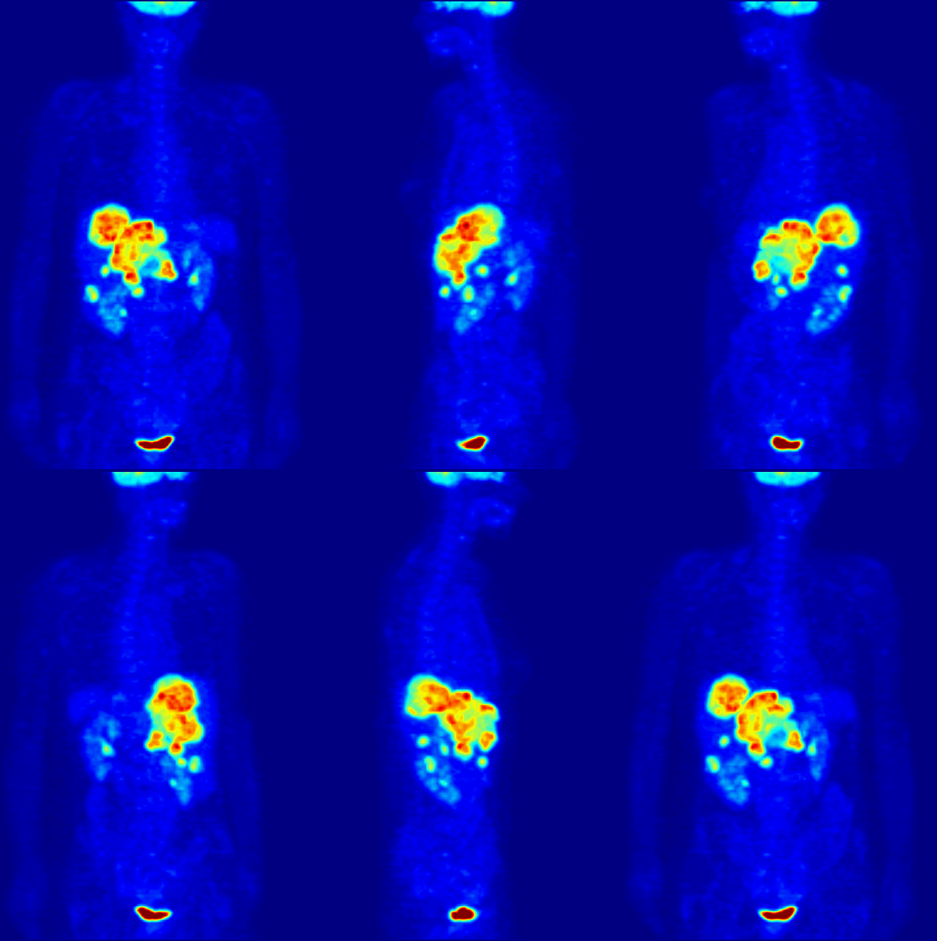
The partial volume effect can be defined as the loss of apparent activity in small objects or regions because of the limited resolution of the imaging system. It occurs in medical imaging and more generally in biological imaging such as positron emission tomography (PET) and single-photon emission computed tomography Single-photon emission computed tomography (SPECT, or less commonly, SPET) is a nuclear medicine tomography, tomographic imaging technique using gamma rays. It is very similar to conventional nuclear medicine planar imaging using a gamma camera ... (SPECT). If the object or region to be imaged is less than twice the full width at half maximum ( FWHM) resolution in x-, y- and z-dimension of the imaging system, the resultant activity in the object or region is underestimated. A higher resolution decreases this effect, as it better resolves the tissue. Partial volume loss alone occurs only when the surrounding activity of the object or region is zero, or less or mo ... [...More Info...] [...Related Items...] OR: [Wikipedia] [Google] [Baidu] |
Medical Imaging
Medical imaging is the technique and process of imaging the interior of a body for clinical analysis and medical intervention, as well as visual representation of the function of some organs or tissues (physiology). Medical imaging seeks to reveal internal structures hidden by the skin and bones, as well as to diagnose and treat disease. Medical imaging also establishes a database of normal anatomy and physiology to make it possible to identify abnormalities. Although imaging of removed organ (anatomy), organs and Tissue (biology), tissues can be performed for medical reasons, such procedures are usually considered part of pathology instead of medical imaging. Measurement and recording techniques that are not primarily designed to produce images, such as electroencephalography (EEG), magnetoencephalography (MEG), electrocardiography (ECG), and others, represent other technologies that produce data susceptible to representation as a parameter graph versus time or maps that contain ... [...More Info...] [...Related Items...] OR: [Wikipedia] [Google] [Baidu] |
Biological Imaging
Biological imaging may refer to any imaging technique used in biology. Typical examples include: * Bioluminescence imaging, a technique for studying laboratory animals using luminescent protein * Calcium imaging, determining the calcium status of a tissue using fluorescent light * Diffuse optical imaging, using near-infrared light to generate images of the body * Diffusion-weighted imaging, a type of MRI that uses water diffusion * Fluorescence lifetime imaging, using the decay rate of a fluorescent sample * Gallium imaging, a nuclear medicine method for the detection of infections and cancers * Imaging agent, a chemical designed to allow clinicians to determine whether a mass is benign or malignant * Imaging studies, which includes many medical imaging techniques * Magnetic resonance imaging (MRI), a non-invasive method to render images of living tissues * Magneto-acousto-electrical tomography (MAET), is an imaging modality to image the electrical conductivity of biological ... [...More Info...] [...Related Items...] OR: [Wikipedia] [Google] [Baidu] |
Positron Emission Tomography
Positron emission tomography (PET) is a functional imaging technique that uses radioactive substances known as radiotracers to visualize and measure changes in metabolic processes, and in other physiological activities including blood flow, regional chemical composition, and absorption. Different tracers are used for various imaging purposes, depending on the target process within the body, such as: * Fluorodeoxyglucose ( 18F">sup>18FDG or FDG) is commonly used to detect cancer; * 18Fodium fluoride">sup>18Fodium fluoride (Na18F) is widely used for detecting bone formation; * Oxygen-15 (15O) is sometimes used to measure blood flow. PET is a common imaging technique, a medical scintillography technique used in nuclear medicine. A radiopharmaceutical—a radioisotope attached to a drug—is injected into the body as a tracer. When the radiopharmaceutical undergoes beta plus decay, a positron is emitted, and when the positron interacts with an ordinary electron, the tw ... [...More Info...] [...Related Items...] OR: [Wikipedia] [Google] [Baidu] |
Single-photon Emission Computed Tomography
Single-photon emission computed tomography (SPECT, or less commonly, SPET) is a nuclear medicine tomography, tomographic imaging technique using gamma rays. It is very similar to conventional nuclear medicine planar imaging using a gamma camera (that is, scintigraphy), but is able to provide true 3D computer graphics, 3D information. This information is typically presented as cross-sectional slices through the patient, but can be freely reformatted or manipulated as required. The technique needs delivery of a gamma-emitting radioisotope (a radionuclide) into the patient, normally through injection into the bloodstream. On occasion, the radioisotope is a simple soluble dissolved ion, such as an Isotopes of gallium, isotope of gallium(III). Usually, however, a marker radioisotope is attached to a specific ligand to create a radioligand, whose properties bind it to certain types of tissues. This marriage allows the combination of ligand and radiopharmaceutical to be carried and bo ... [...More Info...] [...Related Items...] OR: [Wikipedia] [Google] [Baidu] |
Full Width At Half Maximum
In a distribution, full width at half maximum (FWHM) is the difference between the two values of the independent variable at which the dependent variable is equal to half of its maximum value. In other words, it is the width of a spectrum curve measured between those points on the ''y''-axis which are half the maximum amplitude. Half width at half maximum (HWHM) is half of the FWHM if the function is symmetric. The term full duration at half maximum (FDHM) is preferred when the independent variable is time. FWHM is applied to such phenomena as the duration of pulse waveforms and the spectral width of sources used for optical communications and the resolution of spectrometers. The convention of "width" meaning "half maximum" is also widely used in signal processing to define bandwidth as "width of frequency range where less than half the signal's power is attenuated", i.e., the power is at least half the maximum. In signal processing terms, this is at most −3 dB of att ... [...More Info...] [...Related Items...] OR: [Wikipedia] [Google] [Baidu] |
Spillover (imaging)
Spillover effect can be defined as an apparent gain in activity for small objects or regions, as opposed to the partial volume effect. It occurs often in biological imaging modalities such as positron emission tomography (PET) and single-photon emission computed tomography (SPECT) because of their limited spatial resolution. Although partial volume effect and spillover refer to essentially the same physical problem, it is important to distinguish the outcome of these two different effects. For partial volume effect, the apparent loss of activity in the object is distributed across adjacent voxels, which are considered outside the object, resulting in increase in activity in these voxels. This increase in activity is referred to as spillover, whereas loss in activity is referred to as partial volume loss. See also * Partial volume (imaging) The partial volume effect can be defined as the loss of apparent activity in small objects or regions because of the limited resolution of t ... [...More Info...] [...Related Items...] OR: [Wikipedia] [Google] [Baidu] |
Voxel
In computing, a voxel is a representation of a value on a three-dimensional regular grid, akin to the two-dimensional pixel. Voxels are frequently used in the Data visualization, visualization and analysis of medical imaging, medical and scientific data (e.g. geographic information systems (GIS)). Voxels also have technical and artistic applications in video games, largely originating with surface rendering in ''Outcast (video game), Outcast'' (1999). ''Minecraft'' (2011) makes use of an entirely voxelated world to allow for a fully destructable and constructable environment. Voxel art, of the sort used in ''Minecraft'' and elsewhere, is a style and format of 3D art analogous to pixel art. As with pixels in a 2D bitmap, voxels themselves do not typically have their position (i.e. coordinates) explicitly encoded with their values. Instead, Rendering (computer graphics), rendering systems infer the position of a voxel based upon its position relative to other voxels (i.e., its pos ... [...More Info...] [...Related Items...] OR: [Wikipedia] [Google] [Baidu] |
Journal Of Cerebral Blood Flow & Metabolism
The ''Journal of Cerebral Blood Flow & Metabolism'' is a monthly peer-reviewed medical journal the official journal of the International Society for Cerebral Blood Flow & Metabolism and publishes peer-reviewed research and review papers. covering research on experimental, theoretical, and clinical aspects of brain circulation, metabolism and imaging. The editor-in-chief is Jun Chen (University of Pittsburgh). According to the ''Journal Citation Reports'', the journal has a 2020 impact factor The impact factor (IF) or journal impact factor (JIF) of an academic journal is a type of journal ranking. Journals with higher impact factor values are considered more prestigious or important within their field. The Impact Factor of a journa ... of 6.200. References External links * International Society for Cerebral Blood Flow & Metabolism Neuroscience journals Academic journals established in 1981 Nature Research academic journals English-language journals Monthly journals D ... [...More Info...] [...Related Items...] OR: [Wikipedia] [Google] [Baidu] |
Alan C
Alan may refer to: People *Alan (surname), an English and Kurdish surname * Alan (given name), an English given name ** List of people with given name Alan ''Following are people commonly referred to solely by "Alan" or by a homonymous name.'' * Alan (Chinese singer) (born 1987), female Chinese singer of Tibetan ethnicity, active in both China and Japan * Alan (Mexican singer) (born 1973), Mexican singer and actor *Alan (wrestler) (born 1975), a.k.a. Gato Eveready, who wrestles in Asistencia Asesoría y Administración * Alan (footballer, born 1979) (Alan Osório da Costa Silva), Brazilian footballer * Alan (footballer, born 1998) (Alan Cardoso de Andrade), Brazilian footballer *Alan I, King of Brittany (died 907), "the Great" * Alan II, Duke of Brittany (c. 900–952) * Alan III, Duke of Brittany(997–1040) * Alan IV, Duke of Brittany (c. 1063–1119), a.k.a. Alan Fergant ("the Younger" in Breton language) * Alan of Tewkesbury, 12th century abbott * Alan of Lynn (c. 1348–1423) ... [...More Info...] [...Related Items...] OR: [Wikipedia] [Google] [Baidu] |
The Journal Of Nuclear Medicine
''The Journal of Nuclear Medicine'' is a monthly peer-reviewed medical journal published by Society of Nuclear Medicine and Molecular Imaging that covers research on all aspects of nuclear medicine, including molecular imaging. Abstracting and indexing The journal is abstracted and indexed in Science Citation Index, Current Contents/Clinical Medicine, Current Contents/Life Sciences, BIOSIS Previews, and MEDLINE/PubMed. According to the ''Journal Citation Reports'', the journal has a 2020 impact factor The impact factor (IF) or journal impact factor (JIF) of an academic journal is a type of journal ranking. Journals with higher impact factor values are considered more prestigious or important within their field. The Impact Factor of a journa ... of 10.057, ranking it 3rd out of 134 journals in the category "Radiology, Nuclear Medicine & Medical Imaging". References External links * {{DEFAULTSORT:Journal of Nuclear Medicine, The Radiology and medical imaging journals ... [...More Info...] [...Related Items...] OR: [Wikipedia] [Google] [Baidu] |
Neuroimaging
Neuroimaging is the use of quantitative (computational) techniques to study the neuroanatomy, structure and function of the central nervous system, developed as an objective way of scientifically studying the healthy human brain in a non-invasive manner. Increasingly it is also being used for quantitative research studies of brain disease and psychiatric illness. Neuroimaging is highly multidisciplinary involving neuroscience, computer science, psychology and statistics, and is not a medical specialty. Neuroimaging is sometimes confused with neuroradiology. Neuroradiology is a medical specialty that uses non-statistical brain imaging in a clinical setting, practiced by radiologists who are medical practitioners. Neuroradiology primarily focuses on recognizing brain lesions, such as vascular diseases, strokes, tumors, and inflammatory diseases. In contrast to neuroimaging, neuroradiology is qualitative (based on subjective impressions and extensive clinical training) but sometime ... [...More Info...] [...Related Items...] OR: [Wikipedia] [Google] [Baidu] |


