|
Paroxysmal Nocturnal Hemoglobinuria
Paroxysmal nocturnal hemoglobinuria (PNH) is a rare, acquired, life-threatening disease of the blood characterized by destruction of red blood cells by the complement system, a part of the body's innate immune system. This destructive process occurs due to deficiency of the red blood cell surface protein DAF, which normally inhibits such immune reactions. Since the complement cascade attacks the red blood cells within the blood vessels of the circulatory system, the red blood cell destruction (hemolysis) is considered an ''intravascular'' hemolytic anemia. Other key features of the disease, such as the high incidence of venous blood clot formation, are incompletely understood. PNH is the only hemolytic anemia caused by an ''acquired'' (rather than inherited) intrinsic defect in the cell membrane (deficiency of glycophosphatidylinositol or GPI) leading to the absence of protective exterior surface proteins that normally attach via a GPI anchor. It may develop on its own ("primary ... [...More Info...] [...Related Items...] OR: [Wikipedia] [Google] [Baidu] |
Hemolytic Anemia
Hemolytic anemia or haemolytic anaemia is a form of anemia due to hemolysis, the abnormal breakdown of red blood cells (RBCs), either in the blood vessels (intravascular hemolysis) or elsewhere in the human body (extravascular). This most commonly occurs within the spleen, but also can occur in the reticuloendothelial system or mechanically (prosthetic valve damage). Hemolytic anemia accounts for 5% of all existing anemias. It has numerous possible consequences, ranging from general symptoms to life-threatening systemic effects. The general classification of hemolytic anemia is either intrinsic or extrinsic. Treatment depends on the type and cause of the hemolytic anemia. Symptoms of hemolytic anemia are similar to other forms of anemia ( fatigue and shortness of breath), but in addition, the breakdown of red cells leads to jaundice and increases the risk of particular long-term complications, such as gallstones and pulmonary hypertension. Signs and symptoms Symptoms of hemolytic ... [...More Info...] [...Related Items...] OR: [Wikipedia] [Google] [Baidu] |
Dyspnea
Shortness of breath (SOB), also medically known as dyspnea (in AmE) or dyspnoea (in BrE), is an uncomfortable feeling of not being able to breathing, breathe well enough. The American Thoracic Society defines it as "a subjective experience of breathing discomfort that consists of qualitatively distinct sensations that vary in intensity", and recommends evaluating dyspnea by assessing the intensity of its distinct sensations, the degree of distress and discomfort involved, and its burden or impact on the patient's activities of daily living. Distinct sensations include effort/work to breathe, chest tightness or pain, and "air hunger" (the feeling of not enough oxygen). The tripod position is often assumed to be a sign. Dyspnea is a normal symptom of heavy physical exertion but becomes disease, pathological if it occurs in unexpected situations, when resting or during light exertion. In 85% of cases it is due to asthma, pneumonia, cardiac ischemia, interstitial lung disease, congesti ... [...More Info...] [...Related Items...] OR: [Wikipedia] [Google] [Baidu] |
Superior Mesenteric Vein
In human anatomy, the superior mesenteric vein (SMV) is a blood vessel that drains blood from the small intestine (jejunum and ileum). Behind the neck of the pancreas, the superior mesenteric vein combines with the splenic vein to form the hepatic portal vein. The superior mesenteric vein lies to the right of the similarly named artery, the superior mesenteric artery, which originates from the abdominal aorta. Structure Tributaries of the superior mesenteric vein drain the small intestine, large intestine, stomach, pancreas and appendix and include: * Right gastro-omental vein (also known as the right gastro-epiploic vein) * inferior pancreaticoduodenal veins * veins from jejunum * veins from ileum * middle colic vein – drains the transverse colon * right colic vein – drains the ascending colon * ileocolic vein The superior mesenteric vein combines with the splenic vein to form the portal vein. Clinical significance Thrombosis of the superior mesenteric vein is quite rare ... [...More Info...] [...Related Items...] OR: [Wikipedia] [Google] [Baidu] |
Portal Vein Thrombosis
Portal vein thrombosis (PVT) is a vascular disease of the liver that occurs when a blood clot occurs in the hepatic portal vein, which can lead to increased pressure in the portal vein system and reduced blood supply to the liver. The mortality rate is approximately 1 in 10. An equivalent clot in the vasculature that exits the liver carrying deoxygenated blood to the right atrium via the inferior vena cava, is known as hepatic vein thrombosis or Budd-Chiari syndrome. Signs and symptoms Portal vein thrombosis causes upper abdominal pain, possibly accompanied by nausea and an enlarged liver and/or spleen; the abdomen may be filled with fluid (ascites). A persistent fever may result from the generalized inflammation. While abdominal pain may come and go if the thrombus forms suddenly, long-standing clot build-up can also develop without causing symptoms, leading to portal hypertension before it is diagnosed. Other symptoms can develop based on the cause. For example, if portal v ... [...More Info...] [...Related Items...] OR: [Wikipedia] [Google] [Baidu] |
Portal Vein
The portal vein or hepatic portal vein (HPV) is a blood vessel that carries blood from the gastrointestinal tract, gallbladder, pancreas and spleen to the liver. This blood contains nutrients and toxins extracted from digested contents. Approximately 75% of total liver blood flow is through the portal vein, with the remainder coming from the hepatic artery proper. The blood leaves the liver to the heart in the hepatic veins. The portal vein is not a true vein, because it conducts blood to capillary beds in the liver and not directly to the heart. It is a major component of the hepatic portal system, one of only two portal venous systems in the body – with the hypophyseal portal system being the other. The portal vein is usually formed by the confluence of the superior mesenteric, splenic veins, inferior mesenteric, left, right gastric veins and the pancreatic vein. Conditions involving the portal vein cause considerable illness and death. An important example of such a condi ... [...More Info...] [...Related Items...] OR: [Wikipedia] [Google] [Baidu] |
Hepatic Vein
In human anatomy, the hepatic veins are the veins that drain venous blood from the liver into the inferior vena cava (as opposed to the hepatic portal vein which conveys blood from the gastrointestinal organs to the liver). There are usually three large upper hepatic veins draining from the left, middle, and right parts of the liver, as well as a number (6-20) of lower hepatic veins. All hepatic veins are valveless. Structure All the hepatic veins drain into the inferior vena cava. The hepatic veins are divided into an upper and a lower group. Upper group The upper group consists of three hepatic veins - the right, middle, and left hepatic veins - draining the central veins from the right, middle, and left regions of the liver and are larger than the lower group of veins. The veins of the upper group drain into the suprahepatic part of the inferior vena cava (i.e. part superior to the liver). Right hepatic vein The right hepatic vein is the longest and largest of all the h ... [...More Info...] [...Related Items...] OR: [Wikipedia] [Google] [Baidu] |
Pulmonary Embolism
Pulmonary embolism (PE) is a blockage of an pulmonary artery, artery in the lungs by a substance that has moved from elsewhere in the body through the bloodstream (embolism). Symptoms of a PE may include dyspnea, shortness of breath, chest pain particularly upon breathing in, and coughing up blood. Symptoms of a deep vein thrombosis, blood clot in the leg may also be present, such as a erythema, red, warm, swollen, and painful leg. Signs of a PE include low blood oxygen saturation, oxygen levels, tachypnea, rapid breathing, tachycardia, rapid heart rate, and sometimes a mild fever. Severe cases can lead to Syncope (medicine), passing out, shock (circulatory), abnormally low blood pressure, obstructive shock, and cardiac arrest, sudden death. PE usually results from a blood clot in the leg that travels to the lung. The risk of blood clots is increased by advanced age, cancer, prolonged bed rest and immobilization, smoking, stroke, long-haul travel over 4 hours, certain genetics, g ... [...More Info...] [...Related Items...] OR: [Wikipedia] [Google] [Baidu] |
Deep Vein Thrombosis
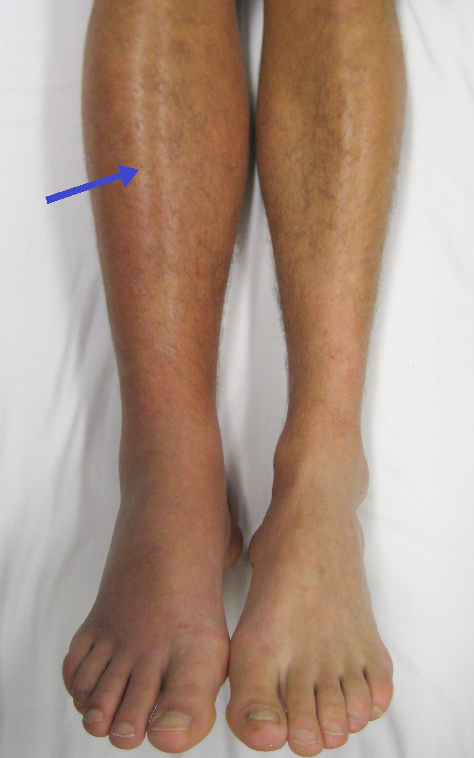
Deep vein thrombosis (DVT) is a type of venous thrombosis involving the formation of a blood clot in a deep vein, most commonly in the legs or pelvis. A minority of DVTs occur in the arms. Symptoms can include pain, swelling, redness, and enlarged veins in the affected area, but some DVTs have no symptoms. The most common life-threatening concern with DVT is the potential for a clot to embolize (detach from the veins), travel as an embolus through the right side of the heart, and become lodged in a pulmonary artery that supplies blood to the lungs. This is called a pulmonary embolism (PE). DVT and PE comprise the cardiovascular disease of venous thromboembolism (VTE). About two-thirds of VTE manifests as DVT only, with one-third manifesting as PE with or without DVT. The most frequent long-term DVT complication is post-thrombotic syndrome, which can cause pain, swelling, a sensation of heaviness, itching, and in severe cases, ulcers. Recurrent VTE occurs in about 30% of those i ... [...More Info...] [...Related Items...] OR: [Wikipedia] [Google] [Baidu] |
Smooth Muscle Tissue
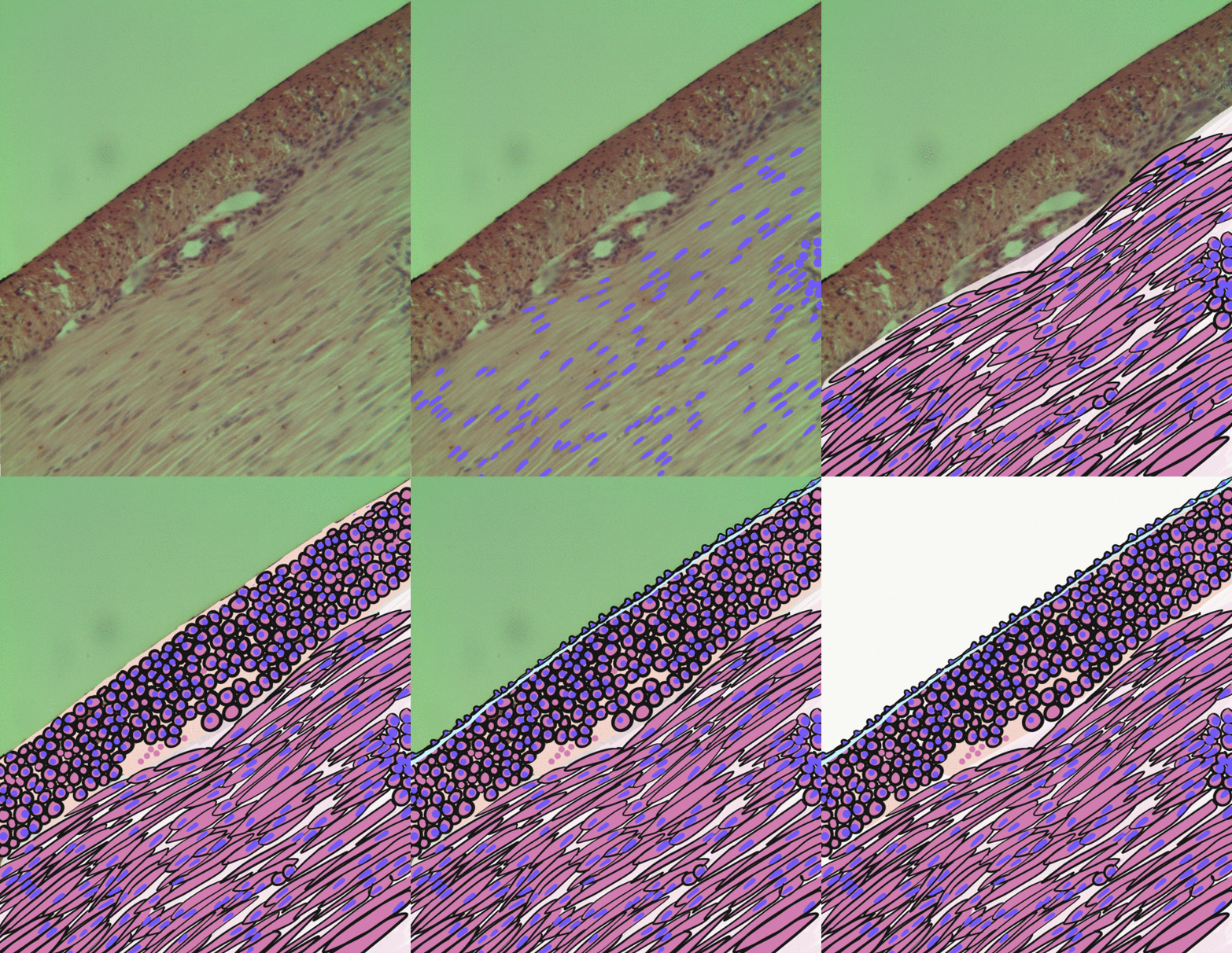
Smooth muscle is an involuntary non-striated muscle, so-called because it has no sarcomeres and therefore no striations (''bands'' or ''stripes''). It is divided into two subgroups, single-unit and multiunit smooth muscle. Within single-unit muscle, the whole bundle or sheet of smooth muscle cells contracts as a syncytium. Smooth muscle is found in the walls of hollow organs, including the stomach, intestines, bladder and uterus; in the walls of passageways, such as blood, and lymph vessels, and in the tracts of the respiratory, urinary, and reproductive systems. In the eyes, the ciliary muscles, a type of smooth muscle, dilate and contract the iris and alter the shape of the lens. In the skin, smooth muscle cells such as those of the arrector pili cause hair to stand erect in response to cold temperature or fear. Structure Gross anatomy Smooth muscle is grouped into two types: single-unit smooth muscle, also known as visceral smooth muscle, and multiunit smooth muscle. M ... [...More Info...] [...Related Items...] OR: [Wikipedia] [Google] [Baidu] |
Erectile Dysfunction
Erectile dysfunction (ED), also called impotence, is the type of sexual dysfunction in which the penis fails to become or stay erect during sexual activity. It is the most common sexual problem in men.Cunningham GR, Rosen RC. Overview of male sexual dysfunction. In: UpToDate, Martin KA (Ed), UpToDate, Waltham, MA, 2018. Through its connection to self-image and to problems in sexual relationships, erectile dysfunction can cause psychological harm. In about 80% of cases, physical causes can be identified. These include cardiovascular disease; diabetes mellitus; neurological problems, such as those following prostatectomy; hypogonadism; and drug side effects. About 10% of cases are psychological impotence, caused by thoughts or feelings; here, there is a strong response to placebo treatment. The term ''erectile dysfunction'' is not used for other disorders of erection, such as priapism. Treatment involves addressing the underlying causes, lifestyle modifications, and addres ... [...More Info...] [...Related Items...] OR: [Wikipedia] [Google] [Baidu] |
Odynophagia
Odynophagia is pain when swallowing. The pain may be felt in the mouth or throat and can occur with or without difficulty swallowing. The pain may be described as an ache, burning sensation, or occasionally a stabbing pain that radiates to the back. Odynophagia often results in inadvertent weight loss. The term is from ''-'' 'pain' and ' 'to eat'. Causes Odynophagia may have environmental or behavioral causes, such as: * Very hot or cold food and drinks (termed cryodynophagia when associated with cold drinks, classically in the setting of cryoglobulinaemia). * Taking certain medications * Using drugs, tobacco, or alcohol * Trauma or injury to the mouth, throat, or tongue It can also be caused by certain medical conditions, such as: * Ulcers * Abscesses * Upper respiratory tract infections * Inflammation or infection of the mouth, tongue, or throat (esophagitis, pharyngitis, tonsillitis, epiglottitis) * Immune disorders * Oral or throat cancer See also * Phagophobia Phagophobia ... [...More Info...] [...Related Items...] OR: [Wikipedia] [Google] [Baidu] |


