|
Paraganglia
A paraganglion (pl. paraganglia) is a group of non-neuronal cells derived of the neural crest. They are named for being generally in close proximity to sympathetic ganglia. They are essentially of two types: (1) chromaffin or sympathetic paraganglia made of chromaffin cells and (2) nonchromaffin or parasympathetic ganglia made of glomus cells. They are neuroendocrine cells, the former with primary endocrine functions and the latter with primary chemoreceptor functions. Chromaffin paraganglia (also called chromaffin bodies) are connected with the ganglia of the sympathetic trunk and the ganglia of the celiac, renal, adrenal, aortic and hypogastric plexuses. They are concentrated near the adrenal glands and essentially function the same way as the adrenal medulla. They are sometimes found in connection with the ganglia of other sympathetic plexuses. None have been found with the sympathetic ganglia associated with the branches of the trigeminal nerve. The largest chromaffin paraga ... [...More Info...] [...Related Items...] OR: [Wikipedia] [Google] [Baidu] |
Chromaffin Cell
Chromaffin cells, also called pheochromocytes (or phaeochromocytes), are neuroendocrine cells found mostly in the medulla of the adrenal glands in mammals. These cells serve a variety of functions such as serving as a response to stress, monitoring carbon dioxide and oxygen concentrations in the body, maintenance of respiration and the regulation of blood pressure. They are in close proximity to pre-synaptic sympathetic ganglia of the sympathetic nervous system, with which they communicate, and structurally they are similar to post-synaptic sympathetic neurons. In order to activate chromaffin cells, the splanchnic nerve of the sympathetic nervous system releases acetylcholine, which then binds to nicotinic acetylcholine receptors on the adrenal medulla. This causes the release of catecholamines. The chromaffin cells release catecholamines: ~80% of adrenaline (epinephrine) and ~20% of noradrenaline (norepinephrine) into systemic circulation for systemic effects on multiple ... [...More Info...] [...Related Items...] OR: [Wikipedia] [Google] [Baidu] |
Paragangliomas
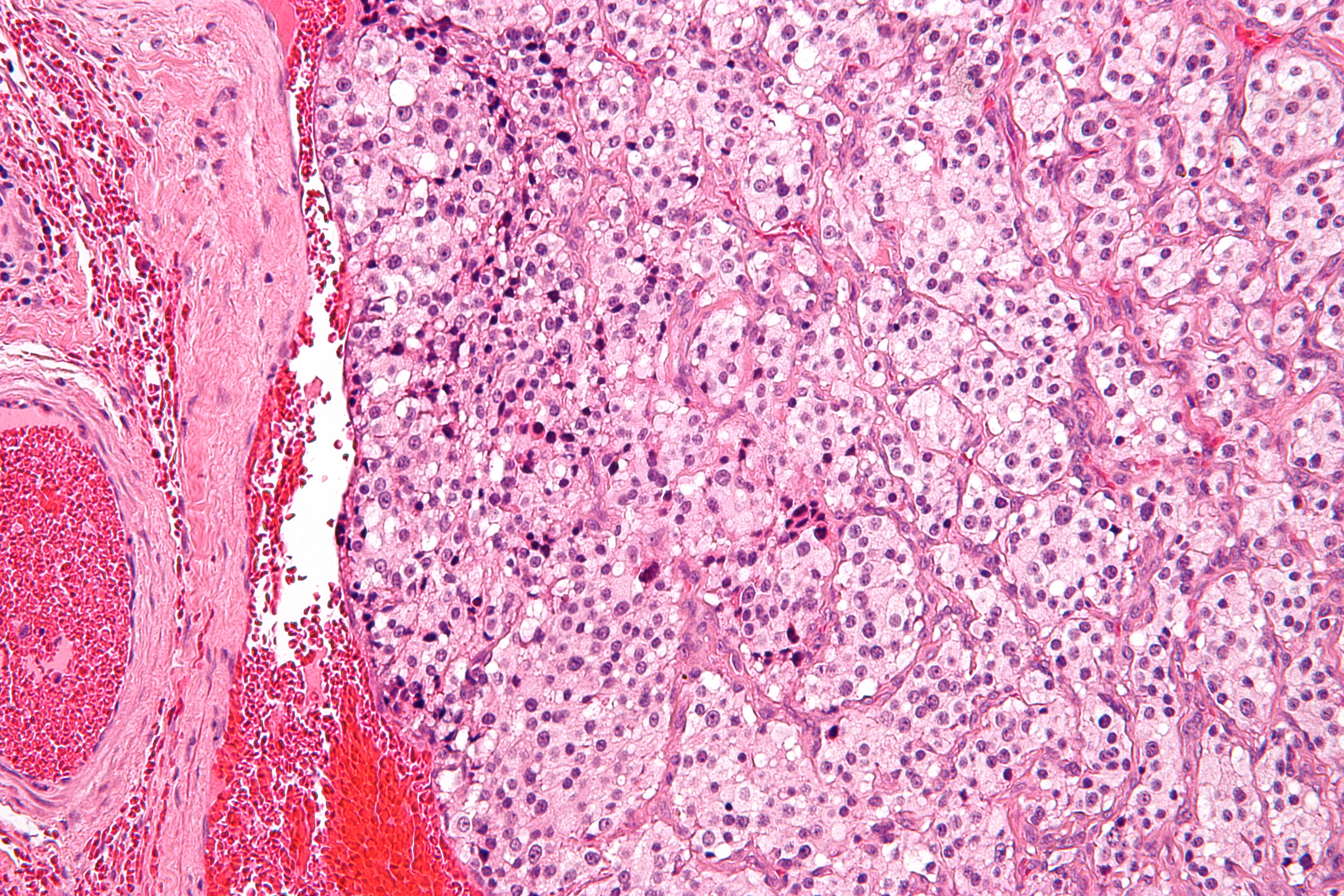
A paraganglioma is a rare neuroendocrine neoplasm that may develop at various body sites (including the head, neck, thorax and abdomen). When the same type of tumor is found in the adrenal gland, they are referred to as a pheochromocytoma. They are rare tumors, with an overall estimated incidence of 1/300,000. There is no test that determines benign from malignant tumors; long-term follow-up is therefore recommended for all individuals with paraganglioma. Signs and symptoms Most paragangliomas are asymptomatic, present as a painless mass, or create symptoms such as hypertension, tachycardia, headache, and palpitations. While all contain neurosecretory granules, only in 1–3% of cases is secretion of hormones such as catecholamines abundant enough to be clinically significant; in that case manifestations often resemble those of pheochromocytomas (intra-medullary paraganglioma). Genetics About 75% of paragangliomas are sporadic; the remaining 25% are hereditary (and have an increa ... [...More Info...] [...Related Items...] OR: [Wikipedia] [Google] [Baidu] |
Glomus Cell
Glomus cells are the cell type mainly located in the carotid bodies and aortic bodies. Glomus type I cells are peripheral chemoreceptors which sense the oxygen, carbon dioxide and pH levels of the blood. When there is a decrease in the blood's pH, a decrease in oxygen (pO2), or an increase in carbon dioxide ( pCO2), the carotid bodies and the aortic bodies signal the dorsal respiratory group in the medulla oblongata to increase the volume and rate of breathing. The glomus cells have a high metabolic rate and good blood perfusion and thus are sensitive to changes in arterial blood gas tension. Glomus type II cells are sustentacular cells having a similar supportive function to glial cells. Structure The signalling within the chemoreceptors is thought to be mediated by the release of neurotransmitters by the glomus cells, including dopamine, noradrenaline, acetylcholine, substance P, vasoactive intestinal peptide and enkephalins. Vasopressin has been found to inhibit the respons ... [...More Info...] [...Related Items...] OR: [Wikipedia] [Google] [Baidu] |
Pheochromocytomas
Pheochromocytoma (PHEO or PCC) is a rare tumor of the adrenal medulla composed of chromaffin cells, also known as pheochromocytes. When a tumor composed of the same cells as a pheochromocytoma develops outside the adrenal gland, it is referred to as a paraganglioma. These neuroendocrine tumors are capable of producing and releasing massive amounts of catecholamines, metanephrines, or methoxytyramine, which result in the most common symptoms, including hypertension (high blood pressure), tachycardia (fast heart rate), and diaphoresis (sweating). However, not all of these tumors will secrete catecholamines. Those that do not are referred to as biochemically silent, and are predominantly located in the head and neck. While patients with biochemically silent disease will not develop the typical disease manifestations described above, the tumors grow and compress the surrounding structures of the head and neck, and can result in pulsatile tinnitus (ringing of the ear), hearing loss, ... [...More Info...] [...Related Items...] OR: [Wikipedia] [Google] [Baidu] |
Organ Of Zuckerkandl
The organ of Zuckerkandl is a chromaffin body derived from neural crest located at the bifurcation of the aorta or at the origin of the inferior mesenteric artery. It can be the source of a paraganglioma. The term para-aortic body is also sometimes used to describe it, as it usually arises near the abdominal aorta, but this term can be the source of confusion, because the term "corpora paraaortica" is also used to describe the aortic body, which arises near the thoracic aorta. This diffuse group of neuroendocrine sympathetic fibres was first described by Emil Zuckerkandl, a professor of anatomy at the University of Vienna, in 1901. Some sources equate the " aortic bodies" and "paraaortic bodies", while other sources explicitly distinguish between the two. When a distinction is made, the "aortic bodies" are chemoreceptors which regulate circulation, while the "paraaortic bodies" are the chromaffin cells which manufacture catecholamines. Structure Function Its physiological r ... [...More Info...] [...Related Items...] OR: [Wikipedia] [Google] [Baidu] |
Gallbladder
In vertebrates, the gallbladder, also known as the cholecyst, is a small hollow organ where bile is stored and concentrated before it is released into the small intestine. In humans, the pear-shaped gallbladder lies beneath the liver, although the structure and position of the gallbladder can vary significantly among animal species. It receives and stores bile, produced by the liver, via the common hepatic duct, and releases it via the common bile duct into the duodenum, where the bile helps in the digestion of fats. The gallbladder can be affected by gallstones, formed by material that cannot be dissolved – usually cholesterol or bilirubin, a product of haemoglobin breakdown. These may cause significant pain, particularly in the upper-right corner of the abdomen, and are often treated with removal of the gallbladder (called a cholecystectomy). Cholecystitis, inflammation of the gallbladder, has a wide range of causes, including result from the impaction of gallstones, inf ... [...More Info...] [...Related Items...] OR: [Wikipedia] [Google] [Baidu] |
Superior Hypogastric Plexus
The superior hypogastric plexus (in older texts, hypogastric plexus or presacral nerve) is a plexus of nerves situated on the vertebral bodies anterior to the bifurcation of the abdominal aorta. Structure From the plexus, sympathetic fibers are carried into the pelvis as two main trunks- the right and left hypogastric nerves- each lying medial to the internal iliac artery and its branches. The right and left hypogastric nerves continues as Inferior hypogastric plexus; these hypogastric nerves send sympathetic fibers to the ovarian and ureteric plexuses, which originate within the renal and abdominal aortic sympathetic plexuses. The superior hypogastric plexus receives contributions from the two lower lumbar splanchnic nerves (L3-L4), which are branches of the chain ganglia. They also contain parasympathetic fibers which arise from pelvic splanchnic nerve (S2-S4) and ascend from Inferior hypogastric plexus; it is more usual for these parasympathetic fibers to ascend to the left-han ... [...More Info...] [...Related Items...] OR: [Wikipedia] [Google] [Baidu] |
Larynx
The larynx (), commonly called the voice box, is an organ in the top of the neck involved in breathing, producing sound and protecting the trachea against food aspiration. The opening of larynx into pharynx known as the laryngeal inlet is about 4–5 centimeters in diameter. The larynx houses the vocal cords, and manipulates pitch and volume, which is essential for phonation. It is situated just below where the tract of the pharynx splits into the trachea and the esophagus. The word ʻlarynxʼ (plural ʻlaryngesʼ) comes from the Ancient Greek word ''lárunx'' ʻlarynx, gullet, throat.ʼ Structure The triangle-shaped larynx consists largely of cartilages that are attached to one another, and to surrounding structures, by muscles or by fibrous and elastic tissue components. The larynx is lined by a ciliated columnar epithelium except for the vocal folds. The cavity of the larynx extends from its triangle-shaped inlet, to the epiglottis, and to the circular outlet at the ... [...More Info...] [...Related Items...] OR: [Wikipedia] [Google] [Baidu] |
Vagus Nerve
The vagus nerve, also known as the tenth cranial nerve, cranial nerve X, or simply CN X, is a cranial nerve that interfaces with the parasympathetic control of the heart, lungs, and digestive tract. It comprises two nerves—the left and right vagus nerves—but they are typically referred to collectively as a single subsystem. The vagus is the longest nerve of the autonomic nervous system in the human body and comprises both sensory and motor fibers. The sensory fibers originate from neurons of the nodose ganglion, whereas the motor fibers come from neurons of the dorsal motor nucleus of the vagus and the nucleus ambiguus. The vagus was also historically called the pneumogastric nerve. Structure Upon leaving the medulla oblongata between the olive and the inferior cerebellar peduncle, the vagus nerve extends through the jugular foramen, then passes into the carotid sheath between the internal carotid artery and the internal jugular vein down to the neck, chest, and ... [...More Info...] [...Related Items...] OR: [Wikipedia] [Google] [Baidu] |
Aortic Bodies
The aortic bodies are one of several small clusters of peripheral chemoreceptors located along the aortic arch. They are important in measuring partial pressures of oxygen and carbon dioxide in the blood, and blood pH. Structure The aortic bodies are collections of chemoreceptors present on the aortic arch. Most are located above the aortic arch, while some are located on the posterior side of the aortic arch between it and the pulmonary artery below. They consist of glomus cells and sustentacular cells. Some sources equate the "aortic bodies" and " paraaortic bodies", while other sources explicitly distinguish between the two. When a distinction is made, the "aortic bodies" are chemoreceptors which regulate the circulatory system, while the "paraaortic bodies" are the chromaffin cells which manufacture catecholamines. Function The aortic bodies measure changes in blood pressure and the composition of arterial blood flowing past it. These changes may include: * oxygen partial ... [...More Info...] [...Related Items...] OR: [Wikipedia] [Google] [Baidu] |
Carotid Bodies
The carotid body is a small cluster of chemoreceptor cells, and supporting sustentacular cells. The carotid body is located in the adventitia, in the bifurcation (fork) of the common carotid artery, which runs along both sides of the neck. The carotid body detects changes in the composition of arterial blood flowing through it, mainly the partial pressure of arterial oxygen, but also of carbon dioxide. It is also sensitive to changes in blood pH, and temperature. Structure The carotid body is made up of two types of cells, called glomus cells: glomus type I cells are peripheral chemoreceptors, and glomus type II cells are sustentacular supportive cells. * Glomus type I cells are derived from the neural crest. They release a variety of neurotransmitters, including acetylcholine, ATP, and dopamine that trigger EPSPs in synapsed neurons leading to the respiratory center. They are innervated by axons of the glossopharyngeal nerve which collectively are called the carotid sinus ... [...More Info...] [...Related Items...] OR: [Wikipedia] [Google] [Baidu] |
Trigeminal Nerve
In neuroanatomy, the trigeminal nerve ( lit. ''triplet'' nerve), also known as the fifth cranial nerve, cranial nerve V, or simply CN V, is a cranial nerve responsible for sensation in the face and motor functions such as biting and chewing; it is the most complex of the cranial nerves. Its name ("trigeminal", ) derives from each of the two nerves (one on each side of the pons) having three major branches: the ophthalmic nerve (V), the maxillary nerve (V), and the mandibular nerve (V). The ophthalmic and maxillary nerves are purely sensory, whereas the mandibular nerve supplies motor as well as sensory (or "cutaneous") functions. Adding to the complexity of this nerve is that autonomic nerve fibers as well as special sensory fibers ( taste) are contained within it. The motor division of the trigeminal nerve derives from the basal plate of the embryonic pons, and the sensory division originates in the cranial neural crest. Sensory information from the face and body i ... [...More Info...] [...Related Items...] OR: [Wikipedia] [Google] [Baidu] |


