|
Palatoquadrate
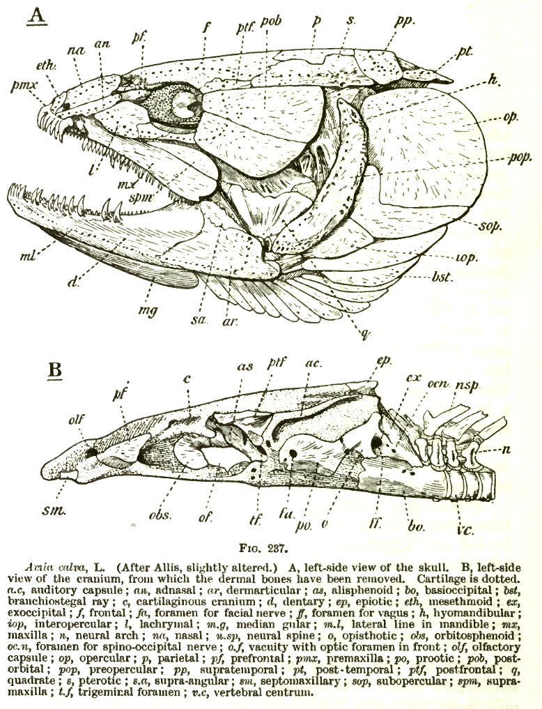
In some fishes, the palatoquadrate is the dorsal component of the mandibular arch, the ventral one being Meckel's cartilage. The palatoquadrate forms from splanchnocranium in various chordates including placoderms and acanthodians. See also * Hyomandibula * Fish anatomy * Helicoprion ''Helicoprion'' is an extinct genus of shark-like eugeneodont fish. Almost all fossil specimens are of spirally arranged clusters of the individuals' teeth, called "tooth whorls", which in life were embedded in the lower jaw. As with most extin ... References * Fish anatomy {{paleo-fish-stub ... [...More Info...] [...Related Items...] OR: [Wikipedia] [Google] [Baidu] |
Helicoprion
''Helicoprion'' is an extinct genus of shark-like eugeneodont fish. Almost all fossil specimens are of spirally arranged clusters of the individuals' teeth, called "tooth whorls", which in life were embedded in the lower jaw. As with most extinct cartilaginous fish, the skeleton is mostly unknown. Fossils of ''Helicoprion'' are known from a 20 million year timespan during the Permian period from the Artinskian stage of the Cisuralian (Early Permian) to the Roadian stage of the Guadalupian (Middle Permian). The closest living relatives of ''Helicoprion'' (and other eugeneodonts) are the chimaeras, though their relationship is very distant. The unusual tooth arrangement is thought to have been an adaption for feeding on soft bodied prey, and may have functioned as a deshelling mechanism for hard bodied cephalopods such as nautiloids and ammonoids. In 2013, systematic revision of ''Helicoprion'' via morphometric analysis of the tooth whorls found only ''H. davisii, H. bessonowi'' ... [...More Info...] [...Related Items...] OR: [Wikipedia] [Google] [Baidu] |
Meckel's Cartilage
In humans, the cartilaginous bar of the mandibular arch is formed by what are known as Meckel's cartilages (right and left) also known as Meckelian cartilages; above this the incus and malleus are developed. Meckel's cartilage arises from the first pharyngeal arch. The dorsal end of each cartilage is connected with the ear-capsule and is ossified to form the malleus; the ventral ends meet each other in the region of the symphysis menti, and are usually regarded as undergoing ossification to form that portion of the mandible which contains the incisor teeth. The intervening part of the cartilage disappears; the portion immediately adjacent to the malleus is replaced by fibrous membrane, which constitutes the sphenomandibular ligament, while from the connective tissue covering the remainder of the cartilage the greater part of the mandible is ossified. Johann Friedrich Meckel, the Younger discovered this cartilage in 1820. Evolution Meckel's cartilage is a piece of cartilage from ... [...More Info...] [...Related Items...] OR: [Wikipedia] [Google] [Baidu] |
Splanchnocranium
The splanchnocranium (or visceral skeleton) is the portion of the cranium that is derived from pharyngeal arches. ''Splanchno'' indicates to the gut because the face forms around the mouth, which is an end of the gut. The splanchnocranium consists of cartilage and endochondral bone. In mammals, the splanchnocranium comprises the three ear ossicles (i.e., incus, malleus, and stapes), as well as the alisphenoid, the styloid process, the hyoid apparatus, and the thyroid cartilage. In other tetrapods, such as amphibians and reptiles, homologous bones to those of mammals, such as the quadrate, articular, columella, and entoglossus are part of the splanchnocranium. See also * Dermatocranium * Endocranium The endocranium in comparative anatomy is a part of the skull base in vertebrates and it represents the basal, inner part of the cranium. The term is also applied to the outer layer of the dura mater in human anatomy. Structure Structurally, t ... References {{anatomy-stub ... [...More Info...] [...Related Items...] OR: [Wikipedia] [Google] [Baidu] |
Placodermi
Placodermi (from Greek πλάξ 'plate' and δέρμα 'skin', literally 'Plate (animal anatomy), plate-skinned') is a Class (biology), class of armoured prehistoric fish, known from fossils, which lived from the Silurian to the end of the Devonian period. Their head and thorax were covered by articulated armoured plates and the rest of the body was scale (zoology), scaled or naked, depending on the species. Placoderms were among the first jawed fish; their Fish jaw, jaws likely evolved from the first of their gill arches. Placoderms are thought to be paraphyly, paraphyletic, consisting of several distinct Outgroup (cladistics), outgroups or sister taxon, sister taxa to all living jawed vertebrates, which originated among their ranks. In contrast, one 2016 analysis concluded that placodermi are likely monophyletic, though these analyses have been further dismissed with more transitional taxa between placoderms and modern gnathosthomes, solidifying their paraphyletic status. Plac ... [...More Info...] [...Related Items...] OR: [Wikipedia] [Google] [Baidu] |
Acanthodii
Acanthodii or acanthodians is an extinct class of gnathostomes (jawed fishes), typically considered a paraphyletic group. They are currently considered to represent a grade of various fish lineages leading up to the extant Chondrichthyes, which includes living sharks, rays, and chimaeras. Acanthodians possess a mosaic of features shared with both osteichthyans (bony fish) and chondrichthyans (cartilaginous fish). In general body shape, they were similar to modern sharks, but their epidermis was covered with tiny rhomboid platelets like the scales of holosteians (gars, bowfins). A lower Silurian species, ''Fanjingshania renovata'', attributed to Climatiiformes is the oldest chondrichthyan with known anatomical features. The popular name "spiny sharks" is because they were superficially shark-shaped, with a streamlined body, paired fins, a strongly upturned tail, and stout, largely immovable bony spines supporting all the fins except the tail—hence, "spiny sharks". However, ... [...More Info...] [...Related Items...] OR: [Wikipedia] [Google] [Baidu] |
Hyomandibula
The hyomandibula, commonly referred to as hyomandibular one( la, os hyomandibulare, from el, hyoeides, "upsilon-shaped" (υ), and Latin: mandibula, "jawbone") is a set of bones that is found in the hyoid region in most fishes. It usually plays a role in suspending the jaws and/or operculum (teleostomi only). It is commonly suggested that in tetrapods (land animals), the hyomandibula evolved into the columella (stapes). Evolutionary context In jawless fishes a series of gills opened behind the mouth, and these gills became supported by cartilaginous elements. The first set of these elements surrounded the mouth to form the jaw. There are ample evidences For example: (1) both sets of bones are made from neural crest cells (rather than mesodermal tissue like most other bones); (2) both structures form the upper and lower bars that bend forward and are hinged in the middle; and (3) the musculature of the jaw seem homologous to the gill arches of jawless fishes. (Gilbert 2000) ... [...More Info...] [...Related Items...] OR: [Wikipedia] [Google] [Baidu] |
Fish Anatomy
Fish anatomy is the study of the form or morphology of fish. It can be contrasted with fish physiology, which is the study of how the component parts of fish function together in the living fish. In practice, fish anatomy and fish physiology complement each other, the former dealing with the structure of a fish, its organs or component parts and how they are put together, such as might be observed on the dissecting table or under the microscope, and the latter dealing with how those components function together in living fish. The anatomy of fish is often shaped by the physical characteristics of water, the medium in which fish live. Water is much denser than air, holds a relatively small amount of dissolved oxygen, and absorbs more light than air does. The body of a fish is divided into a head, trunk and tail, although the divisions between the three are not always externally visible. The skeleton, which forms the support structure inside the fish, is either made of cartilage ( ... [...More Info...] [...Related Items...] OR: [Wikipedia] [Google] [Baidu] |

