|
Palatodonta
''Palatodonta'' is an extinct genus of basal placodontiform marine reptile known from the early Middle Triassic (early Anisian stage) of the Netherlands. It is closely related to a group of marine reptiles called placodonts, characterized by their crushing teeth and shell-like body armor. ''Palatodonta'' is transitional between placodonts and earlier reptiles; like placodonts, it has teeth on its palate, but while these teeth are thick and blunt in placodonts, ''Palatodonta'' has palatal teeth that are thin and pointed (like the teeth that line the jaws of most other reptiles). Discovery ''Palatodonta'' was first named by James M. Neenan, Nicole Klein and Torsten M. Scheyer in 2013 and the type species is ''Palatodonta bleekeri''. The generic name refers to the row of teeth on the palatine bone of the palate. The specific name honours Remco Bleeker, an amateur paleontologist, who discovered the fossil in the summer of 2010 at the Silbeco quarry near Winterswijk. ''Palatodonta' ... [...More Info...] [...Related Items...] OR: [Wikipedia] [Google] [Baidu] |
Placodontiform
Placodontiformes is an extinct clade of sauropterygian marine reptiles that includes placodonts and the non-placodont ''Palatodonta''. It was erected in 2013 with the description of ''Palatodonta''. Placodontiformes is the most basal clade of Sauropterygia and the sister group of Eosauropterygia, which includes all other sauropterygians. Phylogeny Below is a cladogram A cladogram (from Greek ''clados'' "branch" and ''gramma'' "character") is a diagram used in cladistics to show relations among organisms. A cladogram is not, however, an evolutionary tree because it does not show how ancestors are related to d ... from Neenan ''et al.'' (2013) showing the position of Placodontiformes within Sauropterygia: References Placodonts {{triassic-reptile-stub ... [...More Info...] [...Related Items...] OR: [Wikipedia] [Google] [Baidu] |
Placodont
Placodonts (" Tablet teeth") are an extinct order of marine reptiles that lived during the Triassic period, becoming extinct at the end of the period. They were part of Sauropterygia, the group that includes plesiosaurs. Placodonts were generally between in length, with some of the largest measuring long. The first specimen was discovered in 1830. They have been found throughout central Europe, North Africa, the Middle East and China. Palaeobiology The earliest forms, like ''Placodus'', which lived in the early to middle Triassic, resembled barrel-bodied lizards superficially similar to the marine iguana of today, but larger. In contrast to the marine iguana, which feeds on algae, the placodonts ate molluscs and so their teeth were flat and tough to crush shells. In the earliest periods, their size was probably enough to keep away the top sea predators of the time: the sharks. However, as time passed, other kinds of carnivorous reptiles began to colonize the seas, such as ... [...More Info...] [...Related Items...] OR: [Wikipedia] [Google] [Baidu] |
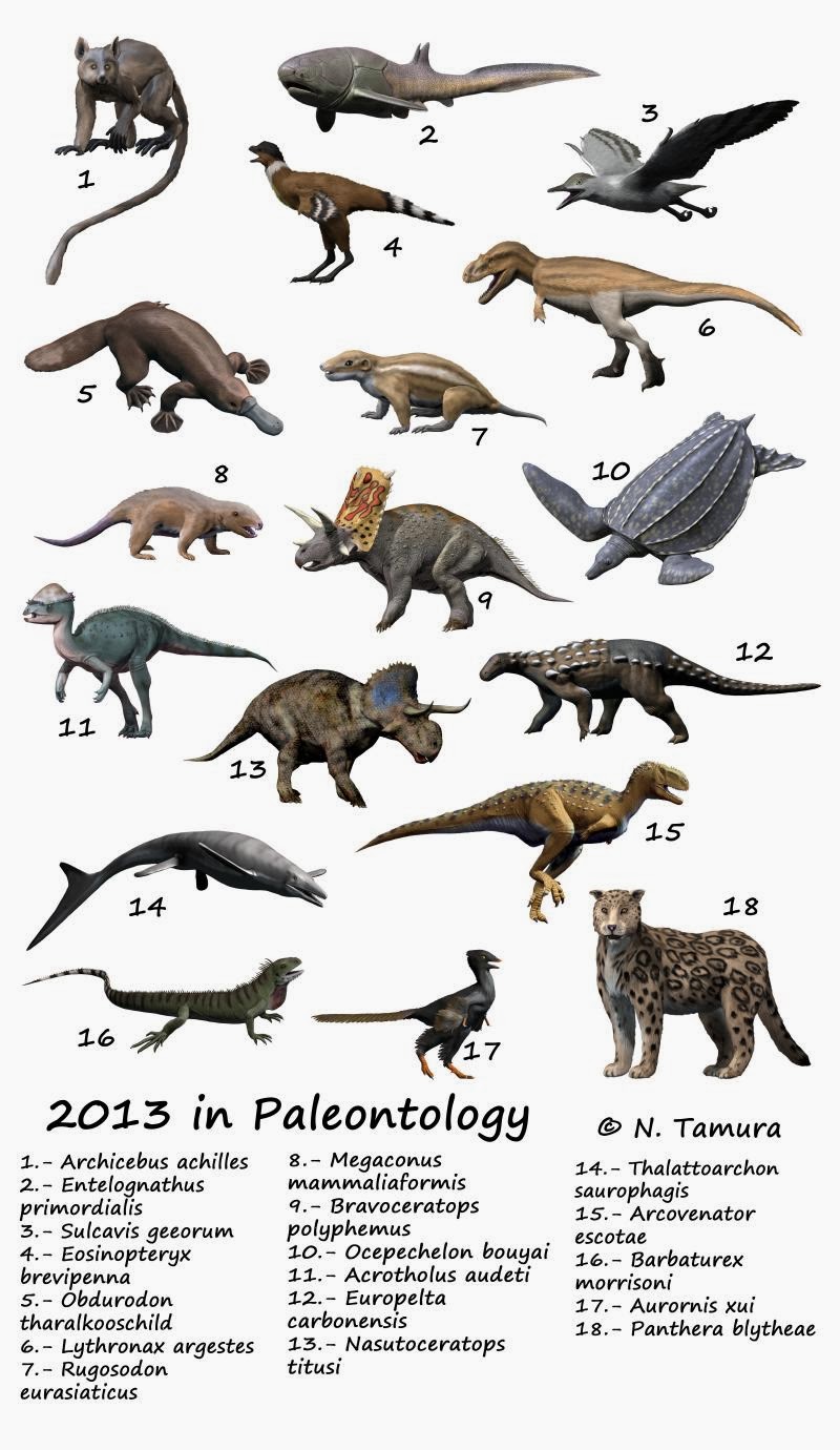
2013 In Paleontology
Plants Cnidarians Arthropods Bryozoans Brachiopods Molluscs Echinoderms Conodonts Fishes Amphibians Research * Laloy ''et al.'' (2013) reinterpret the Eocene frog species ''Rana cadurcorum'' from the Quercy Phosphorites (France) as a junior synonym of '' Thaumastosaurus gezei''. Newly named temnospondyls Newly named lepospondyls Newly named lissamphibians Turtles Research * A study on the anatomy of the brain and inner ear of the Jurassic turtle ''Plesiochelys etalloni'' is published by Paulina Carabajal ''et al.'' (2013). Newly named turtles Thalattosaurs Ichthyopterygians Lepidosauromorphs Newly named sauropterygians Newly named rhynchocephalians Newly named lizards Newly named snakes Archosauromorphs Newly named basal archosauromorphs Archosaurs Other reptiles Synapsids Non-mammalian synapsids Research * The postcranial skeleton of therocephalian '' Ictidosuchoides'' is described by Heidi Fourie (2013). New taxa ... [...More Info...] [...Related Items...] OR: [Wikipedia] [Google] [Baidu] |
Premaxilla
The premaxilla (or praemaxilla) is one of a pair of small cranial bones at the very tip of the upper jaw of many animals, usually, but not always, bearing teeth. In humans, they are fused with the maxilla. The "premaxilla" of therian mammal has been usually termed as the incisive bone. Other terms used for this structure include premaxillary bone or ''os premaxillare'', intermaxillary bone or ''os intermaxillare'', and Goethe's bone. Human anatomy In human anatomy, the premaxilla is referred to as the incisive bone (') and is the part of the maxilla which bears the incisor teeth, and encompasses the anterior nasal spine and alar region. In the nasal cavity, the premaxillary element projects higher than the maxillary element behind. The palatal portion of the premaxilla is a bony plate with a generally transverse orientation. The incisive foramen is bound anteriorly and laterally by the premaxilla and posteriorly by the palatine process of the maxilla. It is formed from the ... [...More Info...] [...Related Items...] OR: [Wikipedia] [Google] [Baidu] |
Maxilla
The maxilla (plural: ''maxillae'' ) in vertebrates is the upper fixed (not fixed in Neopterygii) bone of the jaw formed from the fusion of two maxillary bones. In humans, the upper jaw includes the hard palate in the front of the mouth. The two maxillary bones are fused at the intermaxillary suture, forming the anterior nasal spine. This is similar to the mandible (lower jaw), which is also a fusion of two mandibular bones at the mandibular symphysis. The mandible is the movable part of the jaw. Structure In humans, the maxilla consists of: * The body of the maxilla * Four processes ** the zygomatic process ** the frontal process of maxilla ** the alveolar process ** the palatine process * three surfaces – anterior, posterior, medial * the Infraorbital foramen * the maxillary sinus * the incisive foramen Articulations Each maxilla articulates with nine bones: * two of the cranium: the frontal and ethmoid * seven of the face: the nasal, zygomatic, lacrimal, inferior n ... [...More Info...] [...Related Items...] OR: [Wikipedia] [Google] [Baidu] |
Nasal Bone
The nasal bones are two small oblong bones, varying in size and form in different individuals; they are placed side by side at the middle and upper part of the face and by their junction, form the bridge of the upper one third of the nose. Each has two surfaces and four borders. Structure The two nasal bones are joined at the midline internasal suture and make up the bridge of the nose. Surfaces The ''outer surface'' is concavo-convex from above downward, convex from side to side; it is covered by the procerus and nasalis muscles, and perforated about its center by a foramen, for the transmission of a small vein. The ''inner surface'' is concave from side to side, and is traversed from above downward, by a groove for the passage of a branch of the nasociliary nerve. Articulations The nasal articulates with four bones: two of the cranium, the frontal and ethmoid, and two of the face, the opposite nasal and the maxilla. Other animals In primitive bony fish and tetrapod ... [...More Info...] [...Related Items...] OR: [Wikipedia] [Google] [Baidu] |
Orbit (anatomy)
In anatomy, the orbit is the cavity or socket of the skull in which the eye and its appendages are situated. "Orbit" can refer to the bony socket, or it can also be used to imply the contents. In the adult human, the volume of the orbit is , of which the eye occupies . The orbital contents comprise the eye, the orbital and retrobulbar fascia, extraocular muscles, cranial nerves II, III, IV, V, and VI, blood vessels, fat, the lacrimal gland with its sac and duct, the eyelids, medial and lateral palpebral ligaments, cheek ligaments, the suspensory ligament, septum, ciliary ganglion and short ciliary nerves. Structure The orbits are conical or four-sided pyramidal cavities, which open into the midline of the face and point back into the head. Each consists of a base, an apex and four walls."eye, human."Encyclopædia Britannica from Encyclopædia Britannica 2006 Ultimate Reference Suite DVD 2009 Openings There are two important foramina, or windows, two important fissu ... [...More Info...] [...Related Items...] OR: [Wikipedia] [Google] [Baidu] |
Prefrontal Bone
The prefrontal bone is a bone separating the lacrimal and frontal bones in many tetrapod skulls. It first evolved in the sarcopterygian clade Rhipidistia, which includes lungfish and the Tetrapodomorpha. The prefrontal is found in most modern and extinct lungfish, amphibians and reptiles. The prefrontal is lost in early mammaliaforms and so is not present in modern mammals either. In dinosaurs The prefrontal bone is a very small bone near the top of the skull, which is lost in many groups of coelurosaurian theropod dinosaurs and is completely absent in their modern descendants, the birds. Conversely, a well developed prefrontal is considered to be a primitive feature in dinosaurs. The prefrontal makes contact with several other bones in the skull. The anterior part of the bone articulates with the nasal bone and the lacrimal bone. The posterior part of the bone articulates with the frontal bone and more rarely the palpebral bone The palpebral bone is a small dermal bone found ... [...More Info...] [...Related Items...] OR: [Wikipedia] [Google] [Baidu] |
Middle Triassic
In the geologic timescale, the Middle Triassic is the second of three epochs of the Triassic period or the middle of three series in which the Triassic system is divided in chronostratigraphy. The Middle Triassic spans the time between Ma and Ma (million years ago). It is preceded by the Early Triassic Epoch and followed by the Late Triassic Epoch. The Middle Triassic is divided into the Anisian and Ladinian ages or stages. Formerly the middle series in the Triassic was also known as Muschelkalk. This name is now only used for a specific unit of rock strata with approximately Middle Triassic age, found in western Europe. Middle Triassic fauna Following the Permian–Triassic extinction event, the most devastating of all mass-extinctions, life recovered slowly. In the Middle Triassic, many groups of organisms reached higher diversity again, such as the marine reptiles (e.g. ichthyosaurs, sauropterygians, thallatosaurs), ray-finned fish and many invertebrate groups like ... [...More Info...] [...Related Items...] OR: [Wikipedia] [Google] [Baidu] |
Skull
The skull is a bone protective cavity for the brain. The skull is composed of four types of bone i.e., cranial bones, facial bones, ear ossicles and hyoid bone. However two parts are more prominent: the cranium and the mandible. In humans, these two parts are the neurocranium and the viscerocranium ( facial skeleton) that includes the mandible as its largest bone. The skull forms the anterior-most portion of the skeleton and is a product of cephalisation—housing the brain, and several sensory structures such as the eyes, ears, nose, and mouth. In humans these sensory structures are part of the facial skeleton. Functions of the skull include protection of the brain, fixing the distance between the eyes to allow stereoscopic vision, and fixing the position of the ears to enable sound localisation of the direction and distance of sounds. In some animals, such as horned ungulates (mammals with hooves), the skull also has a defensive function by providing the mount (on the front ... [...More Info...] [...Related Items...] OR: [Wikipedia] [Google] [Baidu] |
Frontal Bone
The frontal bone is a bone in the human skull. The bone consists of two portions.''Gray's Anatomy'' (1918) These are the vertically oriented squamous part, and the horizontally oriented orbital part, making up the bony part of the forehead, part of the bony orbital cavity holding the eye, and part of the bony part of the nose respectively. The name comes from the Latin word ''frons'' (meaning " forehead"). Structure of the frontal bone The frontal bone is made up of two main parts. These are the squamous part, and the orbital part. The squamous part marks the vertical, flat, and also the biggest part, and the main region of the forehead. The orbital part is the horizontal and second biggest region of the frontal bone. It enters into the formation of the roofs of the orbital and nasal cavities. Sometimes a third part is included as the nasal part of the frontal bone, and sometimes this is included with the squamous part. The nasal part is between the brow ridges, and ends in ... [...More Info...] [...Related Items...] OR: [Wikipedia] [Google] [Baidu] |


.jpg)