|
Palate (bones)
In anatomy, the palatine bones () are two irregular bones of the facial skeleton in many animal species, located above the uvula in the throat. Together with the maxillae, they comprise the hard palate. (''Palate'' is derived from the Latin ''palatum''.) Structure The palatine bones are situated at the back of the nasal cavity between the maxilla and the pterygoid process of the sphenoid bone. They contribute to the walls of three cavities: the floor and lateral walls of the nasal cavity, the roof of the mouth, and the floor of the orbits. They help to form the pterygopalatine and pterygoid fossae, and the inferior orbital fissures. Each palatine bone somewhat resembles the letter L, and consists of a horizontal plate, a perpendicular plate, and three projecting processes—the pyramidal process, which is directed backward and lateral from the junction of the two parts, and the orbital and sphenoidal processes, which surmount the vertical part, and are separated by a deep ... [...More Info...] [...Related Items...] OR: [Wikipedia] [Google] [Baidu] |
Anatomy
Anatomy () is the branch of biology concerned with the study of the structure of organisms and their parts. Anatomy is a branch of natural science that deals with the structural organization of living things. It is an old science, having its beginnings in prehistoric times. Anatomy is inherently tied to developmental biology, embryology, comparative anatomy, evolutionary biology, and phylogeny, as these are the processes by which anatomy is generated, both over immediate and long-term timescales. Anatomy and physiology, which study the structure and function (biology), function of organisms and their parts respectively, make a natural pair of related disciplines, and are often studied together. Human anatomy is one of the essential basic research, basic sciences that are applied in medicine. The discipline of anatomy is divided into macroscopic scale, macroscopic and microscopic scale, microscopic. Gross anatomy, Macroscopic anatomy, or gross anatomy, is the examination of an ... [...More Info...] [...Related Items...] OR: [Wikipedia] [Google] [Baidu] |
Sphenoidal Process Of Palatine Bone
The sphenoidal process of the palatine bone In anatomy, the palatine bones () are two irregular bones of the facial skeleton in many animal species, located above the uvula in the throat. Together with the maxillae, they comprise the hard palate. (''Palate'' is derived from the Latin ''pa ... is a thin, compressed plate, much smaller than the orbital, and directed upward and medialward. It presents three surfaces and two borders. * The superior surface articulates with the root of the pterygoid process and the under surface of the sphenoidal concha, its medial border reaching as far as the ala of the vomer; it presents a groove which contributes to the formation of the pharyngeal canal. * The medial surface is concave, and forms part of the lateral wall of the nasal cavity. * The lateral surface is divided into an articular and a non-articular portion: the former is rough, for articulation with the medial pterygoid plate; the latter is smooth, and forms part of the pterygopalat ... [...More Info...] [...Related Items...] OR: [Wikipedia] [Google] [Baidu] |
Salamander
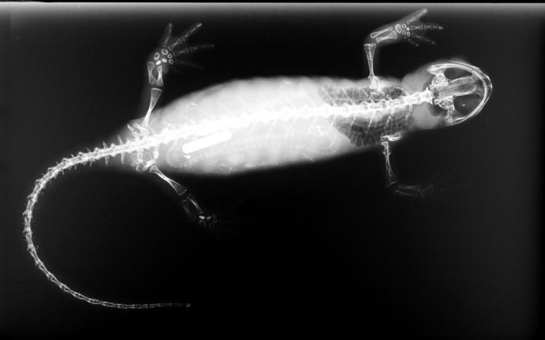
Salamanders are a group of amphibians typically characterized by their lizard-like appearance, with slender bodies, blunt snouts, short limbs projecting at right angles to the body, and the presence of a tail in both larvae and adults. All ten extant salamander families are grouped together under the order Urodela. Salamander diversity is highest in eastern North America, especially in the Appalachian Mountains; most species are found in the Holarctic realm, with some species present in the Neotropical realm. Salamanders rarely have more than four toes on their front legs and five on their rear legs, but some species have fewer digits and others lack hind limbs. Their permeable skin usually makes them reliant on habitats in or near water or other cool, damp places. Some salamander species are fully aquatic throughout their lives, some take to the water intermittently, and others are entirely terrestrial as adults. This group of amphibians is capable of regenerating lost lim ... [...More Info...] [...Related Items...] OR: [Wikipedia] [Google] [Baidu] |
Frog
A frog is any member of a diverse and largely Carnivore, carnivorous group of short-bodied, tailless amphibians composing the order (biology), order Anura (ανοὐρά, literally ''without tail'' in Ancient Greek). The oldest fossil "proto-frog" ''Triadobatrachus'' is known from the Early Triassic of Madagascar, but molecular clock, molecular clock dating suggests their split from other amphibians may extend further back to the Permian, 265 Myr, million years ago. Frogs are widely distributed, ranging from the tropics to subarctic regions, but the greatest concentration of species diversity is in tropical rainforest. Frogs account for around 88% of extant amphibian species. They are also one of the five most diverse vertebrate orders. Warty frog species tend to be called toads, but the distinction between frogs and toads is informal, not from Taxonomy (biology), taxonomy or evolutionary history. An adult frog has a stout body, protruding eyes, anteriorly-attached tongue, limb ... [...More Info...] [...Related Items...] OR: [Wikipedia] [Google] [Baidu] |
Amphibian
Amphibians are tetrapod, four-limbed and ectothermic vertebrates of the Class (biology), class Amphibia. All living amphibians belong to the group Lissamphibia. They inhabit a wide variety of habitats, with most species living within terrestrial animal, terrestrial, fossorial, arboreal or freshwater aquatic ecosystems. Thus amphibians typically start out as larvae living in water, but some species have developed behavioural adaptations to bypass this. The young generally undergo metamorphosis from larva with gills to an adult air-breathing form with lungs. Amphibians use their skin as a secondary respiratory surface and some small terrestrial salamanders and frogs lack lungs and rely entirely on their skin. They are superficially similar to reptiles like lizards but, along with mammals and birds, reptiles are amniotes and do not require water bodies in which to breed. With their complex reproductive needs and permeable skins, amphibians are often ecological indicators; in re ... [...More Info...] [...Related Items...] OR: [Wikipedia] [Google] [Baidu] |
Tetrapod
Tetrapods (; ) are four-limbed vertebrate animals constituting the superclass Tetrapoda (). It includes extant and extinct amphibians, sauropsids ( reptiles, including dinosaurs and therefore birds) and synapsids (pelycosaurs, extinct therapsids and all extant mammals). Tetrapods evolved from a clade of primitive semiaquatic animals known as the Tetrapodomorpha which, in turn, evolved from ancient lobe-finned fish (sarcopterygians) around 390 million years ago in the Middle Devonian period; their forms were transitional between lobe-finned fishes and true four-limbed tetrapods. Limbed vertebrates (tetrapods in the broad sense of the word) are first known from Middle Devonian trackways, and body fossils became common near the end of the Late Devonian but these were all aquatic. The first crown-tetrapods (last common ancestors of extant tetrapods capable of terrestrial locomotion) appeared by the very early Carboniferous, 350 million years ago. The specific aquatic ancestors ... [...More Info...] [...Related Items...] OR: [Wikipedia] [Google] [Baidu] |
Osteichthyes
Osteichthyes (), popularly referred to as the bony fish, is a diverse superclass of fish that have skeletons primarily composed of bone tissue. They can be contrasted with the Chondrichthyes, which have skeletons primarily composed of cartilage. The vast majority of fish are members of Osteichthyes, which is an extremely diverse and abundant group consisting of 45 orders, and over 435 families and 28,000 species. It is the largest class of vertebrates in existence today. The group Osteichthyes is divided into the ray-finned fish (Actinopterygii) and lobe-finned fish (Sarcopterygii). The oldest known fossils of bony fish are about 425 million years old, which are also transitional fossils, showing a tooth pattern that is in between the tooth rows of sharks and bony fishes. Osteichthyes can be compared to Euteleostomi. In paleontology the terms are synonymous. In ichthyology the difference is that Euteleostomi presents a cladistic view which includes the terrestrial tetrap ... [...More Info...] [...Related Items...] OR: [Wikipedia] [Google] [Baidu] |
Sphenopalatine Artery
The sphenopalatine artery (nasopalatine artery) is an artery of the head, commonly known as the artery of epistaxis. Course The sphenopalatine artery is a branch of the maxillary artery which passes through the sphenopalatine foramen into the cavity of the nose, at the back part of the superior meatus. Here it gives off its posterior lateral nasal branches. Crossing the under surface of the sphenoid, the sphenopalatine artery ends on the nasal septum as the posterior septal branches. Here it will anastomose with the branches of the greater palatine artery. Clinical significance The sphenopalatine artery is the artery responsible for the most serious, posterior nosebleeds (also known as epistaxis). It can be ligated surgically or blocked under image guidance with minimally invasive techniques by interventional radiologist using tiny microparticles to control such nosebleeds. See also *Kiesselbach's plexus Kiesselbach's plexus is an anastomotic arterial network (plexus) of four ... [...More Info...] [...Related Items...] OR: [Wikipedia] [Google] [Baidu] |
Sphenopalatine Foramen
The sphenopalatine foramen is a foramen in the skull that connects the nasal cavity with the pterygopalatine fossa. Structure The processes of the superior border of the palatine bone are separated by the ''sphenopalatine notch'', which is converted into the sphenopalatine foramen by the under surface of the body of the sphenoid. In the articulated skull this foramen leads from the pterygopalatine fossa into the posterior part of the superior meatus of the nose, and transmits the sphenopalatine artery and vein and the posterior superior lateral nasal nerve and nasopalatine nerve The nasopalatine nerve (long sphenopalatine nerve) is a nerve of the head. It is a branch of the pterygopalatine ganglion, a continuation from the maxillary nerve (V2). It supplies parts of the palate and nasal septum. Structure The nasopalati ...s. Additional images File:Gray167.png, Articulation of left palatine bone with maxilla. File:Gray168.png, Left palatine bone. Nasal aspect. Enlarged. ... [...More Info...] [...Related Items...] OR: [Wikipedia] [Google] [Baidu] |
Lesser Palatine Foramen
Behind the greater palatine foramen is the pyramidal process of the palatine bone, perforated by one or more lesser palatine foramina which carry the lesser palatine nerve, and marked by the commencement of a transverse ridge, for the attachment of the tendinous expansion of the tensor veli palatini. See also * Greater palatine foramen At either posterior angle of the hard palate is the greater palatine foramen, for the transmission of the descending palatine vessels and greater palatine nerve; and running anteriorly (forward) and medially (towards the center-line) from it is a g ... References External links * Bones of the head and neck {{musculoskeletal-stub ... [...More Info...] [...Related Items...] OR: [Wikipedia] [Google] [Baidu] |
Greater Palatine Foramen
At either posterior angle of the hard palate is the greater palatine foramen, for the transmission of the descending palatine vessels and greater palatine nerve; and running anteriorly (forward) and medially (towards the center-line) from it is a groove, for the same vessels and nerve. Variations The greater palatine foramen (GPF) is related to the upper 3rd molar tooth in most of the skulls (55%), 2nd molar in (12%), between the 2nd and 3rd molar in (19%) and retromolar in (14%). The shape of the foramen is elongated antero-posteriorly; however, an unusually crescent shaped foramen is rare. See also * Greater palatine canal * Lesser palatine foramina Behind the greater palatine foramen is the pyramidal process of the palatine bone, perforated by one or more lesser palatine foramina which carry the lesser palatine nerve, and marked by the commencement of a transverse ridge, for the attachment of ... References External links * * * Bones of the head and neck { ... [...More Info...] [...Related Items...] OR: [Wikipedia] [Google] [Baidu] |
Vomer
The vomer (; lat, vomer, lit=ploughshare) is one of the unpaired facial bones of the skull. It is located in the midsagittal line, and articulates with the sphenoid, the ethmoid, the left and right palatine bones, and the left and right maxillary bones. The vomer forms the inferior part of the nasal septum in humans, with the superior part formed by the perpendicular plate of the ethmoid bone. The name is derived from the Latin word for a ploughshare and the shape of the bone. In humans The vomer is situated in the median plane, but its anterior portion is frequently bent to one side. It is thin, somewhat quadrilateral in shape, and forms the hinder and lower part of the nasal septum; it has two surfaces and four borders. The surfaces are marked by small furrows for blood vessels, and on each is the nasopalatine groove, which runs obliquely downward and forward, and lodges the nasopalatine nerve and vessels. Borders The ''superior border'', the thickest, presents a dee ... [...More Info...] [...Related Items...] OR: [Wikipedia] [Google] [Baidu] |


_Ranomafana.jpg)
.png)

