|
Pulp Stone
Pulp stones (also denticles or endoliths) are nodular, calcified masses appearing in either or both the coronal and root portion of the pulp organ in teeth. Pulp stones are not painful unless they impinge on nerves. They are classified: :A) On the basis of structure ::1) True pulp stones: formed of dentin by odontoblasts ::2) False pulp stones: formed by mineralization of degenerating pulp cells, often in a concentric pattern :B) On the basis of location ::1) Free: entirely surrounded by pulp tissue ::2) Adherent: partly fused with dentin ::3) Embedded: entirely surrounded by dentin Introduction Pulp stones are discrete calcifications found in the pulp chamber of the tooth which may undergo changes to become diffuse pulp calcifications such as dystrophic calcification. They are usually noticed by radiographic examination and appeared as round or ovoid radiopaque lesions. Clinically, a tooth with a pulp stone has normal appearance like any other tooth. The number of pulp stone ... [...More Info...] [...Related Items...] OR: [Wikipedia] [Google] [Baidu] |
X-ray Manual - U
X-rays (or rarely, ''X-radiation'') are a form of high-energy electromagnetic radiation. In many languages, it is referred to as Röntgen radiation, after the German scientist Wilhelm Conrad Röntgen, who discovered it in 1895 and named it ''X-radiation'' to signify an unknown type of radiation.Novelline, Robert (1997). ''Squire's Fundamentals of Radiology''. Harvard University Press. 5th edition. . X-ray wavelengths are shorter than those of ultraviolet rays and longer than those of gamma rays. There is no universally accepted, strict definition of the bounds of the X-ray band. Roughly, X-rays have a wavelength ranging from 10 nanometers to 10 picometers, corresponding to frequencies in the range of 30 petahertz to 30 exahertz ( to ) and photon energies in the range of 100 eV to 100 keV, respectively. X-rays can penetrate many solid substances such as construction materials and living tissue, so X-ray radiography is widely used in medical diagn ... [...More Info...] [...Related Items...] OR: [Wikipedia] [Google] [Baidu] |
Common Carotid Artery
In anatomy, the left and right common carotid arteries (carotids) ( in Merriam-Webster Online Dictionary '.) are arteries that supply the head and neck with oxygenated blood; they divide in the neck to form the external and internal carotid arteries. ... [...More Info...] [...Related Items...] OR: [Wikipedia] [Google] [Baidu] |
Pulp (tooth)
The pulp is the connective tissue, nerves, blood vessels, and odontoblasts that comprise the innermost layer of a tooth. The pulp's activity and signalling processes regulate its behaviour. Anatomy The pulp is the neurovascular bundle central to each tooth, permanent or primary. It is composed of a central pulp chamber, pulp horns, and radicular canals. The large mass of the pulp is contained within the pulp chamber, which is contained in and mimics the overall shape of the crown of the tooth.Illustrated Dental Embryology, Histology, and Anatomy, Bath-Balogh and Fehrenbach, Elsevier, 2011, page 164. Because of the continuous deposition of the dentine, the pulp chamber becomes smaller with the age. This is not uniform throughout the coronal pulp but progresses faster on the floor than on the roof or sidewalls. Radicular pulp canals extend down from the cervical region of the crown to the root apex. They are not always straight but vary in shape, size, and number. They a ... [...More Info...] [...Related Items...] OR: [Wikipedia] [Google] [Baidu] |
Endodontic Therapy
Root canal treatment (also known as endodontic therapy, endodontic treatment, or root canal therapy) is a treatment sequence for the infected pulp of a tooth which is intended to result in the elimination of infection and the protection of the decontaminated tooth from future microbial invasion. Root canals, and their associated pulp chamber, are the physical hollows within a tooth that are naturally inhabited by nerve tissue, blood vessels and other cellular entities. Together, these items constitute the dental pulp. Endodontic therapy involves the ''removal'' of these structures, disinfection and the subsequent shaping, cleaning, and decontamination of the hollows with small files and irrigating solutions, and the ''obturation'' (filling) of the decontaminated canals. Filling of the cleaned and decontaminated canals is done with an inert filling such as gutta-percha and typically a zinc oxide eugenol-based cement. Epoxy resin is employed to bind gutta-percha in some ro ... [...More Info...] [...Related Items...] OR: [Wikipedia] [Google] [Baidu] |
Endodontics
Endodontics (from the Greek roots ''endo-'' "inside" and ''odont-'' "tooth") is the dental specialty concerned with the study and treatment of the dental pulp. Overview Endodontics encompasses the study (practice) of the basic and clinical sciences of normal dental pulp, the etiology, diagnosis, prevention, and treatment of diseases and injuries of the dental pulp along with associated periradicular conditions. In clinical terms, endodontics involves either preserving part, or all of the dental pulp in health, or removing all of the pulp in irreversible disease. This includes teeth with irreversibly inflamed and infected pulpal tissue. Not only does endodontics involve treatment when a dental pulp is present, but also includes preserving teeth which have failed to respond to non-surgical endodontic treatment, or for teeth that have developed new lesions, e.g., when root canal re-treatment is required, or periradicular surgery. Endodontic treatment is one of the most comm ... [...More Info...] [...Related Items...] OR: [Wikipedia] [Google] [Baidu] |
Pulpitis
Pulpitis is inflammation of dental pulp tissue. The pulp contains the blood vessels, the nerves, and connective tissue inside a tooth and provides the tooth’s blood and nutrients. Pulpitis is mainly caused by bacterial infection which itself is a secondary development of caries (tooth decay). It manifests itself in the form of a toothache. Signs and symptoms Increased sensitivity to stimuli, specifically hot and cold, is a common symptom of pulpitis. A prolonged throbbing pain may be associated with the disease. However, pulpitis can also occur without any pain. Reversible pulpitis is characterised by intermittent, brief discomfort initiated by a hot, cold or sweet stimulus. The pain evoked is of short duration and there is no lingering or spontaneous pain. The pain ceases within a short period after removal of the stimulus. With a reversible pulpitis, sleep is usually not affected and no analgesics are necessary. Usually, no atypical change is evident on the radiog ... [...More Info...] [...Related Items...] OR: [Wikipedia] [Google] [Baidu] |
Bleeding
Bleeding, hemorrhage, haemorrhage or blood loss, is blood escaping from the circulatory system from damaged blood vessels. Bleeding can occur internally, or externally either through a natural opening such as the mouth, nose, ear, urethra, vagina or anus, or through a puncture in the skin. Hypovolemia is a massive decrease in blood volume, and death by excessive loss of blood is referred to as exsanguination. Typically, a healthy person can endure a loss of 10–15% of the total blood volume without serious medical difficulties (by comparison, blood donation typically takes 8–10% of the donor's blood volume). The stopping or controlling of bleeding is called hemostasis and is an important part of both first aid and surgery. Types * Upper head ** Intracranial hemorrhage – bleeding in the skull. ** Cerebral hemorrhage – a type of intracranial hemorrhage, bleeding within the brain tissue itself. ** Intracerebral hemorrhage – bleeding in the brain caused by the rupture o ... [...More Info...] [...Related Items...] OR: [Wikipedia] [Google] [Baidu] |
Dentin Dysplasia
Dentin dysplasia (DD) is a rare genetic developmental disorder affecting dentine production of the teeth, commonly exhibiting an autosomal dominant inheritance that causes malformation of the root. It affects both primary and permanent dentitions in approximately 1 in every 100,000 patients. It is characterized by presence of normal enamel but atypical dentin with abnormal pulpal morphology. Witkop in 1972 classified DD into two types which are Type I (DD-1) is the radicular type, and type II (DD-2) is the coronal type. DD-1 has been further divided into 4 different subtypes (DD-1a,1b,1c,1d) based on the radiographic features. Signs and symptoms Clinically the teeth look normal in colour and morphologic appearance; however, they are commonly very mobile and exfoliated prematurely. Both primary and permanent dentitions can be affected by either type I or type II dentin dysplasia. However, deciduous teeth affected by type II dentin dysplasia have a characteristic blue-amber disco ... [...More Info...] [...Related Items...] OR: [Wikipedia] [Google] [Baidu] |
Tumoral Calcinosis
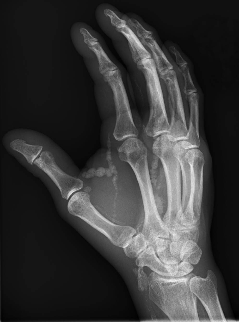
Tumoral calcinosis is a rare condition in which there is calcium deposition in the soft tissue in periarticular location, around joints, outside the joint capsule. They are frequently (0.5–3%) seen in patients undergoing renal dialysis. Clinically also known as hyperphosphatemic familial tumoral calcinosis (HFTC), is often caused by genetic mutations in genes that regulate phosphate physiology in the body (leading to too much phosphate (hyperphosphatemia)). Best described genes that harbour mutations in humans are FGF-23, Klotho (KL), or GALNT3. A zebrafish animal model with reduced GALNT3 expression also showed HFTC-like phenotype, indicating an evolutionary conserved mechanism that is involved in developing tumoral calcinosis. Clinical features The name indicates calcinosis (calcium deposition) which resembles tumor (like a new growth). They are not true neoplasms – they don't have dividing cells. They are just deposition of inorganic calcium with serum exudate. Childre ... [...More Info...] [...Related Items...] OR: [Wikipedia] [Google] [Baidu] |
Calcinosis
Calcinosis is the formation of calcium deposits in any soft tissue. It is a rare condition that has many different causes. These range from infection and injury to systemic diseases like kidney failure. Types Dystrophic calcification The most common type of calcinosis is dystrophic calcification. This type of calcification can occur as a response to any soft tissue damage, including that involved in implantation of medical devices. Metastatic calcification Metastatic calcification involves a systemic calcium excess imbalance, which can be caused by hypercalcemia, kidney failure, milk-alkali syndrome, lack or excess of other minerals, or other causes. Tumoral calcinosis The cause of the rare condition of tumoral calcinosis is not entirely understood. It is generally characterized by large, globular calcifications near joints. See also * Calcification * Calcinosis cutis * Dermatomyositis * Fahr's syndrome * Hyperphosphatemia * Primrose syndrome * Scleroderma Scleroderma ... [...More Info...] [...Related Items...] OR: [Wikipedia] [Google] [Baidu] |
Ehlers–Danlos Syndromes
Ehlers–Danlos syndromes (EDS) are a group of 13 genetic connective-tissue disorders in the current classification, with the latest type discovered in 2018. Symptoms include loose joints, joint pain, stretchy velvety skin, and abnormal scar formation. These may be noticed at birth or in early childhood. Complications may include aortic dissection, joint dislocations, scoliosis, chronic pain, or early osteoarthritis. EDS occurs due to variations of more than 19 genes that are present at birth. The specific gene affected determines the type of EDS. Some cases result from a new variation occurring during early development, while others are inherited in an autosomal dominant or recessive manner. Typically, these variations result in defects in the structure or processing of the protein collagen. Diagnosis is often based on symptoms and confirmed by genetic testing or skin biopsy, but people may initially be misdiagnosed with hypochondriasis, depression, or chronic fatigue synd ... [...More Info...] [...Related Items...] OR: [Wikipedia] [Google] [Baidu] |
Marfan Syndrome
Marfan syndrome (MFS) is a multi-systemic genetic disorder that affects the connective tissue. Those with the condition tend to be tall and thin, with long arms, legs, fingers, and toes. They also typically have exceptionally flexible joints and abnormally curved spines. The most serious complications involve the heart and aorta, with an increased risk of mitral valve prolapse and aortic aneurysm. The lungs, eyes, bones, and the covering of the spinal cord are also commonly affected. The severity of the symptoms is variable. MFS is caused by a mutation in '' FBN1'', one of the genes that makes fibrillin, which results in abnormal connective tissue. It is an autosomal dominant disorder. In about 75% of cases, it is inherited from a parent with the condition, while in about 25% it is a new mutation. Diagnosis is often based on the Ghent criteria. There is no known cure for MFS. Many of those with the disorder have a normal life expectancy with proper treatment. Management ... [...More Info...] [...Related Items...] OR: [Wikipedia] [Google] [Baidu] |
_(14734336166).jpg)


