|
Pudendal Canal
The pudendal canal (also called Alcock's canal) is an anatomical structure in the pelvis through which the internal pudendal artery, internal pudendal veins, and the pudendal nerve pass. Structure The pudendal canal is formed by the fascia of the obturator internus muscle, or obturator fascia. It encloses the following: * Internal pudendal artery. * Internal pudendal veins. * Pudendal nerve. These vessels and nerve cross the pelvic surface of the obturator internus. Clinical significance Pudendal nerve entrapment can occur when the pudendal nerve is compressed while it passes through the pudendal canal. History The pudendal canal is also known as Alcock's canal, named after Benjamin Alcock. Additional images Image:Gray542.png , The superficial branches of the internal pudendal artery. (Canal not labeled, but pudendal nerve and internal pudendal artery labeled at bottom right.) See also * Femoral canal * Inguinal canal The inguinal canals are the two passages i ... [...More Info...] [...Related Items...] OR: [Wikipedia] [Google] [Baidu] |
Pelvis
The pelvis (plural pelves or pelvises) is the lower part of the trunk, between the abdomen and the thighs (sometimes also called pelvic region), together with its embedded skeleton (sometimes also called bony pelvis, or pelvic skeleton). The pelvic region of the trunk includes the bony pelvis, the pelvic cavity (the space enclosed by the bony pelvis), the pelvic floor, below the pelvic cavity, and the perineum, below the pelvic floor. The pelvic skeleton is formed in the area of the back, by the sacrum and the coccyx and anteriorly and to the left and right sides, by a pair of hip bones. The two hip bones connect the spine with the lower limbs. They are attached to the sacrum posteriorly, connected to each other anteriorly, and joined with the two femurs at the hip joints. The gap enclosed by the bony pelvis, called the pelvic cavity, is the section of the body underneath the abdomen and mainly consists of the reproductive organs (sex organs) and the rectum, while the pelvic f ... [...More Info...] [...Related Items...] OR: [Wikipedia] [Google] [Baidu] |
Fasciæ
A fascia (; plural fasciae or fascias; adjective fascial; from Latin: "band") is a band or sheet of connective tissue, primarily collagen, beneath the skin that attaches to, stabilizes, encloses, and separates muscles and other internal organs. Fascia is classified by layer, as superficial fascia, deep fascia, and ''visceral'' or ''parietal'' fascia, or by its function and anatomical location. Like ligaments, aponeuroses, and tendons, fascia is made up of fibrous connective tissue containing closely packed bundles of collagen fibers oriented in a wavy pattern parallel to the direction of pull. Fascia is consequently flexible and able to resist great unidirectional tension forces until the wavy pattern of fibers has been straightened out by the pulling force. These collagen fibers are produced by fibroblasts located within the fascia. Fasciae are similar to ligaments and tendons as they have collagen as their major component. They differ in their location and function: li ... [...More Info...] [...Related Items...] OR: [Wikipedia] [Google] [Baidu] |
Pudendal Nerve
The pudendal nerve is the main nerve of the perineum. It carries sensation from the external genitalia of both sexes and the skin around the anus and perineum, as well as the motor supply to various pelvic muscles, including the male or female external urethral sphincter and the external anal sphincter. If damaged, most commonly by childbirth, lesions may cause sensory loss or fecal incontinence. The nerve may be temporarily blocked as part of an anaesthetic procedure. The pudendal canal that carries the pudendal nerve is also known by the eponymous term "Alcock's canal", after Benjamin Alcock, an Irish anatomist who documented the canal in 1836. Structure The pudendal nerve is paired, meaning there are two nerves, one on the left and one on the right side of the body. Each is formed as three roots immediately converge above the upper border of the sacrotuberous ligament and the coccygeus muscle. The three roots become two cords when the middle and lower root join to fo ... [...More Info...] [...Related Items...] OR: [Wikipedia] [Google] [Baidu] |
Internal Pudendal Artery
The internal pudendal artery is one of the three pudendal arteries. It branches off the internal iliac artery, and provides blood to the external genitalia. Structure The internal pudendal artery is the terminal branch of the anterior trunk of the internal iliac artery. It is smaller in the female than in the male. Path It arises from the anterior division of internal iliac artery. It runs on the lateral pelvic wall. It exits the pelvic cavity through the greater sciatic foramen, inferior to the piriformis muscle, to enter the gluteal region. It then curves around the sacrospinous ligament to enter the perineum through the lesser sciatic foramen. It travels through the pudendal canal with the internal pudendal veins and the pudendal nerve. Branches The internal pudendal artery gives off the following branches: The deep artery of clitoris is a branch of the internal pudendal artery and supplies the clitoral crura. Another branch of the internal pudendal artery is the dorsal ... [...More Info...] [...Related Items...] OR: [Wikipedia] [Google] [Baidu] |
Internal Pudendal Veins
The internal pudendal veins (internal pudic veins) are a set of veins in the pelvis. They are the venae comitantes of the internal pudendal artery. Internal pudendal veins are enclosed by pudendal canal, with internal pudendal artery and pudendal nerve. They begin in the deep veins of the vulva and of the penis, issuing from the bulb of the vestibule and the bulb of the penis, respectively. They accompany the internal pudendal artery, and unite to form a single vessel, which ends in the internal iliac vein. They receive the veins from the urethral bulb, the perineal and inferior hemorrhoidal veins. The deep dorsal vein of the penis Deep or The Deep may refer to: Places United States * Deep Creek (Appomattox River tributary), Virginia * Deep Creek (Great Salt Lake), Idaho and Utah * Deep Creek (Mahantango Creek tributary), Pennsylvania * Deep Creek (Mojave River tributary), ... communicates with the internal pudendal veins, but ends mainly in the pudendal plexus. Reference ... [...More Info...] [...Related Items...] OR: [Wikipedia] [Google] [Baidu] |
Pudendal Nerve
The pudendal nerve is the main nerve of the perineum. It carries sensation from the external genitalia of both sexes and the skin around the anus and perineum, as well as the motor supply to various pelvic muscles, including the male or female external urethral sphincter and the external anal sphincter. If damaged, most commonly by childbirth, lesions may cause sensory loss or fecal incontinence. The nerve may be temporarily blocked as part of an anaesthetic procedure. The pudendal canal that carries the pudendal nerve is also known by the eponymous term "Alcock's canal", after Benjamin Alcock, an Irish anatomist who documented the canal in 1836. Structure The pudendal nerve is paired, meaning there are two nerves, one on the left and one on the right side of the body. Each is formed as three roots immediately converge above the upper border of the sacrotuberous ligament and the coccygeus muscle. The three roots become two cords when the middle and lower root join to fo ... [...More Info...] [...Related Items...] OR: [Wikipedia] [Google] [Baidu] |
Internal Obturator Muscle
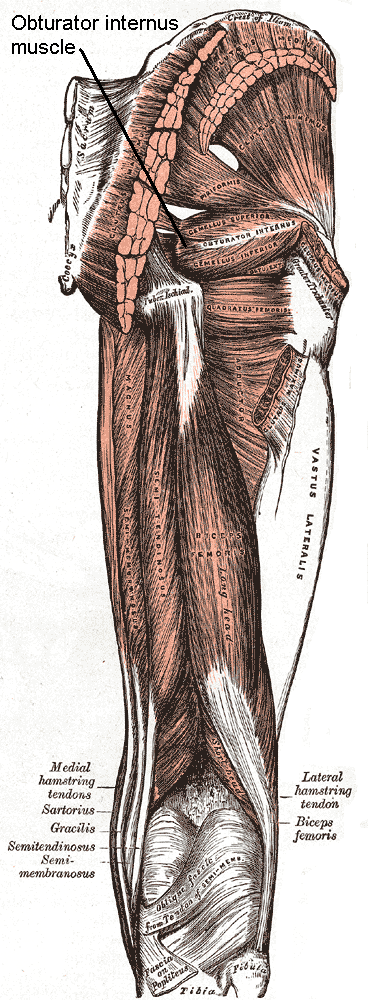
The internal obturator muscle or obturator internus muscle originates on the medial surface of the obturator membrane, the ischium near the membrane, and the rim of the pubis. It exits the pelvic cavity through the lesser sciatic foramen. The internal obturator is situated partly within the lesser pelvis, and partly at the back of the hip-joint. It functions to help laterally rotate femur with hip extension and abduct femur with hip flexion, as well as to steady the femoral head in the acetabulum. Structure Origin The internal obturator muscle arises from the inner surface of the antero-lateral wall of the pelvis. It surrounds the obturator foramen. It is attached to the inferior pubic ramus and ischium, and at the side to the inner surface of the hip bone below and behind the pelvic brim. It reaches from the upper part of the greater sciatic foramen above and behind to the obturator foramen below and in front. It also arises from the pelvic surface of the obturator membran ... [...More Info...] [...Related Items...] OR: [Wikipedia] [Google] [Baidu] |
Obturator Fascia
The obturator fascia, or fascia of the internal obturator muscle, covers the pelvic surface of that muscle and is attached around the margin of its origin. Above, it is loosely connected to the back part of the arcuate line, and here it is continuous with the iliac fascia. In front of this, as it follows the line of origin of the internal obturator, it gradually separates from the iliac fascia and the continuity between the two is retained only through the periosteum. It arches beneath the obturator vessels and nerve, completing the obturator canal, and at the front of the pelvis is attached to the back of the superior ramus of the pubis. Below, the obturator fascia is attached to the falciform process of the sacrotuberous ligament and to the pubic arch, where it becomes continuous with the superior fascia of the urogenital diaphragm. Behind, it is prolonged into the gluteal region. The internal pudendal vessels and pudendal nerve cross the pelvic surface of the internal o ... [...More Info...] [...Related Items...] OR: [Wikipedia] [Google] [Baidu] |
Internal Pudendal Artery
The internal pudendal artery is one of the three pudendal arteries. It branches off the internal iliac artery, and provides blood to the external genitalia. Structure The internal pudendal artery is the terminal branch of the anterior trunk of the internal iliac artery. It is smaller in the female than in the male. Path It arises from the anterior division of internal iliac artery. It runs on the lateral pelvic wall. It exits the pelvic cavity through the greater sciatic foramen, inferior to the piriformis muscle, to enter the gluteal region. It then curves around the sacrospinous ligament to enter the perineum through the lesser sciatic foramen. It travels through the pudendal canal with the internal pudendal veins and the pudendal nerve. Branches The internal pudendal artery gives off the following branches: The deep artery of clitoris is a branch of the internal pudendal artery and supplies the clitoral crura. Another branch of the internal pudendal artery is the dorsal ... [...More Info...] [...Related Items...] OR: [Wikipedia] [Google] [Baidu] |
Internal Pudendal Veins
The internal pudendal veins (internal pudic veins) are a set of veins in the pelvis. They are the venae comitantes of the internal pudendal artery. Internal pudendal veins are enclosed by pudendal canal, with internal pudendal artery and pudendal nerve. They begin in the deep veins of the vulva and of the penis, issuing from the bulb of the vestibule and the bulb of the penis, respectively. They accompany the internal pudendal artery, and unite to form a single vessel, which ends in the internal iliac vein. They receive the veins from the urethral bulb, the perineal and inferior hemorrhoidal veins. The deep dorsal vein of the penis Deep or The Deep may refer to: Places United States * Deep Creek (Appomattox River tributary), Virginia * Deep Creek (Great Salt Lake), Idaho and Utah * Deep Creek (Mahantango Creek tributary), Pennsylvania * Deep Creek (Mojave River tributary), ... communicates with the internal pudendal veins, but ends mainly in the pudendal plexus. Reference ... [...More Info...] [...Related Items...] OR: [Wikipedia] [Google] [Baidu] |
Obturator Internus
The internal obturator muscle or obturator internus muscle originates on the medial surface of the obturator membrane, the ischium near the membrane, and the rim of the pubis. It exits the pelvic cavity through the lesser sciatic foramen. The internal obturator is situated partly within the lesser pelvis, and partly at the back of the hip-joint. It functions to help laterally rotate femur with hip extension and abduct femur with hip flexion, as well as to steady the femoral head in the acetabulum. Structure Origin The internal obturator muscle arises from the inner surface of the antero-lateral wall of the pelvis. It surrounds the obturator foramen. It is attached to the inferior pubic ramus and ischium, and at the side to the inner surface of the hip bone below and behind the pelvic brim. It reaches from the upper part of the greater sciatic foramen above and behind to the obturator foramen below and in front. It also arises from the pelvic surface of the obturator membra ... [...More Info...] [...Related Items...] OR: [Wikipedia] [Google] [Baidu] |
Pudendal Nerve Entrapment
Pudendal nerve entrapment (PNE), also known as Alcock canal syndrome, is an uncommon source of chronic pain in which the pudendal nerve (located in the pelvis) is entrapped or compressed in Alcock's canal. There are several different types of PNE based on the site of entrapment anatomically (see Anatomy). Pain is positional and is worsened by sitting. Other symptoms include genital numbness, fecal incontinence and urinary incontinence. The term pudendal neuralgia (PN) is often used interchangeably with "pudendal nerve entrapment". This condition can greatly affect a persons’ quality of life. Pudendal neuralgia can be caused by many factors including inflammation, extreme cycling, and can be a "secondary condition to childbirth". A 2009 review study found both that "prevalence of PN is unknown and it seems to be a rare event" and that "there is no evidence to support equating the presence of this syndrome with a diagnosis of pudendal nerve entrapment," meaning that it is poss ... [...More Info...] [...Related Items...] OR: [Wikipedia] [Google] [Baidu] |


