|
Pudenda
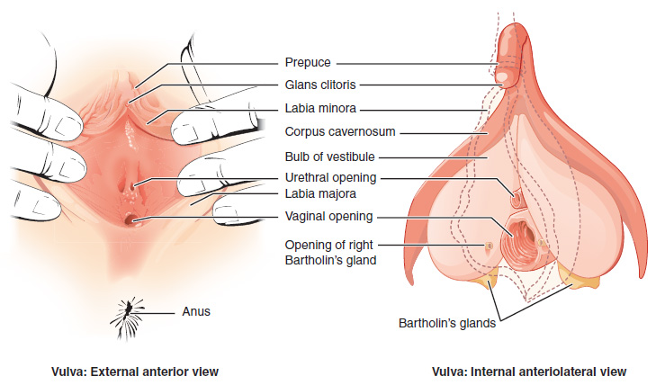
The vulva (plural: vulvas or vulvae; derived from Latin for wrapper or covering) consists of the external female sex organs. The vulva includes the mons pubis (or mons veneris), labia majora, labia minora, clitoris, vestibular bulbs, vulval vestibule, urinary meatus, the vaginal opening, hymen, and Bartholin's and Skene's vestibular glands. The urinary meatus is also included as it opens into the vulval vestibule. Other features of the vulva include the pudendal cleft, sebaceous glands, the urogenital triangle (anterior part of the perineum), and pubic hair. The vulva includes the entrance to the vagina, which leads to the uterus, and provides a double layer of protection for this by the folds of the outer and inner labia. Pelvic floor muscles support the structures of the vulva. Other muscles of the urogenital triangle also give support. Blood supply to the vulva comes from the three pudendal arteries. The internal pudendal veins give drainage. Afferent lymph vessels ca ... [...More Info...] [...Related Items...] OR: [Wikipedia] [Google] [Baidu] |
Pudendal Nerve
The pudendal nerve is the main nerve of the perineum. It carries sensation from the external genitalia of both sexes and the skin around the anus and perineum, as well as the motor supply to various pelvic muscles, including the male or female external urethral sphincter and the external anal sphincter. If damaged, most commonly by childbirth, lesions may cause sensory loss or fecal incontinence. The nerve may be temporarily blocked as part of an anaesthetic procedure. The pudendal canal that carries the pudendal nerve is also known by the eponymous term "Alcock's canal", after Benjamin Alcock, an Irish anatomist who documented the canal in 1836. Structure The pudendal nerve is paired, meaning there are two nerves, one on the left and one on the right side of the body. Each is formed as three roots immediately converge above the upper border of the sacrotuberous ligament and the coccygeus muscle. The three roots become two cords when the middle and lower root jo ... [...More Info...] [...Related Items...] OR: [Wikipedia] [Google] [Baidu] |
Internal Pudendal Veins
The internal pudendal veins (internal pudic veins) are a set of veins in the pelvis. They are the venae comitantes of the internal pudendal artery. Internal pudendal veins are enclosed by pudendal canal, with internal pudendal artery and pudendal nerve. They begin in the deep veins of the vulva and of the penis, issuing from the bulb of the vestibule and the bulb of the penis, respectively. They accompany the internal pudendal artery, and unite to form a single vessel, which ends in the internal iliac vein. They receive the veins from the urethral bulb, the perineal and inferior hemorrhoidal veins. The deep dorsal vein of the penis Deep or The Deep may refer to: Places United States * Deep Creek (Appomattox River tributary), Virginia * Deep Creek (Great Salt Lake), Idaho and Utah * Deep Creek (Mahantango Creek tributary), Pennsylvania * Deep Creek (Mojave River tributary) ... communicates with the internal pudendal veins, but ends mainly in the pudendal plexus. Ref ... [...More Info...] [...Related Items...] OR: [Wikipedia] [Google] [Baidu] |
Internal Pudendal Artery
The internal pudendal artery is one of the three pudendal arteries. It branches off the internal iliac artery, and provides blood to the external genitalia. Structure The internal pudendal artery is the terminal branch of the anterior trunk of the internal iliac artery. It is smaller in the female than in the male. Path It arises from the anterior division of internal iliac artery. It runs on the lateral pelvic wall. It exits the pelvic cavity through the greater sciatic foramen, inferior to the piriformis muscle, to enter the gluteal region. It then curves around the sacrospinous ligament to enter the perineum through the lesser sciatic foramen. It travels through the pudendal canal with the internal pudendal veins and the pudendal nerve. Branches The internal pudendal artery gives off the following branches: The deep artery of clitoris is a branch of the internal pudendal artery and supplies the clitoral crura. Another branch of the internal pudendal arter ... [...More Info...] [...Related Items...] OR: [Wikipedia] [Google] [Baidu] |
Pudendum Muliebre
The vulva (plural: vulvas or vulvae; derived from Latin for wrapper or covering) consists of the external female sex organs. The vulva includes the mons pubis (or mons veneris), labia majora, labia minora, clitoris, vestibular bulbs, vulval vestibule, urinary meatus, the vaginal opening, hymen, and Bartholin's and Skene's vestibular glands. The urinary meatus is also included as it opens into the vulval vestibule. Other features of the vulva include the pudendal cleft, sebaceous glands, the urogenital triangle (anterior part of the perineum), and pubic hair. The vulva includes the entrance to the vagina, which leads to the uterus, and provides a double layer of protection for this by the folds of the outer and inner labia. Pelvic floor muscles support the structures of the vulva. Other muscles of the urogenital triangle also give support. Blood supply to the vulva comes from the three pudendal arteries. The internal pudendal veins give drainage. Afferent lymph vessels carr ... [...More Info...] [...Related Items...] OR: [Wikipedia] [Google] [Baidu] |
Pubic Hair
Pubic hair is terminal body hair that is found in the genital area of adolescent and adult humans. The hair is located on and around the sex organs and sometimes at the top of the inside of the thighs. In the pubic region around the pubis bone, it is known as a ''pubic patch''. Pubic hair is also found on the scrotum in the male and on the vulva in the female. Although fine vellus hair is present in the area in childhood, pubic hair is considered to be the heavier, longer and coarser hair that develops during puberty as an effect of rising levels of androgens in males and estrogens in females. Pubic hair differs from other hair on the body and is a secondary sex characteristic. Many cultures regard pubic hair as erotic, and in most cultures it is associated with the genitals, which both men and women are expected to keep covered at all times. In some cultures, it is the norm for pubic hair to be removed, especially of females; the practice is regarded as part of personal hy ... [...More Info...] [...Related Items...] OR: [Wikipedia] [Google] [Baidu] |
Vagina
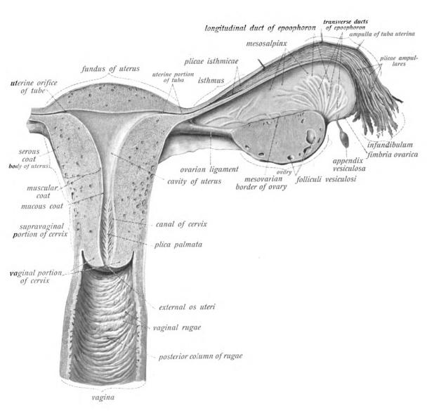
In mammals, the vagina is the elastic, muscular part of the female genital tract. In humans, it extends from the vestibule to the cervix. The outer vaginal opening is normally partly covered by a thin layer of mucosal tissue called the hymen. At the deep end, the cervix (neck of the uterus) bulges into the vagina. The vagina allows for sexual intercourse and birth. It also channels menstrual flow, which occurs in humans and closely related primates as part of the menstrual cycle. Although research on the vagina is especially lacking for different animals, its location, structure and size are documented as varying among species. Female mammals usually have two external openings in the vulva; these are the urethral opening for the urinary tract and the vaginal opening for the genital tract. This is different from male mammals, who usually have a single urethral opening for both urination and reproduction. The vaginal opening is much larger than the nearby urethral ... [...More Info...] [...Related Items...] OR: [Wikipedia] [Google] [Baidu] |
Skene's Gland
In female human anatomy, Skene's glands or the Skene glands ( , also known as the lesser vestibular glands, paraurethral glands) are glands located around the lower end of the urethra. The glands are surrounded by tissue that swells with blood during sexual arousal, and secrete a fluid from openings near the urethra, particularly during orgasm. Structure and function The Skene's glands are located in the vestibule of the vulva, around the lower end of the urethra. The two Skene's ducts lead from the Skene's glands to the vulvar vestibule, to the left and right of the urethral opening, from which they are structurally capable of secreting fluid. Although there remains debate about the function of the Skene's glands, one purpose is to secrete a fluid that helps lubricate the urethral opening. Skene's glands produce a milk-like ultrafiltrate of blood plasma. The glands may be the source of female ejaculation, but this has not been proven. Because they and the male prostate act ... [...More Info...] [...Related Items...] OR: [Wikipedia] [Google] [Baidu] |
Vagina
In mammals, the vagina is the elastic, muscular part of the female genital tract. In humans, it extends from the vestibule to the cervix. The outer vaginal opening is normally partly covered by a thin layer of mucosal tissue called the hymen. At the deep end, the cervix (neck of the uterus) bulges into the vagina. The vagina allows for sexual intercourse and birth. It also channels menstrual flow, which occurs in humans and closely related primates as part of the menstrual cycle. Although research on the vagina is especially lacking for different animals, its location, structure and size are documented as varying among species. Female mammals usually have two external openings in the vulva; these are the urethral opening for the urinary tract and the vaginal opening for the genital tract. This is different from male mammals, who usually have a single urethral opening for both urination and reproduction. The vaginal opening is much larger than the nearby urethral ... [...More Info...] [...Related Items...] OR: [Wikipedia] [Google] [Baidu] |
Mons Pubis
In human anatomy, and in mammals in general, the ''mons pubis'' or pubic mound (also known simply as the mons, and known specifically in females as the ''mons Venus'' or ''mons veneris'') is a rounded mass of fatty tissue found over the pubic symphysis of the pubic bones. Anatomy For females, the ''mons pubis'' forms the anterior portion of the vulva. It divides into the labia majora (literally "larger lips"), on either side of the furrow known as the pudendal cleft, that surrounds the labia minora, clitoris, urethra, vaginal opening, and other structures of the vulval vestibule. Although present in both men and women, the ''mons pubis'' tends to be larger in women. Its fatty tissue is sensitive to estrogen, causing a distinct mound to form with the onset of female puberty. This pushes the forward portion of the labia majora out and away from the pubic bone. The mound also becomes covered with pubic hair. It often becomes less prominent with the decrease in bodily estrog ... [...More Info...] [...Related Items...] OR: [Wikipedia] [Google] [Baidu] |
Sebaceous Gland
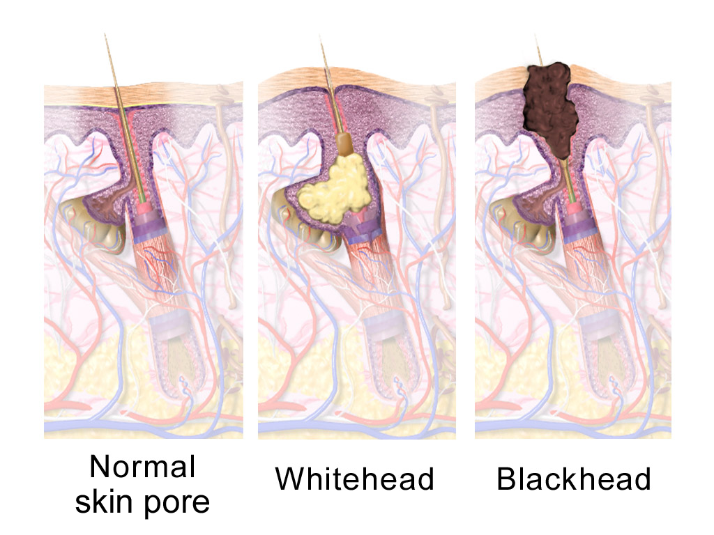
A sebaceous gland is a microscopic exocrine gland in the skin that opens into a hair follicle to secrete an oily or waxy matter, called sebum, which lubricates the hair and skin of mammals. In humans, sebaceous glands occur in the greatest number on the face and scalp, but also on all parts of the skin except the palms of the hands and soles of the feet. In the eyelids, meibomian glands, also called tarsal glands, are a type of sebaceous gland that secrete a special type of sebum into tears. Surrounding the female nipple, areolar glands are specialized sebaceous glands for lubricating the nipple. Fordyce spots are benign, visible, sebaceous glands found usually on the lips, gums and inner cheeks, and genitals. Structure Location Sebaceous glands are found throughout all areas of the skin, except the palms of the hands and soles of the feet. There are two types of sebaceous glands, those connected to hair follicles and those that exist independently. Sebaceous gland ... [...More Info...] [...Related Items...] OR: [Wikipedia] [Google] [Baidu] |
Lymphatic Vessel
The lymphatic vessels (or lymph vessels or lymphatics) are thin-walled vessels (tubes), structured like blood vessels, that carry lymph. As part of the lymphatic system, lymph vessels are complementary to the cardiovascular system. Lymph vessels are lined by endothelial cells, and have a thin layer of smooth muscle, and adventitia that binds the lymph vessels to the surrounding tissue. Lymph vessels are devoted to the propulsion of the lymph from the lymph capillaries, which are mainly concerned with the absorption of interstitial fluid from the tissues. Lymph capillaries are slightly bigger than their counterpart capillaries of the vascular system. Lymph vessels that carry lymph to a lymph node are called afferent lymph vessels, and those that carry it from a lymph node are called efferent lymph vessels, from where the lymph may travel to another lymph node, may be returned to a vein, or may travel to a larger lymph duct. Lymph ducts drain the lymph into one of the subcla ... [...More Info...] [...Related Items...] OR: [Wikipedia] [Google] [Baidu] |


_1.png)


