|
Puborectalis
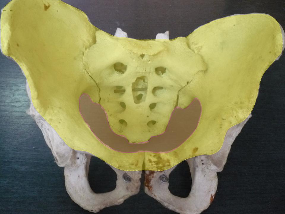
The levator ani is a broad, thin muscle group, situated on either side of the pelvis. It is formed from three muscle components: the pubococcygeus, the iliococcygeus, and the puborectalis. It is attached to the inner surface of each side of the lesser pelvis, and these unite to form the greater part of the pelvic floor. The coccygeus muscle completes the pelvic floor, which is also called the ''pelvic diaphragm''. It supports the viscera in the pelvic cavity, and surrounds the various structures that pass through it. The levator ani is the main pelvic floor muscle and painfully contracts during vaginismus. It also contracts rhythmically during orgasm. Structure The levator ani is made up of 3 parts: * Iliococcygeus muscle * Pubococcygeus muscle * Puborectalis muscle The iliococcygeus arises from the inner side of the ischium (the lower and back part of the hip bone) and from the posterior part of the tendinous arch of the obturator fascia, and is attached to the coccyx an ... [...More Info...] [...Related Items...] OR: [Wikipedia] [Google] [Baidu] |
Iliococcygeus
The levator ani is a broad, thin muscle group, situated on either side of the pelvis. It is formed from three muscle components: the pubococcygeus, the iliococcygeus, and the puborectalis. It is attached to the inner surface of each side of the lesser pelvis, and these unite to form the greater part of the pelvic floor. The coccygeus muscle completes the pelvic floor, which is also called the ''pelvic diaphragm''. It supports the viscera in the pelvic cavity, and surrounds the various structures that pass through it. The levator ani is the main pelvic floor muscle and painfully contracts during vaginismus. It also contracts rhythmically during orgasm. Structure The levator ani is made up of 3 parts: * Iliococcygeus muscle * Pubococcygeus muscle * Puborectalis muscle The iliococcygeus arises from the inner side of the ischium (the lower and back part of the hip bone) and from the posterior part of the tendinous arch of the obturator fascia, and is attached to the coccyx an ... [...More Info...] [...Related Items...] OR: [Wikipedia] [Google] [Baidu] |
Puborectalis
The levator ani is a broad, thin muscle group, situated on either side of the pelvis. It is formed from three muscle components: the pubococcygeus, the iliococcygeus, and the puborectalis. It is attached to the inner surface of each side of the lesser pelvis, and these unite to form the greater part of the pelvic floor. The coccygeus muscle completes the pelvic floor, which is also called the ''pelvic diaphragm''. It supports the viscera in the pelvic cavity, and surrounds the various structures that pass through it. The levator ani is the main pelvic floor muscle and painfully contracts during vaginismus. It also contracts rhythmically during orgasm. Structure The levator ani is made up of 3 parts: * Iliococcygeus muscle * Pubococcygeus muscle * Puborectalis muscle The iliococcygeus arises from the inner side of the ischium (the lower and back part of the hip bone) and from the posterior part of the tendinous arch of the obturator fascia, and is attached to the coccyx an ... [...More Info...] [...Related Items...] OR: [Wikipedia] [Google] [Baidu] |
Pubococcygeus
The levator ani is a broad, thin muscle group, situated on either side of the pelvis. It is formed from three muscle components: the pubococcygeus, the iliococcygeus, and the puborectalis. It is attached to the inner surface of each side of the lesser pelvis, and these unite to form the greater part of the pelvic floor. The coccygeus muscle completes the pelvic floor, which is also called the ''pelvic diaphragm''. It supports the viscera in the pelvic cavity, and surrounds the various structures that pass through it. The levator ani is the main pelvic floor muscle and painfully contracts during vaginismus. It also contracts rhythmically during orgasm. Structure The levator ani is made up of 3 parts: * Iliococcygeus muscle * Pubococcygeus muscle * Puborectalis muscle The iliococcygeus arises from the inner side of the ischium (the lower and back part of the hip bone) and from the posterior part of the tendinous arch of the obturator fascia, and is attached to the coccyx an ... [...More Info...] [...Related Items...] OR: [Wikipedia] [Google] [Baidu] |
Sacral Spinal Nerve 3
The sacral spinal nerve 3 (S3) is a spinal nerve of the sacral segment. Nervous System -- Groups of Nerves It originates from the from below the 3rd body of the sacrum. 
Muscles S3 supplies many muscles, either directly or through nerves originating from S3. They are not innervated with S3 as single origin, but partly by S3 and partly by other spinal nerves. The muscles are: *[...More Info...] [...Related Items...] OR: [Wikipedia] [Google] [Baidu] |
Sacral Spinal Nerve 4
The sacral spinal nerve 4 (S4) is a spinal nerve of the sacral segment. Nervous System -- Groups of Nerves It originates from the from below the 4th body of the 
Muscles S4 supplies many muscles, either directly or through nerves originating from S4. They are not innervated with S4 as single origin, but partly by S4 and partly by other spinal nerves. The muscles a ...[...More Info...] [...Related Items...] OR: [Wikipedia] [Google] [Baidu] |
External Anal Sphincter
The external anal sphincter (or sphincter ani externus ) is a flat plane of skeletal muscle fibers, elliptical in shape and intimately adherent to the skin surrounding the margin of the anus. Anatomy The external anal sphincter measures about 8 to 10 cm in length, from its anterior to its posterior extremity, and is about 2.5 cm opposite the anus, the sphincter muscle retracts on defecating. It consists of two layers: ''superficial'' and ''deep''. * The superficial layer, constitutes the main portion of the muscle, and arises from a narrow tendinous band, the anococcygeal raphe, which stretches from the tip of the coccyx to the posterior margin of the anus; it forms two flattened planes of muscular tissue, which encircle the anus and meet in front to be inserted into the central tendinous point of the perineum, joining with the superficial transverse perineal muscle, the levator ani, and the bulbospongiosus muscle also known as the bulbocavernosus. * The deeper laye ... [...More Info...] [...Related Items...] OR: [Wikipedia] [Google] [Baidu] |
Viscera
In biology, an organ is a collection of tissues joined in a structural unit to serve a common function. In the hierarchy of life, an organ lies between tissue and an organ system. Tissues are formed from same type cells to act together in a function. Tissues of different types combine to form an organ which has a specific function. The intestinal wall for example is formed by epithelial tissue and smooth muscle tissue. Two or more organs working together in the execution of a specific body function form an organ system, also called a biological system or body system. An organ's tissues can be broadly categorized as parenchyma, the functional tissue, and stroma, the structural tissue with supportive, connective, or ancillary functions. For example, the gland's tissue that makes the hormones is the parenchyma, whereas the stroma includes the nerves that innervate the parenchyma, the blood vessels that oxygenate and nourish it and carry away its metabolic wastes, and the conne ... [...More Info...] [...Related Items...] OR: [Wikipedia] [Google] [Baidu] |
Pelvic Cavity
The pelvic cavity is a body cavity that is bounded by the bones of the pelvis. Its oblique roof is the pelvic inlet (the superior opening of the pelvis). Its lower boundary is the pelvic floor. The pelvic cavity primarily contains the reproductive organs, urinary bladder, distal ureters, proximal urethra, terminal sigmoid colon, rectum, and anal canal. In females, the uterus, Fallopian tubes, ovaries and upper vagina occupy the area between the other viscera. The rectum is located at the back of the pelvis, in the curve of the sacrum and coccyx; the bladder is in front, behind the pubic symphysis. The pelvic cavity also contains major arteries, veins, muscles, and nerves. These structures coexist in a crowded space, and disorders of one pelvic component may impact upon another; for example, constipation may overload the rectum and compress the urinary bladder, or childbirth might damage the pudendal nerves and later lead to anal weakness. Structure The pelvis has a ... [...More Info...] [...Related Items...] OR: [Wikipedia] [Google] [Baidu] |
3D Medical Animation Levator Ani Structure
3-D, 3D, or 3d may refer to: Science, technology, and mathematics Relating to three-dimensionality * Three-dimensional space ** 3D computer graphics, computer graphics that use a three-dimensional representation of geometric data ** 3D film, a motion picture that gives the illusion of three-dimensional perception ** 3D modeling, developing a representation of any three-dimensional surface or object ** 3D printing, making a three-dimensional solid object of a shape from a digital model ** 3D display, a type of information display that conveys depth to the viewer ** 3D television, television that conveys depth perception to the viewer ** Stereoscopy, any technique capable of recording three-dimensional visual information or creating the illusion of depth in an image Other uses in science and technology or commercial products * 3D projection * 3D rendering * 3D scanning, making a digital representation of three-dimensional objects * 3D video game (other) * 3-D Secure, a ... [...More Info...] [...Related Items...] OR: [Wikipedia] [Google] [Baidu] |
Coronal Section
The coronal plane (also known as the frontal plane) is an anatomical plane that divides the body into dorsal and ventral sections. It is perpendicular to the sagittal and transverse planes. Details The coronal plane is an example of a longitudinal plane. For a human, the mid-coronal plane would transect a standing body into two halves (front and back, or anterior and posterior) in an imaginary line that cuts through both shoulders. The description of the coronal plane applies to most animals as well as humans even though humans walk upright and the various planes are usually shown in the vertical orientation. The sternal plane (''planum sternale'') is a coronal plane which transects the front of the sternum. Etymology The term is derived from Latin ''corona'' ('garland, crown'), from Ancient Greek κορώνη (''korōnē'', 'garland, wreath'). The coronal plane is so-called because it lies in the direction of Coronal suture. Additional images File:Coronal plane CT scan ... [...More Info...] [...Related Items...] OR: [Wikipedia] [Google] [Baidu] |
Muscle
Skeletal muscles (commonly referred to as muscles) are organs of the vertebrate muscular system and typically are attached by tendons to bones of a skeleton. The muscle cells of skeletal muscles are much longer than in the other types of muscle tissue, and are often known as muscle fibers. The muscle tissue of a skeletal muscle is striated – having a striped appearance due to the arrangement of the sarcomeres. Skeletal muscles are voluntary muscles under the control of the somatic nervous system. The other types of muscle are cardiac muscle which is also striated and smooth muscle which is non-striated; both of these types of muscle tissue are classified as involuntary, or, under the control of the autonomic nervous system. A skeletal muscle contains multiple fascicles – bundles of muscle fibers. Each individual fiber, and each muscle is surrounded by a type of connective tissue layer of fascia. Muscle fibers are formed from the fusion of developmental myoblasts ... [...More Info...] [...Related Items...] OR: [Wikipedia] [Google] [Baidu] |
Pelvic Floor
The pelvic floor or pelvic diaphragm is composed of muscle fibers of the levator ani, the coccygeus muscle, and associated connective tissue which span the area underneath the pelvis. The pelvic diaphragm is a muscular partition formed by the levatores ani and coccygei, with which may be included the parietal pelvic fascia on their upper and lower aspects. The pelvic floor separates the pelvic cavity above from the perineal region (including perineum) below. Both males and females have a pelvic floor. To accommodate the birth canal, a female's pelvic cavity is larger than a male's. Structure The right and left levator ani lie almost horizontally in the floor of the pelvis, separated by a narrow gap that transmits the urethra, vagina, and anal canal. The levator ani is usually considered in three parts: pubococcygeus, puborectalis, and iliococcygeus. The pubococcygeus, the main part of the levator, runs backward from the body of the pubis toward the coccyx and may be damaged duri ... [...More Info...] [...Related Items...] OR: [Wikipedia] [Google] [Baidu] |


.png)