|
Prorenin
Prorenin () is a protein that constitutes a precursor for renin, the hormone that activates the renin–angiotensin system, which serves to raise blood pressure. Prorenin is converted into renin by the juxtaglomerular cells, which are specialised smooth muscle cells present mainly in the afferent, but also the efferent, arterioles of the glomerular capillary bed. Prorenin is a relatively large molecule, weighing approximately 46 KDa. History Prorenin was discovered by Eugenie Lumbers in 1971. Synthesis In addition to juxtaglomerular cells, prorenin is also synthesised by other organs, such as the adrenal glands, the ovaries, the testis and the pituitary gland, which is why it is found in the plasma of anephric individuals. Concentration Blood concentration levels of prorenin are between 5 and 10 times higher than those of renin. There is evidence to suggest that, in diabetes mellitus, prorenin levels are even higher. One study using relatively newer technology found that blood ... [...More Info...] [...Related Items...] OR: [Wikipedia] [Google] [Baidu] |
Renin
Renin (etymology and pronunciation), also known as an angiotensinogenase, is an aspartic protease protein and enzyme secreted by the kidneys that participates in the body's renin–angiotensin–aldosterone system (RAAS)—also known as the renin–angiotensin–aldosterone axis—that increases the volume of extracellular fluid (blood plasma, lymph and interstitial fluid) and causes arterial vasoconstriction. Thus, it increases the body's mean arterial blood pressure. Renin is not commonly referred to as a hormone, albeit it having a receptor, the (pro)renin receptor, also known as the renin receptor and prorenin receptor (see also below), as well as enzymatic activity with which it hydrolyzes angiotensinogen to angiotensin I. Biochemistry and physiology Structure The primary structure of renin precursor consists of 406 amino acids with a pre- and a pro-segment carrying 20 and 46 amino acids, respectively. Mature renin contains 340 amino acids and has a mass of 37 kDa. ... [...More Info...] [...Related Items...] OR: [Wikipedia] [Google] [Baidu] |
Renin–angiotensin System
The renin–angiotensin system (RAS), or renin–angiotensin–aldosterone system (RAAS), is a hormone system that regulates blood pressure, fluid and electrolyte balance, and systemic vascular resistance. When renal blood flow is reduced, juxtaglomerular cells in the kidneys convert the precursor prorenin (already present in the blood) into renin and secrete it directly into the circulation. Plasma renin then carries out the conversion of angiotensinogen, released by the liver, to a decapeptide called angiotensin I. Angiotensin I is subsequently converted to angiotensin II (an octapeptide) by the angiotensin-converting enzyme (ACE) found on the surface of vascular endothelial cells, predominantly those of the lungs. Angiotensin II has a short life of about 1 to 2 minutes. Then, it is rapidly degraded into a heptapeptide called angiotensin III by angiotensinases which are present in red blood cells and vascular beds in many tissues. Angiotensin III increases blood pressure and ... [...More Info...] [...Related Items...] OR: [Wikipedia] [Google] [Baidu] |
Blood Pressure
Blood pressure (BP) is the pressure of circulating blood against the walls of blood vessels. Most of this pressure results from the heart pumping blood through the circulatory system. When used without qualification, the term "blood pressure" refers to the pressure in the large arteries. Blood pressure is usually expressed in terms of the systolic pressure (maximum pressure during one heartbeat) over diastolic pressure (minimum pressure between two heartbeats) in the cardiac cycle. It is measured in millimeters of mercury ( mmHg) above the surrounding atmospheric pressure. Blood pressure is one of the vital signs—together with respiratory rate, heart rate, oxygen saturation, and body temperature—that healthcare professionals use in evaluating a patient's health. Normal resting blood pressure, in an adult is approximately systolic over diastolic, denoted as "120/80 mmHg". Globally, the average blood pressure, age standardized, has remained about the same since 1 ... [...More Info...] [...Related Items...] OR: [Wikipedia] [Google] [Baidu] |
Juxtaglomerular Cell
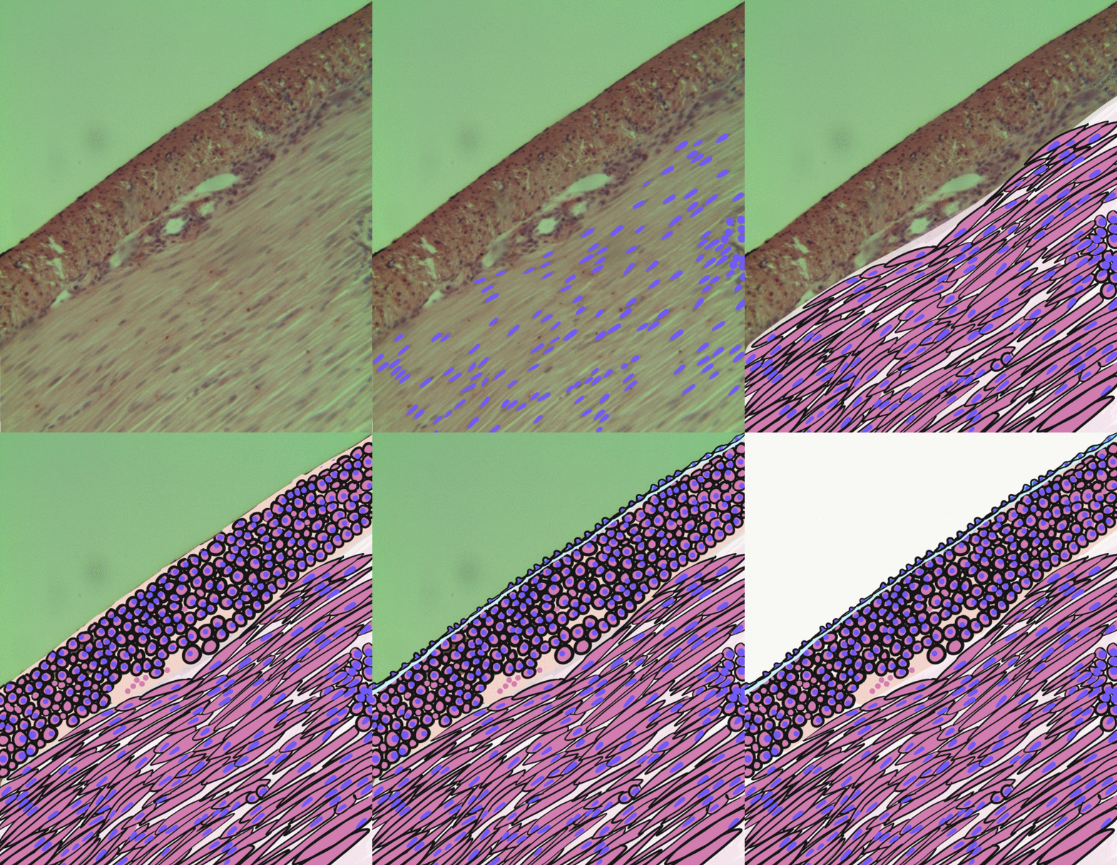
Juxtaglomerular cells (JG cells), also known as juxtaglomerular granular cells are cells in the kidney that synthesize, store, and secrete the enzyme renin. They are specialized smooth muscle cells mainly in the walls of the afferent arterioles (and some in the efferent arterioles) that deliver blood to the glomerulus. In synthesizing renin, they play a critical role in the renin–angiotensin system and thus in autoregulation of the kidney. Juxtaglomerular cells secrete renin in response to a drop in pressure detected by stretch receptors in the vascular walls, or when stimulated by macula densa cells. Macula densa cells are located in the distal convoluted tubule, and stimulate juxtaglomerular cells to release renin when they detect a drop in chloride concentration in tubular fluid. Together, juxtaglomerular cells, extraglomerular mesangial cells and macula densa cells comprise the juxtaglomerular apparatus. In appropriately stained tissue sections, juxtaglomerular cells a ... [...More Info...] [...Related Items...] OR: [Wikipedia] [Google] [Baidu] |
Smooth Muscle
Smooth muscle is an involuntary non-striated muscle, so-called because it has no sarcomeres and therefore no striations (''bands'' or ''stripes''). It is divided into two subgroups, single-unit and multiunit smooth muscle. Within single-unit muscle, the whole bundle or sheet of smooth muscle cells contracts as a syncytium. Smooth muscle is found in the walls of hollow organs, including the stomach, intestines, bladder and uterus; in the walls of passageways, such as blood, and lymph vessels, and in the tracts of the respiratory, urinary, and reproductive systems. In the eyes, the ciliary muscles, a type of smooth muscle, dilate and contract the iris and alter the shape of the lens. In the skin, smooth muscle cells such as those of the arrector pili cause hair to stand erect in response to cold temperature or fear. Structure Gross anatomy Smooth muscle is grouped into two types: single-unit smooth muscle, also known as visceral smooth muscle, and multiunit smooth muscle. ... [...More Info...] [...Related Items...] OR: [Wikipedia] [Google] [Baidu] |
Arteriole
An arteriole is a small-diameter blood vessel in the microcirculation that extends and branches out from an artery and leads to capillaries. Arterioles have muscular walls (usually only one to two layers of smooth muscle cells) and are the primary site of vascular resistance. The greatest change in blood pressure and velocity of blood flow occurs at the transition of arterioles to capillaries.This function is extremely important because it prevents the thin, one-layer capillaries from exploding upon pressure. The arterioles achieve this decrease in pressure, as they are the site with the highest resistance (a large contributor to total peripheral resistance) which translates to a large decrease in the pressure. Structure Microanatomy In a healthy vascular system the endothelium lines all blood-contacting surfaces, including arteries, arterioles, veins, venules, capillaries, and heart chambers. This healthy condition is promoted by the ample production of nitric oxide by the end ... [...More Info...] [...Related Items...] OR: [Wikipedia] [Google] [Baidu] |
Glomerulus
''Glomerulus'' () is a common term used in anatomy to describe globular structures of entwined vessels, fibers, or neurons. ''Glomerulus'' is the diminutive of the Latin ''glomus'', meaning "ball of yarn". ''Glomerulus'' may refer to: * the filtering unit of the kidney; see Glomerulus (kidney). * a structure in the olfactory bulb; see Glomerulus (olfaction). * the contact between specific cells in the cerebellum; see Glomerulus (cerebellum) The cerebellar glomerulus is a small, intertwined mass of nerve fiber terminals in the granular layer of the cerebellar cortex. It consists of post-synaptic granule cell dendrites and pre-synaptic Golgi cell axon terminals surrounding the pre- .... See also * Glomerulation, a hemorrhage of the bladder {{SIA ... [...More Info...] [...Related Items...] OR: [Wikipedia] [Google] [Baidu] |
Eugenie Lumbers
Eugenie Ruth Lumbers (also known as Eugenie Forbes) is an Australian medical researcher whose work has focused on the role of the renin-angiotensin system in fetal development and in women's health. Career She earned her MBBS medical degrees and her MD doctorate from the University of Adelaide. She was the first woman to be awarded a CJ Martin Fellowship by the National Health and Medical Research Council of Australia, and with that funding she studied fetal physiology at Oxford University. In 1974 she joined the faculty of University of New South Wales (UNSW). She was awarded the degree of DSc in 1986 and became the first woman appointed as a Scientia Professor at UNSW in 1999. She was elected as Fellow to the Australian Academy of Science and received the Centenary Medal in 2002. In 2010, she was elected a Fellow of the Royal Society of New South Wales. In 2012, she was appointed a Member of the Order of Australia. She received a joint appointment at University of Queensl ... [...More Info...] [...Related Items...] OR: [Wikipedia] [Google] [Baidu] |
Diabetes Mellitus
Diabetes, also known as diabetes mellitus, is a group of metabolic disorders characterized by a high blood sugar level ( hyperglycemia) over a prolonged period of time. Symptoms often include frequent urination, increased thirst and increased appetite. If left untreated, diabetes can cause many health complications. Acute complications can include diabetic ketoacidosis, hyperosmolar hyperglycemic state, or death. Serious long-term complications include cardiovascular disease, stroke, chronic kidney disease, foot ulcers, damage to the nerves, damage to the eyes, and cognitive impairment. Diabetes is due to either the pancreas not producing enough insulin, or the cells of the body not responding properly to the insulin produced. Insulin is a hormone which is responsible for helping glucose from food get into cells to be used for energy. There are three main types of diabetes mellitus: * Type 1 diabetes results from failure of the pancreas to produce enough insulin due to lo ... [...More Info...] [...Related Items...] OR: [Wikipedia] [Google] [Baidu] |
Amniotic Fluid
The amniotic fluid is the protective liquid contained by the amniotic sac of a gravid amniote. This fluid serves as a cushion for the growing fetus, but also serves to facilitate the exchange of nutrients, water, and biochemical products between mother and fetus. For humans, the amniotic fluid is commonly called water or waters (Latin liquor amnii). Development Amniotic fluid is present from the formation of the gestational sac. Amniotic fluid is in the amniotic sac. It is generated from maternal plasma, and passes through the fetal membranes by osmotic and hydrostatic forces. When fetal kidneys begin to function around week 16, fetal urine also contributes to the fluid. In earlier times, it was believed that the amniotic fluid was composed entirely of fetal urine. The fluid is absorbed through the fetal tissue and skin. After 22 to 25 week of pregnancy, keratinization of an embryo's skin occurs. When this process completes around the 25th week, the fluid is primarily absor ... [...More Info...] [...Related Items...] OR: [Wikipedia] [Google] [Baidu] |
Amnion
The amnion is a membrane that closely covers the human and various other embryos when first formed. It fills with amniotic fluid, which causes the amnion to expand and become the amniotic sac that provides a protective environment for the developing embryo. The amnion, along with the chorion, the yolk sac and the allantois protect the embryo. In birds, reptiles and monotremes, the protective sac is enclosed in a shell. In marsupials and placental mammals, it is enclosed in a uterus. The term is from Ancient Greek ἀμνίον 'little lamb', diminutive of ἀμνός 'lamb'. it is cognate with the English verb 'yean', bring forth young (usually lambs). The amnion is a feature of the vertebrate clade ''Amniota'', which includes reptiles, birds, and mammals. Amphibians and fish are not amniotes and thus lack the amnion. The amnion stems from the extra-embryonic somatic mesoderm on the outer side and the extra-embryonic ectoderm or trophoblast on the inner side. In humans In the ... [...More Info...] [...Related Items...] OR: [Wikipedia] [Google] [Baidu] |
Placenta
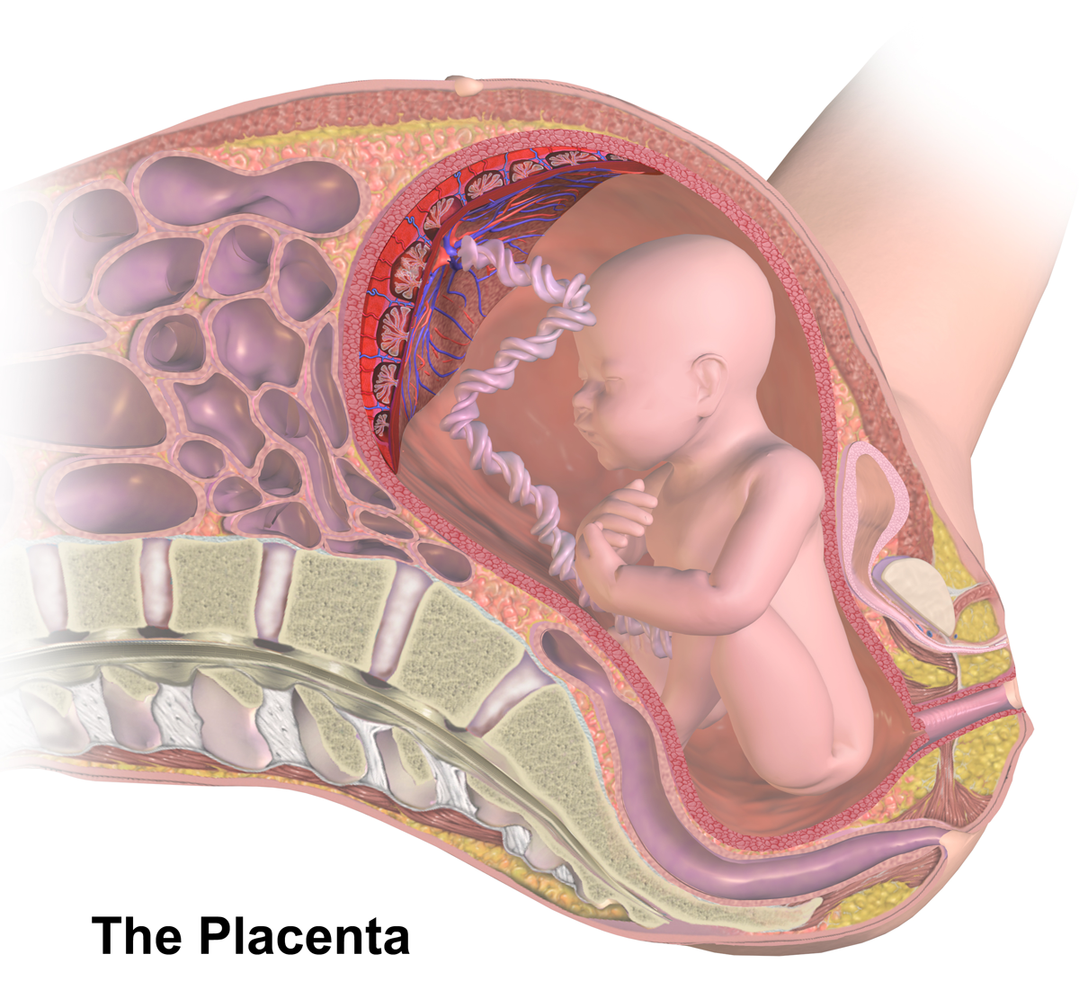
The placenta is a temporary embryonic and later fetal organ that begins developing from the blastocyst shortly after implantation. It plays critical roles in facilitating nutrient, gas and waste exchange between the physically separate maternal and fetal circulations, and is an important endocrine organ, producing hormones that regulate both maternal and fetal physiology during pregnancy. The placenta connects to the fetus via the umbilical cord, and on the opposite aspect to the maternal uterus in a species-dependent manner. In humans, a thin layer of maternal decidual (endometrial) tissue comes away with the placenta when it is expelled from the uterus following birth (sometimes incorrectly referred to as the 'maternal part' of the placenta). Placentas are a defining characteristic of placental mammals, but are also found in marsupials and some non-mammals with varying levels of development. Mammalian placentas probably first evolved about 150 million to 200 million years ... [...More Info...] [...Related Items...] OR: [Wikipedia] [Google] [Baidu] |


