|
Pronephros
Pronephros is the most basic of the three excretory organs that develop in vertebrates, corresponding to the first stage of kidney development. It is succeeded by the mesonephros, which in fish and amphibians remains as the adult kidney. In amniotes the mesonephros is the embryonic kidney and a more complex metanephros acts as the adult kidney. Once a more advanced kidney forms, the previous version typically degenerates by apoptosis or becomes part of the male reproductive system. The pronephros develops from the intermediate mesoderm, as do the later kidneys. It is a paired organ, consisting of a single giant nephron that processes blood filtrate produced from glomeruli or glomera- large embryonic glomeruli. The filtrate is deposited into the coelom. It then passes through thin ciliated tubules into the pronephric nephron where it is processed for solute recovery. The organ is active in adult forms of some primitive fish, like lampreys or hagfish. It is present at the embryo ... [...More Info...] [...Related Items...] OR: [Wikipedia] [Google] [Baidu] |
Intermediate Mesoderm
Intermediate mesoderm or intermediate mesenchyme is a narrow section of the mesoderm (one of the three primary germ layers) located between the paraxial mesoderm and the lateral plate of the developing embryo. The intermediate mesoderm develops into vital parts of the urogenital system (kidneys, gonads and respective tracts). Early formation Factors regulating the formation of the intermediate mesoderm are not fully understood. It is believed that bone morphogenic proteins, or BMPs, specify regions of growth along the dorsal-ventral axis of the mesoderm and plays a central role in formation of the intermediate mesoderm. Vg1/ Nodal signalling is an identified regulator of intermediate mesoderm formation acting through BMP signalling. Excess Vg1/Nodal signalling during early gastrulation stages results in expansion of the intermediate mesoderm at the expense of the adjacent paraxial mesoderm, whereas inhibition of Vg1/Nodal signalling represses intermediate mesoderm formation. A li ... [...More Info...] [...Related Items...] OR: [Wikipedia] [Google] [Baidu] |
Metanephros
Kidney development, or nephrogenesis, describes the embryologic origins of the kidney, a major organ in the urinary system. This article covers a 3 part developmental process that is observed in most reptiles, birds and mammals, including humans. Nephrogenesis is often considered in the broader context of the development of the urinary and reproductive organs. Phases The development of the kidney proceeds through a series of successive phases, each marked by the development of a more advanced kidney: the archinephros, pronephros, mesonephros, and metanephros. The pronephros is the most immature form of kidney, while the metanephros is most developed. The metanephros persists as the definitive adult kidney. Archinephros The archinephros is considered as hypothetical or primitive kidney. Pronephros The pronephros develops in the cervical region of the embryo. During approximately day 22 of human gestation, the paired pronephri appears towards the cranial end of the intermediate ... [...More Info...] [...Related Items...] OR: [Wikipedia] [Google] [Baidu] |
Nephrotome
In earlier conceptions of kidney biology, the nephrotome was a section of the mesoderm that gives rise to the pronephros and eventually to the rest of the kidney. Older texts describe the pronephros as forming through the fusion of multiple nephrotomes. Modern visualization methods, such as scanning electron microscopy, in situ hybridization, and confocal microscopy, combined with the simple approach of peeling off the epidermis to see the forming kidney, have shown that this is not the case. Nephric mesenchyme Mesenchyme () is a type of loosely organized animal embryonic connective tissue of undifferentiated cells that give rise to most tissues, such as skin, blood or bone. The interactions between mesenchyme and epithelium help to form nearly every o ... separates from the intermediate mesoderm as a single elongated primordia. As this primordia begins to undergo epithelialization the anterior portions of the pronephros form rounded protrusions, which then later became th ... [...More Info...] [...Related Items...] OR: [Wikipedia] [Google] [Baidu] |
Mesonephros
The mesonephros ( el, middle kidney) is one of three excretory organs that develop in vertebrates. It serves as the main excretory organ of aquatic vertebrates and as a temporary kidney in reptiles, birds, and mammals. The mesonephros is included in the Wolffian body after Caspar Friedrich Wolff who described it in 1759. (The Wolffian body is composed of: mesonephros + paramesonephrotic blastema) Structure The mesonephros acts as a structure similar to the kidney that, in humans, functions between the sixth and tenth weeks of embryological life. Despite the similarity in structure, function, and terminology, however, the mesonephric nephrons do not form any part of the mature kidney or nephrons. In humans, the mesonephros consists of units which are similar in structure and function to nephrons of the adult kidney. Each of these consists of a glomerulus, a tuft of capillaries which arises from lateral branches of dorsal aorta and drains into the inferior cardinal vein; a Bowman' ... [...More Info...] [...Related Items...] OR: [Wikipedia] [Google] [Baidu] |
Kidney
The kidneys are two reddish-brown bean-shaped organs found in vertebrates. They are located on the left and right in the retroperitoneal space, and in adult humans are about in length. They receive blood from the paired renal arteries; blood exits into the paired renal veins. Each kidney is attached to a ureter, a tube that carries excreted urine to the bladder. The kidney participates in the control of the volume of various body fluids, fluid osmolality, acid–base balance, various electrolyte concentrations, and removal of toxins. Filtration occurs in the glomerulus: one-fifth of the blood volume that enters the kidneys is filtered. Examples of substances reabsorbed are solute-free water, sodium, bicarbonate, glucose, and amino acids. Examples of substances secreted are hydrogen, ammonium, potassium and uric acid. The nephron is the structural and functional unit of the kidney. Each adult human kidney contains around 1 million nephrons, while a mouse kidney ... [...More Info...] [...Related Items...] OR: [Wikipedia] [Google] [Baidu] |
Fish
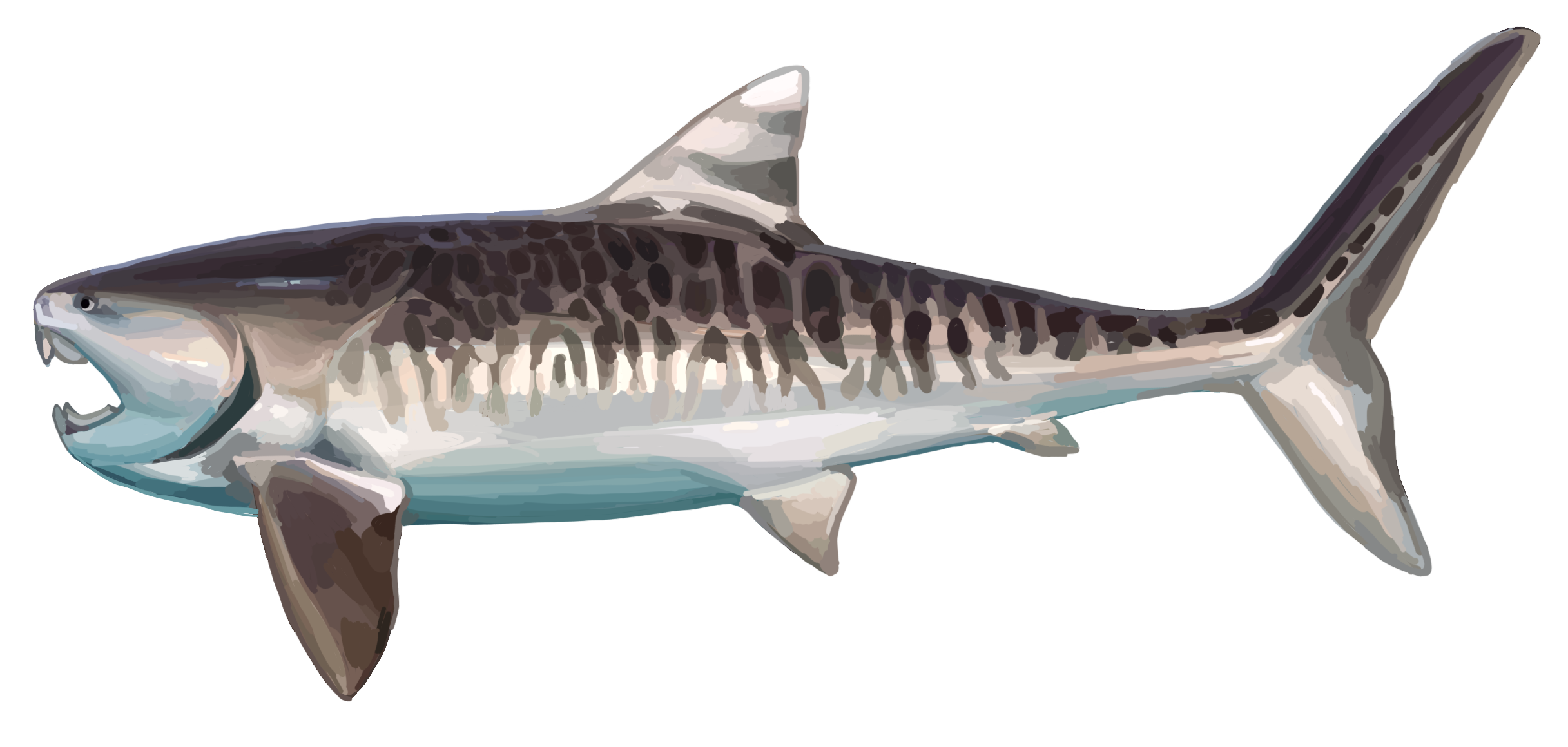
Fish are aquatic, craniate, gill-bearing animals that lack limbs with digits. Included in this definition are the living hagfish, lampreys, and cartilaginous and bony fish as well as various extinct related groups. Approximately 95% of living fish species are ray-finned fish, belonging to the class Actinopterygii, with around 99% of those being teleosts. The earliest organisms that can be classified as fish were soft-bodied chordates that first appeared during the Cambrian period. Although they lacked a true spine, they possessed notochords which allowed them to be more agile than their invertebrate counterparts. Fish would continue to evolve through the Paleozoic era, diversifying into a wide variety of forms. Many fish of the Paleozoic developed external armor that protected them from predators. The first fish with jaws appeared in the Silurian period, after which many (such as sharks) became formidable marine predators rather than just the prey of arthrop ... [...More Info...] [...Related Items...] OR: [Wikipedia] [Google] [Baidu] |
Hagfish
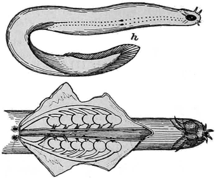
Hagfish, of the class Myxini (also known as Hyperotreti) and order Myxiniformes , are eel-shaped, slime-producing marine fish (occasionally called slime eels). They are the only known living animals that have a skull but no vertebral column, although hagfish do have rudimentary vertebrae. Along with lampreys, hagfish are jawless; the two form the sister group to jawed vertebrates, and living hagfish remain similar to hagfish from around 300 million years ago. The classification of hagfish had been controversial. The issue was whether the hagfish was a degenerate type of vertebrate-fish that through evolution had lost its vertebrae (the original scheme) and was most closely related to lampreys, or whether hagfish represent a stage that precedes the evolution of the vertebral column (the alternative scheme) as is the case with lancelets. Recent DNA evidence has supported the original scheme. The original scheme groups hagfish and lampreys together as cyclostomes (or histori ... [...More Info...] [...Related Items...] OR: [Wikipedia] [Google] [Baidu] |
Metanephroi
The kidneys are two reddish-brown bean-shaped organs found in vertebrates. They are located on the left and right in the retroperitoneal space, and in adult humans are about in length. They receive blood from the paired renal arteries; blood exits into the paired renal veins. Each kidney is attached to a ureter, a tube that carries excreted urine to the bladder. The kidney participates in the control of the volume of various body fluids, fluid osmolality, acid–base balance, various electrolyte concentrations, and removal of toxins. Filtration occurs in the glomerulus: one-fifth of the blood volume that enters the kidneys is filtered. Examples of substances reabsorbed are solute-free water, sodium, bicarbonate, glucose, and amino acids. Examples of substances secreted are hydrogen, ammonium, potassium and uric acid. The nephron is the structural and functional unit of the kidney. Each adult human kidney contains around 1 million nephrons, while a mouse kid ... [...More Info...] [...Related Items...] OR: [Wikipedia] [Google] [Baidu] |
Mesonephroi
The mesonephros ( el, middle kidney) is one of three excretory organs that develop in vertebrates. It serves as the main excretory organ of aquatic vertebrates and as a temporary kidney in reptiles, birds, and mammals. The mesonephros is included in the Wolffian body after Caspar Friedrich Wolff who described it in 1759. (The Wolffian body is composed of: mesonephros + paramesonephrotic blastema) Structure The mesonephros acts as a structure similar to the kidney that, in humans, functions between the sixth and tenth weeks of embryological life. Despite the similarity in structure, function, and terminology, however, the mesonephric nephrons do not form any part of the mature kidney or nephrons. In humans, the mesonephros consists of units which are similar in structure and function to nephrons of the adult kidney. Each of these consists of a glomerulus, a tuft of capillaries which arises from lateral branches of dorsal aorta and drains into the inferior cardinal vein; a Bowman' ... [...More Info...] [...Related Items...] OR: [Wikipedia] [Google] [Baidu] |
Lateral Plate
The lateral plate mesoderm is the mesoderm that is found at the periphery of the embryo. It is to the side of the paraxial mesoderm, and further to the axial mesoderm. The lateral plate mesoderm is separated from the paraxial mesoderm by a narrow region of intermediate mesoderm. The mesoderm is the middle layer of the three germ layers, between the outer ectoderm and inner endoderm. During the third week of embryonic development the lateral plate mesoderm splits into two layers forming the intraembryonic coelom. The outer layer of lateral plate mesoderm adheres to the ectoderm to become the somatic or parietal layer known as the somatopleure. The inner layer adheres to the endoderm to become the splanchnic or visceral layer known as the splanchnopleure. Development The lateral plate mesoderm will split into two layers, the somatopleuric mesenchyme, and the splanchnopleuric mesenchyme. * The ''somatopleuric layer'' forms the future body wall. * The ''splanchnopleuric layer'' form ... [...More Info...] [...Related Items...] OR: [Wikipedia] [Google] [Baidu] |
Wolffian Duct
The mesonephric duct (also known as the Wolffian duct, archinephric duct, Leydig's duct or nephric duct) is a paired organ that forms during the embryonic development of humans and other mammals and gives rise to male reproductive organs. Structure The mesonephric duct connects the primitive kidney, the ''mesonephros'', to the cloaca. It also serves as the primordium for male urogenital structures including the epididymis, vas deferens, and seminal vesicles. Development In both male and female the mesonephric duct develops into the trigone of urinary bladder, a part of the bladder wall, but the sexes differentiate in other ways during development of the urinary and reproductive organs. Male In a male, it develops into a system of connected organs between the efferent ducts of the testis and the prostate, namely the epididymis, the vas deferens, and the seminal vesicle. The prostate forms from the urogenital sinus and the efferent ducts form from the mesonephric tubules. ... [...More Info...] [...Related Items...] OR: [Wikipedia] [Google] [Baidu] |


