|
Probst Bundle
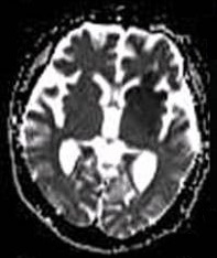
Longitudinal callosal fascicles, or Probst bundles, are aberrant bundles of axons that run in a front-back (antero-posterior) direction rather than a left-right direction between the cerebral hemispheres. They are characteristic of patients with agenesis of the corpus callosum and are due to failure of the callosally-projecting neurons (mostly layer 2/3 pyramidal neurons) to extend axons across the midline and therefore form the corpus callosum. The inability of these axons to cross the midline results in anomalous axonal guidance and front-to-back projections within each hemisphere, rather than connecting between the hemispheres in the normal corpus callosum. These longitudinal callosal fascicles were originally described by Moriz Probst in 1901 by gross anatomical observation. More recently, these anomalies are detected by magnetic resonance imaging or diffusion tensor imaging Diffusion-weighted magnetic resonance imaging (DWI or DW-MRI) is the use of specific MRI sequences a ... [...More Info...] [...Related Items...] OR: [Wikipedia] [Google] [Baidu] |
Axon
An axon (from Greek ἄξων ''áxōn'', axis), or nerve fiber (or nerve fibre: see spelling differences), is a long, slender projection of a nerve cell, or neuron, in vertebrates, that typically conducts electrical impulses known as action potentials away from the nerve cell body. The function of the axon is to transmit information to different neurons, muscles, and glands. In certain sensory neurons (pseudounipolar neurons), such as those for touch and warmth, the axons are called afferent nerve fibers and the electrical impulse travels along these from the periphery to the cell body and from the cell body to the spinal cord along another branch of the same axon. Axon dysfunction can be the cause of many inherited and acquired neurological disorders that affect both the peripheral and central neurons. Nerve fibers are classed into three typesgroup A nerve fibers, group B nerve fibers, and group C nerve fibers. Groups A and B are myelinated, and group C are unmyelinated. ... [...More Info...] [...Related Items...] OR: [Wikipedia] [Google] [Baidu] |
Agenesis Of The Corpus Callosum
Agenesis of the corpus callosum (ACC) is a rare birth defect in which there is a complete or partial absence of the corpus callosum. It occurs when the development of the corpus callosum, the band of white matter connecting the two hemispheres in the brain, in the embryo is disrupted. The result of this is that the fibers that would otherwise form the corpus callosum are instead longitudinally oriented along the ipsilateral ventricular wall and form structures called Probst bundles. In addition to agenesis, other degrees of callosal defects exist, including hypoplasia (underdevelopment or thinness), hypogenesis (partial agenesis) or dysgenesis (malformation). ACC is found in many syndromes and can often present alongside hypoplasia of the cerebellar vermis. When this is the case, there can also be an enlarged fourth ventricle or hydrocephalus; this is called Dandy–Walker malformation. Signs and symptoms Signs and symptoms of ACC and other callosal disorders vary greatly ... [...More Info...] [...Related Items...] OR: [Wikipedia] [Google] [Baidu] |
Pyramidal Neuron
Pyramidal cells, or pyramidal neurons, are a type of multipolar neuron found in areas of the brain including the cerebral cortex, the hippocampus, and the amygdala. Pyramidal neurons are the primary excitation units of the mammalian prefrontal cortex and the corticospinal tract. Pyramidal neurons are also one of two cell types where the characteristic sign, Negri bodies, are found in post-mortem rabies infection. Pyramidal neurons were first discovered and studied by Santiago Ramón y Cajal. Since then, studies on pyramidal neurons have focused on topics ranging from neuroplasticity to cognition. Structure File:GFPneuron.png, Pyramidal neuron visualized by green fluorescent protein (gfp) File:Hippocampal-pyramidal-cell.png, A hippocampal pyramidal cell One of the main structural features of the pyramidal neuron is the conic shaped soma, or cell body, after which the neuron is named. Other key structural features of the pyramidal cell are a single axon, a large apical dendrite, ... [...More Info...] [...Related Items...] OR: [Wikipedia] [Google] [Baidu] |
Axonal Guidance
Axon guidance (also called axon pathfinding) is a subfield of neural development concerning the process by which neurons send out axons to reach their correct targets. Axons often follow very precise paths in the nervous system, and how they manage to find their way so accurately is an area of ongoing research. Axon growth takes place from a region called the growth cone and reaching the axon target is accomplished with relatively few guidance molecules. Growth cone receptors respond to the guidance cues. Mechanisms Growing axons have a highly motile structure at the growing tip called the growth cone, which responds to signals in the extracellular environment that instruct the axon in which direction to grow. These signals, called guidance cues, can be fixed in place or diffusible; they can attract or repel axons. Growth cones contain receptors that recognize these guidance cues and interpret the signal into a chemotropic response. The general theoretical framework is that wh ... [...More Info...] [...Related Items...] OR: [Wikipedia] [Google] [Baidu] |
Corpus Callosum
The corpus callosum (Latin for "tough body"), also callosal commissure, is a wide, thick nerve tract, consisting of a flat bundle of commissural fibers, beneath the cerebral cortex in the brain. The corpus callosum is only found in placental mammals. It spans part of the longitudinal fissure, connecting the left and right cerebral hemispheres, enabling communication between them. It is the largest white matter structure in the human brain, about in length and consisting of 200–300 million axonal projections. A number of separate nerve tracts, classed as subregions of the corpus callosum, connect different parts of the hemispheres. The main ones are known as the genu, the rostrum, the trunk or body, and the splenium. Structure The corpus callosum forms the floor of the longitudinal fissure that separates the two cerebral hemispheres. Part of the corpus callosum forms the roof of the lateral ventricles. The corpus callosum has four main parts – individual nerve tracts ... [...More Info...] [...Related Items...] OR: [Wikipedia] [Google] [Baidu] |
Moriz Probst
Moriz Probst (1 October 1867 in Deutschlandsberg - 21 March 1923 in Vienna) was an Austrian psychiatrist and neuroanatomist. He is best known for first description of so-called Probst bundles, the anomalous brain structures found in dysgenesis of corpus callosum. He studied medicine in Graz, after graduation he worked as an assistant in Neuropsychiatric Clinic in Graz under Gabriel Anton Gabriel Anton (28 July 1858 – 3 January 1933) was an Austrian neurology, neurologist and psychiatry, psychiatrist. He is primarily remembered for his studies of psychiatric conditions arising from damage to the cerebral cortex and the basal gang .... Later he was affiliated with Wiener Irrenanstalt. Since 1900 he practised as forensic psychiatrist in Vienna. He worked also in Landesirrenanstalt neuroanatomical laboratory. Selected works * ''Physiologische, anatomische und pathologisch-anatomische Untersuchungen des Sehhügels''. Archiv für Psychiatrie und Nervenkrankheiten 33: 721-817 (19 ... [...More Info...] [...Related Items...] OR: [Wikipedia] [Google] [Baidu] |
Magnetic Resonance Imaging
Magnetic resonance imaging (MRI) is a medical imaging technique used in radiology to form pictures of the anatomy and the physiological processes of the body. MRI scanners use strong magnetic fields, magnetic field gradients, and radio waves to generate images of the organs in the body. MRI does not involve X-rays or the use of ionizing radiation, which distinguishes it from CT and PET scans. MRI is a medical application of nuclear magnetic resonance (NMR) which can also be used for imaging in other NMR applications, such as NMR spectroscopy. MRI is widely used in hospitals and clinics for medical diagnosis, staging and follow-up of disease. Compared to CT, MRI provides better contrast in images of soft-tissues, e.g. in the brain or abdomen. However, it may be perceived as less comfortable by patients, due to the usually longer and louder measurements with the subject in a long, confining tube, though "Open" MRI designs mostly relieve this. Additionally, implants and oth ... [...More Info...] [...Related Items...] OR: [Wikipedia] [Google] [Baidu] |
Diffusion Tensor Imaging
Diffusion-weighted magnetic resonance imaging (DWI or DW-MRI) is the use of specific MRI sequences as well as software that generates images from the resulting data that uses the diffusion of water molecules to generate contrast in MR images. It allows the mapping of the diffusion process of molecules, mainly water, in biological tissues, in vivo and non-invasively. Molecular diffusion in tissues is not random, but reflects interactions with many obstacles, such as macromolecules, fibers, and membranes. Water molecule diffusion patterns can therefore reveal microscopic details about tissue architecture, either normal or in a diseased state. A special kind of DWI, diffusion tensor imaging (DTI), has been used extensively to map white matter tractography in the brain. Introduction In diffusion weighted imaging (DWI), the intensity of each image element (voxel) reflects the best estimate of the rate of water diffusion at that location. Because the mobility of water is driven by t ... [...More Info...] [...Related Items...] OR: [Wikipedia] [Google] [Baidu] |
Central Nervous System Disorders
Central nervous system diseases, also known as central nervous system disorders, are a group of neurological disorders that affect the structure or function of the brain or spinal cord, which collectively form the central nervous system (CNS). These disorders may be caused by such things as infection, injury, blood clots, age related degeneration, cancer, autoimmune disfunction, and birth defects. The symptoms vary widely, as do the treatments. Central nervous system tumors are the most common forms of pediatric cancer. Brain tumors are the most frequent and have the highest mortality. Some disorders, such as substance addiction, autism, and ADHD may be regarded as CNS disorders, though the classifications are not without dispute. Signs and symptoms Every disease has different signs and symptoms. Some of them are persistent headache; pain in the face, back, arms, or legs; an inability to concentrate; loss of feeling; memory loss; loss of muscle strength; tremors; seizures; incr ... [...More Info...] [...Related Items...] OR: [Wikipedia] [Google] [Baidu] |


