|
Prethalamus
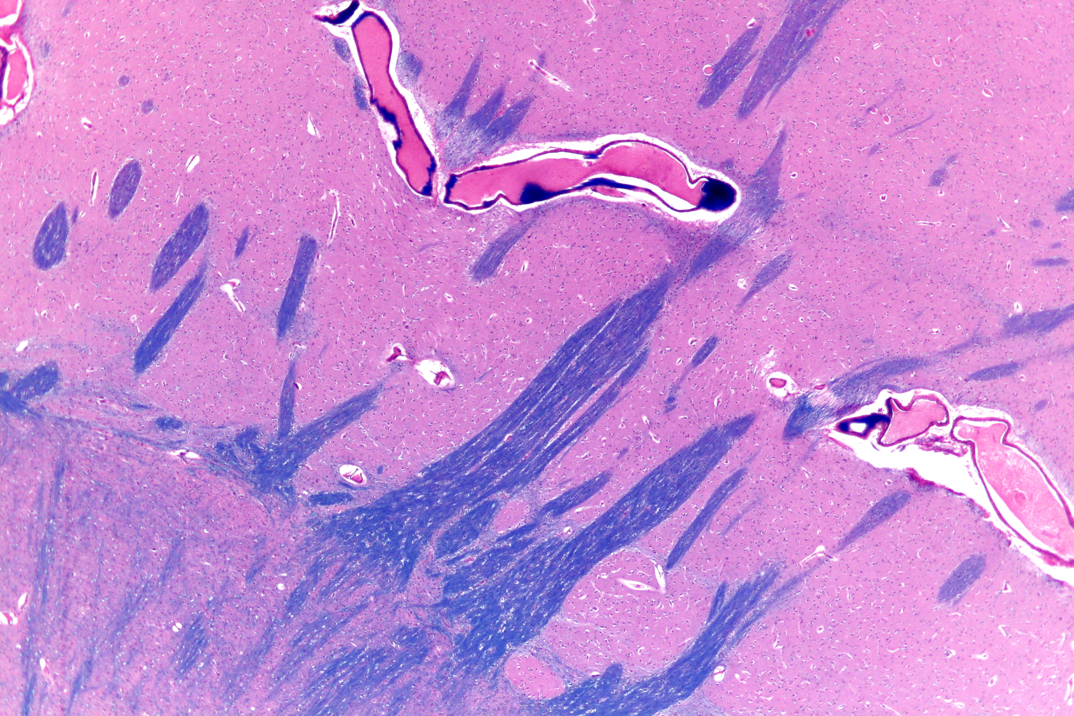
The subthalamus or prethalamus is a part of the diencephalon. Its most prominent structure is the subthalamic nucleus. The subthalamus connects to the globus pallidus, a basal nucleus of the telencephalon. Structure The subthalamus is located ventral to the thalamus, medial to the internal capsule and lateral to the hypothalamus. It is a region formed by several grey matter nuclei and their associated white matter structures, namely: *The subthalamic nucleus, whose neurons contain glutamate and have excitatory effects over neurons of globus pallidus and substantia nigra *Zona incerta, located between fields of Forel H1 and H2. It is continuous with the thalamic reticular nucleus and receives input from the precentral cortex. *Subthalamic fasciculus, formed by fibers that connect the globus pallidus with the subthalamic nucleus *Fields of Forel *Ansa lenticularis During development the subthalamus is continuous with the hypothalamus, but is separated by white matter fibres mai ... [...More Info...] [...Related Items...] OR: [Wikipedia] [Google] [Baidu] |
Basal Ganglia
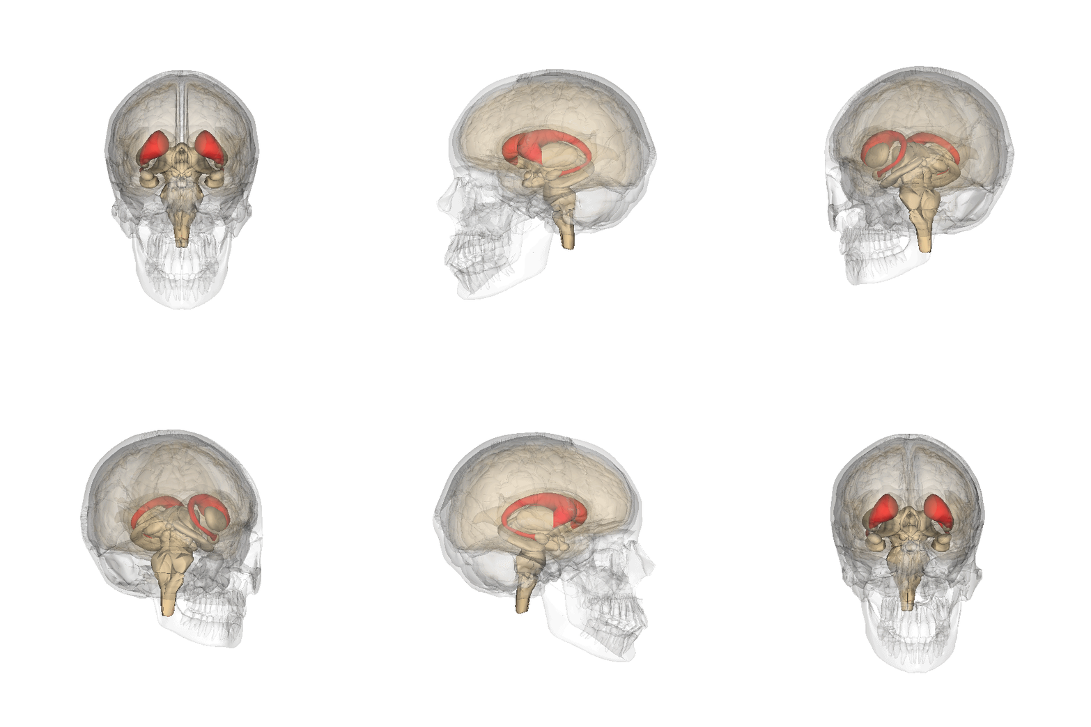
The basal ganglia (BG), or basal nuclei, are a group of subcortical nuclei, of varied origin, in the brains of vertebrates. In humans, and some primates, there are some differences, mainly in the division of the globus pallidus into an external and internal region, and in the division of the striatum. The basal ganglia are situated at the base of the forebrain and top of the midbrain. Basal ganglia are strongly interconnected with the cerebral cortex, thalamus, and brainstem, as well as several other brain areas. The basal ganglia are associated with a variety of functions, including control of voluntary motor movements, procedural learning, habit learning, conditional learning, eye movements, cognition, and emotion. The main components of the basal ganglia – as defined functionally – are the striatum, consisting of both the dorsal striatum (caudate nucleus and putamen) and the ventral striatum (nucleus accumbens and olfactory tubercle), the globus pallidus, ... [...More Info...] [...Related Items...] OR: [Wikipedia] [Google] [Baidu] |
Primary Motor Cortex
The primary motor cortex (Brodmann area 4) is a brain region that in humans is located in the dorsal portion of the frontal lobe. It is the primary region of the motor system and works in association with other motor areas including premotor cortex, the supplementary motor area, posterior parietal cortex, and several subcortical brain regions, to plan and execute voluntary movements. Primary motor cortex is defined anatomically as the region of cortex that contains large neurons known as Betz cells, which, along with other cortical neurons, send long axons down the spinal cord to synapse onto the interneuron circuitry of the spinal cord and also directly onto the alpha motor neurons in the spinal cord which connect to the muscles. At the primary motor cortex, motor representation is orderly arranged (in an inverted fashion) from the toe (at the top of the cerebral hemisphere) to mouth (at the bottom) along a fold in the cortex called the central sulcus. However, some body parts ... [...More Info...] [...Related Items...] OR: [Wikipedia] [Google] [Baidu] |
Subthalamic Nucleus
The subthalamic nucleus (STN) is a small lens-shaped nucleus in the brain where it is, from a functional point of view, part of the basal ganglia system. In terms of anatomy, it is the major part of the subthalamus. As suggested by its name, the subthalamic nucleus is located ventral to the thalamus. It is also dorsal to the substantia nigra and medial to the internal capsule. It was first described by Jules Bernard Luys in 1865, and the term ''corpus Luysi'' or ''Luys' body'' is still sometimes used. Anatomy Structure The principal type of neuron found in the subthalamic nucleus has rather long, sparsely spiny dendrites. In the more centrally located neurons, the dendritic arbors have a more ellipsoidal shape. The dimensions of these arbors (1200 μm, 600 μm, and 300 μm) are similar across many species—including rat, cat, monkey and human—which is unusual. However, the number of neurons increases with brain size as well as the external dimensions of th ... [...More Info...] [...Related Items...] OR: [Wikipedia] [Google] [Baidu] |
Afferent Nerve
A sensory nerve, or afferent nerve, is a general anatomic term for a nerve which contains predominantly somatic afferent nerve fibers. Afferent nerve fibers in a sensory nerve carry sensory information toward the central nervous system (CNS) from different sensory receptors of sensory neurons in the peripheral nervous system. A motor nerve carries information from the CNS to the PNS, and both types of nerve are called peripheral nerves. Afferent nerve fibers link the sensory neurons throughout the body, in pathways to the relevant processing circuits in the central nervous system. Afferent nerve fibers are often paired with efferent nerve fibers from the motor neurons (that travel from the CNS to the PNS), in mixed nerves. Stimuli cause nerve impulses in the receptors and alter the potentials, which is known as sensory transduction. Spinal cord entry Afferent nerve fibers leave the sensory neuron from the dorsal root ganglia of the spinal cord, and motor commands carried ... [...More Info...] [...Related Items...] OR: [Wikipedia] [Google] [Baidu] |
Mesencephalon
The midbrain or mesencephalon is the forward-most portion of the brainstem and is associated with vision, hearing, motor control, sleep and wakefulness, arousal (alertness), and temperature regulation. The name comes from the Greek ''mesos'', "middle", and ''enkephalos'', "brain". Structure The principal regions of the midbrain are the tectum, the cerebral aqueduct, tegmentum, and the cerebral peduncles. Rostrally the midbrain adjoins the diencephalon (thalamus, hypothalamus, etc.), while caudally it adjoins the hindbrain (pons, medulla and cerebellum). In the rostral direction, the midbrain noticeably splays laterally. Sectioning of the midbrain is usually performed axially, at one of two levels – that of the superior colliculi, or that of the inferior colliculi. One common technique for remembering the structures of the midbrain involves visualizing these cross-sections (especially at the level of the superior colliculi) as the upside-down face of a bea ... [...More Info...] [...Related Items...] OR: [Wikipedia] [Google] [Baidu] |
Red Nucleus
The red nucleus or nucleus ruber is a structure in the rostral midbrain involved in motor coordination. The red nucleus is pale pink, which is believed to be due to the presence of iron in at least two different forms: hemoglobin and ferritin. The structure is located in the tegmentum of the midbrain next to the substantia nigra and comprises caudal magnocellular and rostral parvocellular components. The red nucleus and substantia nigra are subcortical centers of the extrapyramidal motor system. Function In a vertebrate without a significant corticospinal tract, gait is mainly controlled by the red nucleus. However, in primates, where the corticospinal tract is dominant, the rubrospinal tract may be regarded as vestigial in motor function. Therefore, the red nucleus is less important in primates than in many other mammals. Nevertheless, the crawling of babies is controlled by the red nucleus, as is arm swinging in typical walking. The red nucleus may play an additional role ... [...More Info...] [...Related Items...] OR: [Wikipedia] [Google] [Baidu] |
Putamen
The putamen (; from Latin, meaning "nutshell") is a round structure located at the base of the forebrain (telencephalon). The putamen and caudate nucleus together form the dorsal striatum. It is also one of the structures that compose the basal nuclei. Through various pathways, the putamen is connected to the substantia nigra, the globus pallidus, the claustrum, and the thalamus, in addition to many regions of the cerebral cortex. A primary function of the putamen is to regulate movements at various stages (e.g. preparation and execution) and influence various types of learning. It employs GABA, acetylcholine, and enkephalin to perform its functions. The putamen also plays a role in degenerative neurological disorders, such as Parkinson's disease. History The word "putamen" is from Latin, referring to that which "falls off in pruning", from "putare", meaning "to prune, to think, or to consider". Until recently, most MRI research focused broadly on the basal ganglia as a who ... [...More Info...] [...Related Items...] OR: [Wikipedia] [Google] [Baidu] |
Caudate Nucleus
The caudate nucleus is one of the structures that make up the corpus striatum, which is a component of the basal ganglia in the human brain. While the caudate nucleus has long been associated with motor processes due to its role in Parkinson's disease, it plays important roles in various other nonmotor functions as well, including procedural learning, associative learning and inhibitory control of action, among other functions. The caudate is also one of the brain structures which compose the reward system and functions as part of the cortico–basal ganglia– thalamic loop. Structure Together with the putamen, the caudate forms the dorsal striatum, which is considered a single functional structure; anatomically, it is separated by a large white matter tract, the internal capsule, so it is sometimes also referred to as two structures: the medial dorsal striatum (the caudate) and the lateral dorsal striatum (the putamen). In this vein, the two are functionally distinct not a ... [...More Info...] [...Related Items...] OR: [Wikipedia] [Google] [Baidu] |
Striatum
The striatum, or corpus striatum (also called the striate nucleus), is a nucleus (a cluster of neurons) in the subcortical basal ganglia of the forebrain. The striatum is a critical component of the motor and reward systems; receives glutamatergic and dopaminergic inputs from different sources; and serves as the primary input to the rest of the basal ganglia. Functionally, the striatum coordinates multiple aspects of cognition, including both motor and action planning, decision-making, motivation, reinforcement, and reward perception. The striatum is made up of the caudate nucleus and the lentiform nucleus. The lentiform nucleus is made up of the larger putamen, and the smaller globus pallidus. Strictly speaking the globus pallidus is part of the striatum. It is common practice, however, to implicitly exclude the globus pallidus when referring to striatal structures. In primates, the striatum is divided into a ventral striatum, and a dorsal striatum, subdivisions that are ... [...More Info...] [...Related Items...] OR: [Wikipedia] [Google] [Baidu] |
Efferent Nerve
Efferent nerve fibers refer to axonal projections that ''exit'' a particular region; as opposed to afferent projections that ''arrive'' at the region. These terms have a slightly different meaning in the context of the peripheral nervous system (PNS) and central nervous system (CNS). The efferent fiber is a long process projecting far from the neuron's body that carries nerve impulses away from the central nervous system toward the peripheral effector organs (mainly muscles and glands). A bundle of these fibers is called an efferent nerve (if it connects to muscles, then it is a motor nerve). The opposite direction of neural activity is afferent conduction, which carries impulses by way of the afferent nerve fibers of sensory neurons. In the nervous system there is a "closed loop" system of sensation, decision, and reactions. This process is carried out through the activity of sensory neurons, interneurons, and motor neurons. In the CNS, afferent and efferent projections c ... [...More Info...] [...Related Items...] OR: [Wikipedia] [Google] [Baidu] |
Zona Limitans Intrathalamica
The ''zona limitans intrathalamica'' (ZLI) is a lineage-restriction compartment and primary developmental boundary in the vertebrate forebrain (which is analogous to the human cerebrum) that serves as a signaling center and a restrictive border between the thalamus (also known as the dorsal thalamus) and the prethalamus (ventral thalamus). Sonic hedgehog (shh) signaling from the ZLI is crucial in the development of the '' diencephalon'', which develops into the thalamus, the pretectum, and the anterior tegmental structures. The ZLI together with the prethalamus and thalamus make up the mid-diencephalic territory (MDT). Discovery Cell lineage restriction boundaries, across which replicating cells cannot migrate, were first discovered in invertebrates, where the expression of various Hox genes in each compartment confer the differentiation of observable segments in the adult body of Drosophila melanogaster. Analogous structures were discovered in the developing vertebrate brain ... [...More Info...] [...Related Items...] OR: [Wikipedia] [Google] [Baidu] |
Ansa Lenticularis
The ansa lenticularis (''ansa lentiformis'' in older texts) is a part of the brain, making up the superior layer of the substantia innominata. Its fibers, derived from the medullary lamina of the lentiform nucleus, pass medially to end in the thalamus and subthalamic region, while others are said to end in the tegmentum and red nucleus. It is classified by NeuroNames as part of the subthalamus The subthalamus or prethalamus is a part of the diencephalon. Its most prominent structure is the subthalamic nucleus. The subthalamus connects to the globus pallidus, a basal nucleus of the telencephalon. Structure The subthalamus is locate .... References External links * {{Authority control Thalamic connections Basal ganglia connections ... [...More Info...] [...Related Items...] OR: [Wikipedia] [Google] [Baidu] |


