|
Premature Junctional Contractions
Premature junctional contractions (PJCs), also called atrioventricular junctional premature complexes or junctional extrasystole, are premature cardiac electrical impulses originating from the atrioventricular node of the heart or "junction". This area is not the normal but only a secondary source of cardiac electrical impulse formation. These premature beats can be found occasionally in healthy people and more commonly in some pathologic conditions, typically in the case of drug cardiotoxicity, electrolyte imbalance, mitral valve surgery, and cold water immersion. If more than two such beats are seen, then the condition is termed junctional rhythm. On the surface ECG, premature junctional contractions will appear as a normally shaped ventricular complex or QRS complex, not preceded by any atrial complex or P wave or preceded by an abnormal P wave with a shorter PR interval. Rarely, the abnormal P wave can follow the QRS. See also * Premature atrial contraction Premature atri ... [...More Info...] [...Related Items...] OR: [Wikipedia] [Google] [Baidu] |
Atrioventricular Node
The atrioventricular node or AV node electrically connects the heart's atria and ventricles to coordinate beating in the top of the heart; it is part of the electrical conduction system of the heart. The AV node lies at the lower back section of the interatrial septum near the opening of the coronary sinus, and conducts the normal electrical impulse from the atria to the ventricles. The AV node is quite compact (~1 x 3 x 5 mm).Full Size Picture triangle of-Koch.jpg Retrieved on 2008-12-22 Structure Location The AV node lies at the lower back section of the |
Ectopic Beat
Ectopic beat is a disturbance of the cardiac rhythm frequently related to the electrical conduction system of the heart, in which beats arise from fibers or group of fibers outside the region in the heart muscle ordinarily responsible for impulse formation (''i.e.'', the sinoatrial node). An ectopic beat can be further classified as either a premature ventricular contraction (PVC), or a premature atrial contraction (PAC). Some patients describe this experience as a "flip" or a "jolt" in the chest, or a "heart hiccup", while others report dropped or missed beats. Ectopic beats are more common during periods of psychological stress, exercise or debility; they may also be triggered by consumption of some food like carbohydrates, strong cheese, or chocolate. It is a form of cardiac arrhythmia in which ectopic foci within either ventricular or atrial myocardium, or from finer branches of the electric transduction system, cause additional beats of the heart. Some medications may wors ... [...More Info...] [...Related Items...] OR: [Wikipedia] [Google] [Baidu] |
Cardiac Pacemaker
350px, Image showing the cardiac pacemaker or SA node, the primary pacemaker within the electrical_conduction_system_of_the_heart">SA_node,_the_primary_pacemaker_within_the_electrical_conduction_system_of_the_heart. The_muscle_contraction.html" "title="electrical conduction system of the heart.">electrical conduction system of the heart">SA node, the primary pacemaker within the electrical conduction system of the heart. The muscle contraction">contraction of cardiac muscle (heart muscle) in all animals is initiated by electrical impulses known as action potentials that in the heart are known as cardiac action potentials. The rate at which these impulses fire controls the rate of cardiac contraction, that is, the heart rate. The cells that create these rhythmic impulses, setting the pace for blood pumping, are called pacemaker cells, and they directly control the heart rate. They make up the cardiac pacemaker, that is, the natural pacemaker of the heart. In most humans, the h ... [...More Info...] [...Related Items...] OR: [Wikipedia] [Google] [Baidu] |
Disease
A disease is a particular abnormal condition that negatively affects the structure or function of all or part of an organism, and that is not immediately due to any external injury. Diseases are often known to be medical conditions that are associated with specific signs and symptoms. A disease may be caused by external factors such as pathogens or by internal dysfunctions. For example, internal dysfunctions of the immune system can produce a variety of different diseases, including various forms of immunodeficiency, hypersensitivity, allergies and autoimmune disorders. In humans, ''disease'' is often used more broadly to refer to any condition that causes pain, dysfunction, distress, social problems, or death to the person affected, or similar problems for those in contact with the person. In this broader sense, it sometimes includes injuries, disabilities, disorders, syndromes, infections, isolated symptoms, deviant behaviors, and atypical variations of structur ... [...More Info...] [...Related Items...] OR: [Wikipedia] [Google] [Baidu] |
Cardiotoxicity
Cardiotoxicity is the occurrence of heart dysfunction as electric or muscle damage, resulting in heart toxicity. The heart becomes weaker and is not as efficient in pumping blood. Cardiotoxicity may be caused by chemotherapy (a usual example is the class of anthracyclines) treatment and/or radiotherapy; complications from anorexia nervosa; adverse effects of heavy metals intake; the long-term abuse of or ingestion at high doses of certain strong stimulants such as cocaine; or an incorrectly administered drug such as bupivacaine. One of the ways to detect cardiotoxicity at early stages when there is a subclinical dysfunction is by measuring changes in regional function of the heart using strains. See also * Cardiotoxin III * Batrachotoxin * Heart failure * Drug interaction Drug interactions occur when a drug's mechanism of action is disturbed by the concomitant administration of substances such as foods, beverages, or other drugs. The cause is often the inhibition of the sp ... [...More Info...] [...Related Items...] OR: [Wikipedia] [Google] [Baidu] |
Water–electrolyte Imbalance
Electrolyte imbalance, or water-electrolyte imbalance, is an abnormality in the concentration of electrolytes in the body. Electrolytes play a vital role in maintaining homeostasis in the body. They help to regulate heart and neurological function, fluid balance, oxygen delivery, acid–base balance and much more. Electrolyte imbalances can develop by consuming too little or too much electrolyte as well as excreting too little or too much electrolyte. Examples of electrolytes include: calcium, chloride, magnesium, phosphate, potassium, and sodium. Electrolyte disturbances are involved in many disease processes, and are an important part of patient management in medicine. The causes, severity, treatment, and outcomes of these disturbances can differ greatly depending on the implicated electrolyte. The most serious electrolyte disturbances involve abnormalities in the levels of sodium, potassium or calcium. Other electrolyte imbalances are less common and often occur in conjunctio ... [...More Info...] [...Related Items...] OR: [Wikipedia] [Google] [Baidu] |
Mitral Valve
The mitral valve (), also known as the bicuspid valve or left atrioventricular valve, is one of the four heart valves. It has two cusps or flaps and lies between the left atrium and the left ventricle of the heart. The heart valves are all one-way valves allowing blood flow in just one direction. The mitral valve and the tricuspid valve are known as the atrioventricular valves because they lie between the atria and the ventricles. In normal conditions, blood flows through an open mitral valve during diastole with contraction of the left atrium, and the mitral valve closes during systole with contraction of the left ventricle. The valve opens and closes because of pressure differences, opening when there is greater pressure in the left atrium than ventricle and closing when there is greater pressure in the left ventricle than atrium. In abnormal conditions, blood may flow backward through the valve ( mitral regurgitation) or the mitral valve may be narrowed (mitral stenosis). Rh ... [...More Info...] [...Related Items...] OR: [Wikipedia] [Google] [Baidu] |
Mammalian Diving Reflex
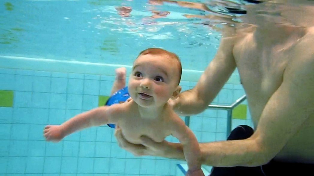
The diving reflex, also known as the diving response and mammalian diving reflex, is a set of physiological responses to immersion that overrides the basic homeostatic reflexes, and is found in all air-breathing vertebrates studied to date. It optimizes respiration by preferentially distributing oxygen stores to the heart and brain, enabling submersion for an extended time. The diving reflex is exhibited strongly in aquatic mammals, such as seals, otters, dolphins, and muskrats, and exists as a lesser response in other animals, including human babies up to 6 months old (see infant swimming), and diving birds, such as ducks and penguins. Adult humans generally exhibit a mild response, the dive-hunting Sama-Bajau people being a notable outlier. The diving reflex is triggered specifically by chilling and wetting the nostrils and face while breath-holding, and is sustained via neural processing originating in the carotid chemoreceptors. The most noticeable effects are on the cardio ... [...More Info...] [...Related Items...] OR: [Wikipedia] [Google] [Baidu] |
Institute Of Naval Medicine
The Institute of Naval Medicine is the main research centre and training facility of the Royal Navy Medical Service. History The site was established in 1969 to research environmental health conditions for submariners in the Royal Navy. At a safety conference on Saturday 25 March 1972 at the University of Birmingham, organised by the National Council of British Mountaineering, with around five hundred climbing experts present, Surgeon Commander Duncan Walters (August 1927 - August 2021) showed a film entitled ''Give Him Air'', about a swimmer in Malta that was accidentally speared in the lung by a harpoon gun. The film showed the gruesome after-effects of the harpoon incident, which caused eight conference attendees to faint, and had to be carried outside. In November 1973 a £200,000 environmental medical centre opened, which simulated life inside a submarine. From 12 November 1973, four sailors (medical ratings) were shut inside this for thirty days, to test atmospheric poll ... [...More Info...] [...Related Items...] OR: [Wikipedia] [Google] [Baidu] |
Junctional Rhythm
Junctional rhythm describes an abnormal heart rhythm resulting from impulses coming from a locus of tissue in the area of the atrioventricular node(AV node), the "junction" between atria and ventricles. Under normal conditions, the heart's sinoatrial node(SA node) determines the rate by which the organ beats – in other words, it is the heart's "pacemaker". The electrical activity of sinus rhythm originates in the sinoatrial node and depolarizes the atria. Current then passes from the atria through the atrioventricular node and into the bundle of His, from which it travels along Purkinje fibers to reach and depolarize the ventricles. This sinus rhythm is important because it ensures that the heart's atria reliably contract before the ventricles. In junctional rhythm, however, the sinoatrial node does not control the heart's rhythm – this can happen in the case of a block in conduction somewhere along the pathway described above, or in sick sinus syndrome, or many other situatio ... [...More Info...] [...Related Items...] OR: [Wikipedia] [Google] [Baidu] |
QRS Complex
The QRS complex is the combination of three of the graphical deflections seen on a typical electrocardiogram (ECG or EKG). It is usually the central and most visually obvious part of the tracing. It corresponds to the depolarization of the right and left ventricles of the heart and contraction of the large ventricular muscles. In adults, the QRS complex normally lasts ; in children it may be shorter. The Q, R, and S waves occur in rapid succession, do not all appear in all leads, and reflect a single event and thus are usually considered together. A Q wave is any downward deflection immediately following the P wave. An R wave follows as an upward deflection, and the S wave is any downward deflection after the R wave. The T wave follows the S wave, and in some cases, an additional U wave follows the T wave. To measure the QRS interval start at the end of the PR interval (or beginning of the Q wave) to the end of the S wave. Normally this interval is 0.08 to 0.10 seconds. When ... [...More Info...] [...Related Items...] OR: [Wikipedia] [Google] [Baidu] |
P Wave (electrocardiography)
The P wave on the ECG represents atrial depolarization, which results in atrial contraction, or atrial systole. Physiology The P wave is a summation wave generated by the depolarization front as it transits the atria. Normally the right atrium depolarizes slightly earlier than left atrium since the depolarization wave originates in the sinoatrial node, in the high right atrium and then travels to and through the left atrium. The depolarization front is carried through the atria along semi-specialized conduction pathways including Bachmann's bundle resulting in uniform shaped waves. Depolarization originating elsewhere in the atria (atrial ectopics) result in P waves with a different morphology from normal. Pathology Peaked P waves (> 0.25 mV) suggest right atrial enlargement, cor pulmonale, (''P pulmonale'' rhythm), but have a low predictive value (~20%). A P wave with increased amplitude can indicate hypokalemia. It can also indicate right atrial enlargement. A P wave ... [...More Info...] [...Related Items...] OR: [Wikipedia] [Google] [Baidu] |


