|
Posterior Superior Alveolar Artery
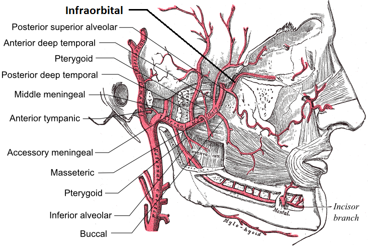
The posterior superior alveolar artery (posterior dental artery) is given off from the maxillary, frequently in conjunction with the infraorbital artery just as the trunk of the vessel is passing into the pterygopalatine fossa. Branches Descending upon the tuberosity of the maxilla, it divides into numerous branches, it descends on the posterior surface of the maxilla and gives branches that supply the molar and premolar teeth and the lining of the maxillary sinus, while others are continued forward on the alveolar process to supply the gingiva. See also * Anterior superior alveolar arteries * Posterior superior alveolar nerve The posterior superior alveolar branches (posterior superior dental branches) arise from the trunk of the maxillary nerve just before it enters the infraorbital groove; they are generally two in number, but sometimes arise by a single trunk. They ... Additional images File:Gray157.png, Left maxilla. Outer surface. References External links * ( ... [...More Info...] [...Related Items...] OR: [Wikipedia] [Google] [Baidu] |
Maxillary Artery
The maxillary artery supplies deep structures of the face. It branches from the external carotid artery just deep to the neck of the mandible. Structure The maxillary artery, the larger of the two terminal branches of the external carotid artery, arises behind the neck of the mandible, and is at first imbedded in the substance of the parotid gland; it passes forward between the ramus of the mandible and the sphenomandibular ligament, and then runs, either superficial or deep to the lateral pterygoid muscle, to the pterygopalatine fossa. It supplies the deep structures of the face, and may be divided into mandibular, pterygoid, and pterygopalatine portions. First portion The ''first'' or ''mandibular '' or ''bony'' portion passes horizontally forward, between the neck of the mandible and the sphenomandibular ligament, where it lies parallel to and a little below the auriculotemporal nerve; it crosses the inferior alveolar nerve, and runs along the lower border of the lateral pte ... [...More Info...] [...Related Items...] OR: [Wikipedia] [Google] [Baidu] |
Alveolar Canals
The alveolar canals are apertures in the center of the infratemporal surface of the maxilla. The alveolar canals transmit the posterior superior alveolar vessels and nerves A nerve is an enclosed, cable-like bundle of nerve fibers (called axons) in the peripheral nervous system. A nerve transmits electrical impulses. It is the basic unit of the peripheral nervous system. A nerve provides a common pathway for the e .... References Foramina of the skull {{musculoskeletal-stub ... [...More Info...] [...Related Items...] OR: [Wikipedia] [Google] [Baidu] |
Gingiva
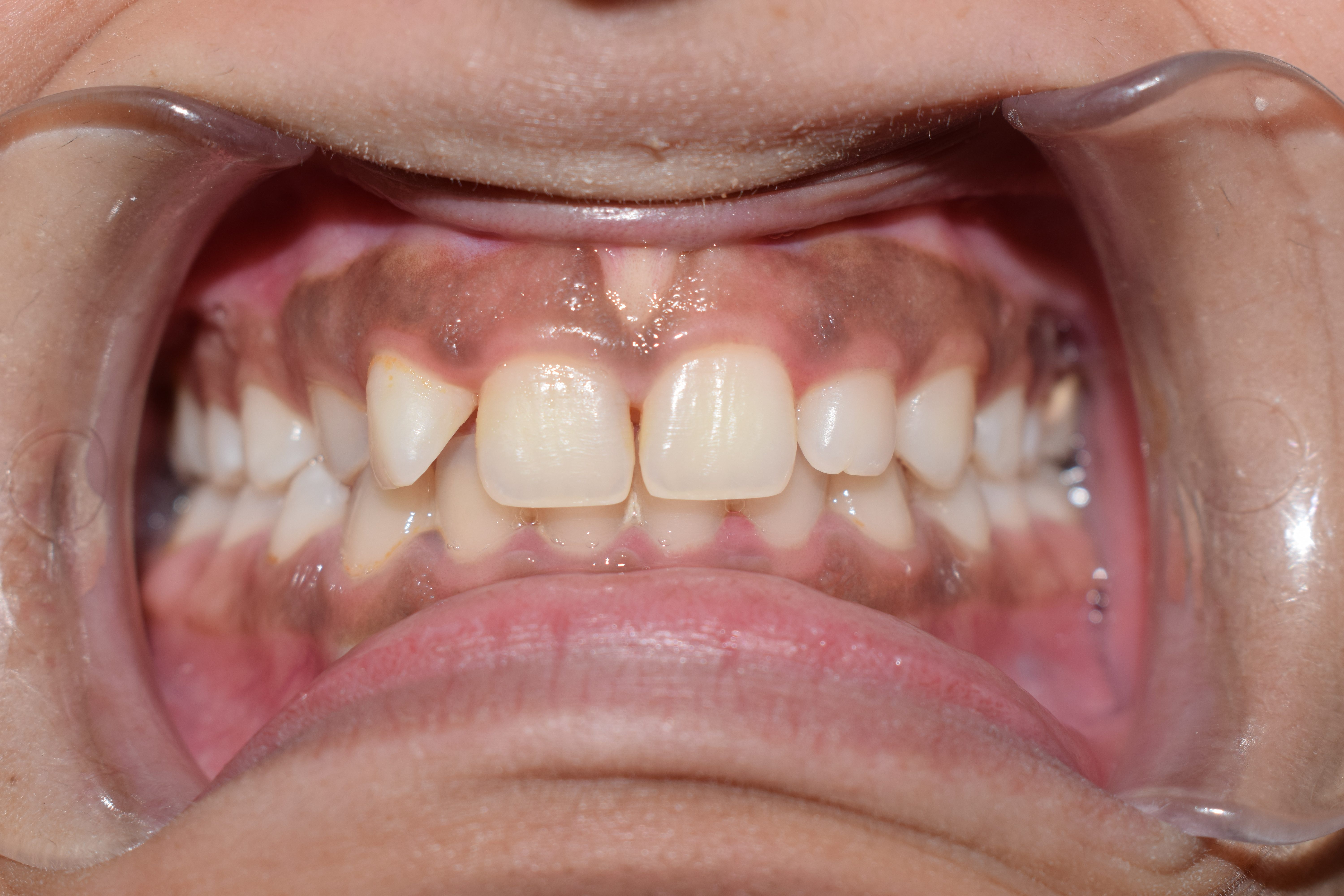
The gums or gingiva (plural: ''gingivae'') consist of the mucosal tissue that lies over the mandible and maxilla inside the mouth. Gum health and disease can have an effect on general health. Structure The gums are part of the soft tissue lining of the mouth. They surround the teeth and provide a seal around them. Unlike the soft tissue linings of the lips and cheeks, most of the gums are tightly bound to the underlying bone which helps resist the friction of food passing over them. Thus when healthy, it presents an effective barrier to the barrage of periodontal insults to deeper tissue. Healthy gums are usually coral pink in light skinned people, and may be naturally darker with melanin pigmentation. Changes in color, particularly increased redness, together with swelling and an increased tendency to bleed, suggest an inflammation that is possibly due to the accumulation of bacterial plaque. Overall, the clinical appearance of the tissue reflects the underlying histology, b ... [...More Info...] [...Related Items...] OR: [Wikipedia] [Google] [Baidu] |
Molar (tooth)
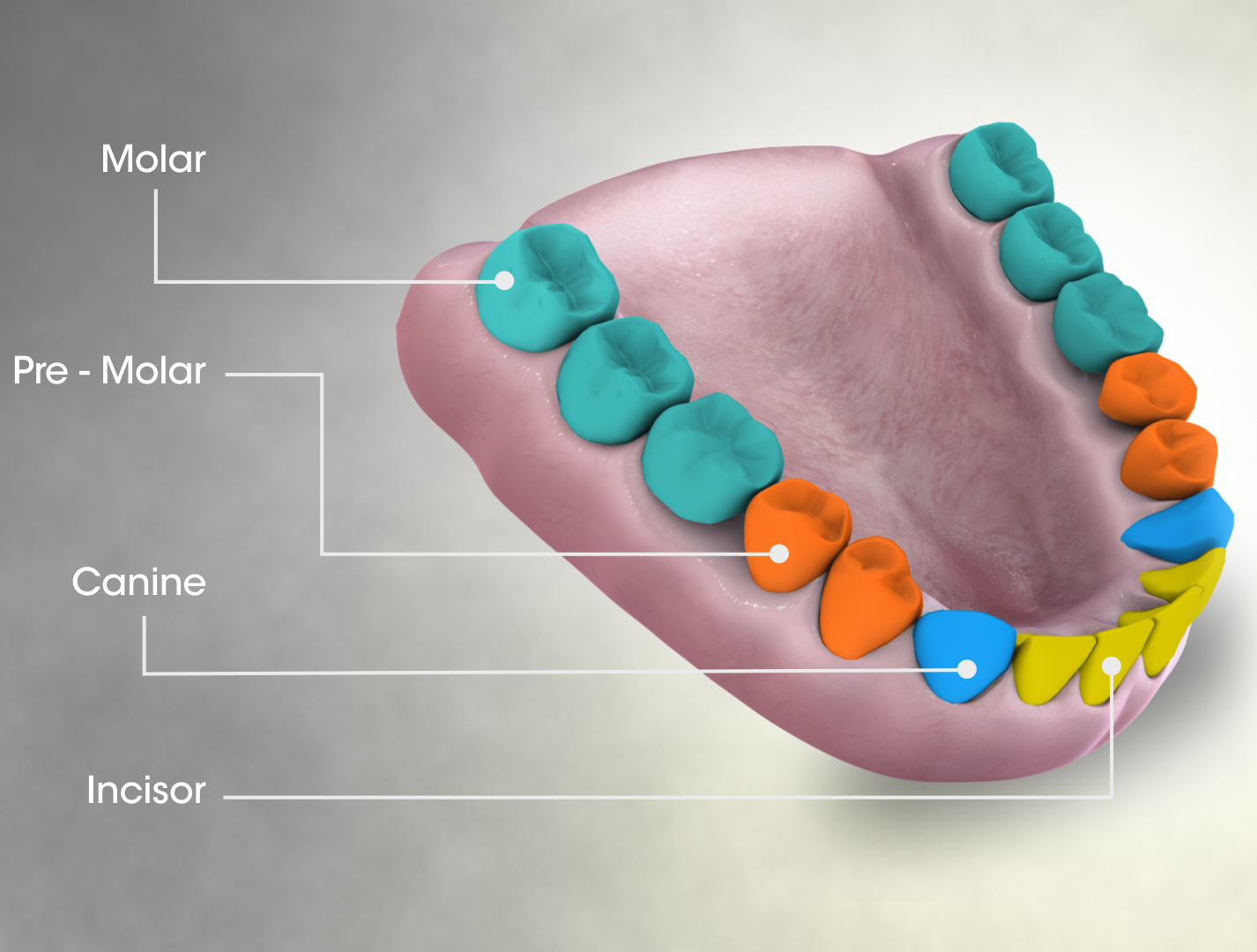
The molars or molar teeth are large, flat teeth at the back of the mouth. They are more developed in mammals. They are used primarily to grind food during chewing. The name ''molar'' derives from Latin, ''molaris dens'', meaning "millstone tooth", from ''mola'', millstone and ''dens'', tooth. Molars show a great deal of diversity in size and shape across mammal groups. The third molar of humans is sometimes vestigial. Human anatomy In humans, the molar teeth have either four or five cusps. Adult humans have 12 molars, in four groups of three at the back of the mouth. The third, rearmost molar in each group is called a wisdom tooth. It is the last tooth to appear, breaking through the front of the gum at about the age of 20, although this varies from individual to individual. Race can also affect the age at which this occurs, with statistical variations between groups. In some cases, it may not even erupt at all. The human mouth contains upper (maxillary) and lower (mandib ... [...More Info...] [...Related Items...] OR: [Wikipedia] [Google] [Baidu] |
Premolar
The premolars, also called premolar teeth, or bicuspids, are transitional teeth located between the canine and molar teeth. In humans, there are two premolars per quadrant in the permanent set of teeth, making eight premolars total in the mouth. They have at least two cusps. Premolars can be considered transitional teeth during chewing, or mastication. They have properties of both the canines, that lie anterior and molars that lie posterior, and so food can be transferred from the canines to the premolars and finally to the molars for grinding, instead of directly from the canines to the molars. Human anatomy The premolars in humans are the maxillary first premolar, maxillary second premolar, mandibular first premolar, and the mandibular second premolar. Premolar teeth by definition are permanent teeth distal to the canines, preceded by deciduous molars. Morphology There is always one large buccal cusp, especially so in the mandibular first premolar. The lower second ... [...More Info...] [...Related Items...] OR: [Wikipedia] [Google] [Baidu] |
Maxillary Sinus
The pyramid-shaped maxillary sinus (or antrum of Highmore) is the largest of the paranasal sinuses, and drains into the middle meatus of the nose through the osteomeatal complex.Human Anatomy, Jacobs, Elsevier, 2008, page 209-210 Structure It is the largest air sinus in the body. Found in the body of the maxilla, this sinus has three recesses: an alveolar recess pointed inferiorly, bounded by the alveolar process of the maxilla; a zygomatic recess pointed laterally, bounded by the zygomatic bone; and an infraorbital recess pointed superiorly, bounded by the inferior orbital surface of the maxilla. The medial wall is composed primarily of cartilage. The ostia for drainage are located high on the medial wall and open into the semilunar hiatus of the lateral nasal cavity; because of the position of the ostia, gravity cannot drain the maxillary sinus contents when the head is erect (see pathology). The ostium of the maxillary sinus is high up on the medial wall and on average is 2. ... [...More Info...] [...Related Items...] OR: [Wikipedia] [Google] [Baidu] |
Maxillary Artery
The maxillary artery supplies deep structures of the face. It branches from the external carotid artery just deep to the neck of the mandible. Structure The maxillary artery, the larger of the two terminal branches of the external carotid artery, arises behind the neck of the mandible, and is at first imbedded in the substance of the parotid gland; it passes forward between the ramus of the mandible and the sphenomandibular ligament, and then runs, either superficial or deep to the lateral pterygoid muscle, to the pterygopalatine fossa. It supplies the deep structures of the face, and may be divided into mandibular, pterygoid, and pterygopalatine portions. First portion The ''first'' or ''mandibular '' or ''bony'' portion passes horizontally forward, between the neck of the mandible and the sphenomandibular ligament, where it lies parallel to and a little below the auriculotemporal nerve; it crosses the inferior alveolar nerve, and runs along the lower border of the lateral pte ... [...More Info...] [...Related Items...] OR: [Wikipedia] [Google] [Baidu] |
Infraorbital Artery
The infraorbital artery is an artery in the head that branches off the maxillary artery, emerging through the infraorbital foramen, just under the orbit of the eye. Course The infraorbital artery appears, from its direction, to be the continuation of the trunk of the maxillary artery, but often arises in conjunction with the posterior superior alveolar artery. It runs along the infraorbital groove and canal with the infraorbital nerve, and emerges on the face through the infraorbital foramen, beneath the infraorbital head of the levator labii superioris muscle. Branches While in the canal, it gives off * (a) orbital branches which assist in supplying the inferior rectus and inferior oblique and the lacrimal sac, and * (b) anterior superior alveolar arteries - branches which descend through the anterior alveolar canals to supply the upper incisor and canine teeth and the mucous membrane of the maxillary sinus. On the face, some branches pass upward to the medial angle of the o ... [...More Info...] [...Related Items...] OR: [Wikipedia] [Google] [Baidu] |
Pterygopalatine Fossa
In human anatomy, the pterygopalatine fossa (sphenopalatine fossa) is a fossa in the skull. A human skull contains two pterygopalatine fossae—one on the left side, and another on the right side. Each fossa is a cone-shaped paired depression deep to the infratemporal fossa and posterior to the maxilla on each side of the skull, located between the pterygoid process and the maxillary tuberosity close to the apex of the orbit. It is the indented area medial to the pterygomaxillary fissure leading into the sphenopalatine foramen. It communicates with the nasal and oral cavities, infratemporal fossa, orbit, pharynx, and middle cranial fossa through eight foramina. Structure Boundaries It has the following boundaries: * ''anterior'': superomedial part of the infratemporal surface of maxilla * ''posterior'': root of the pterygoid process and adjoining anterior surface of the greater wing of sphenoid bone * ''medial'': perpendicular plate of the palatine bone and its orbital and sph ... [...More Info...] [...Related Items...] OR: [Wikipedia] [Google] [Baidu] |
Anterior Superior Alveolar Arteries
The anterior superior alveolar arteries originate from the infraorbital artery; they supply the upper incisors and canines; they also supply the mucous membrane of the maxillary sinus. See also * Anterior superior alveolar nerve * Posterior superior alveolar artery The posterior superior alveolar artery (posterior dental artery) is given off from the maxillary, frequently in conjunction with the infraorbital artery just as the trunk of the vessel is passing into the pterygopalatine fossa. Branches Descendin ... Arteries of the head and neck {{Circulatory-stub ... [...More Info...] [...Related Items...] OR: [Wikipedia] [Google] [Baidu] |
Posterior Superior Alveolar Nerve
The posterior superior alveolar branches (posterior superior dental branches) arise from the trunk of the maxillary nerve just before it enters the infraorbital groove; they are generally two in number, but sometimes arise by a single trunk. They descend on the tuberosity of the maxilla and give off several twigs to the gums and neighboring parts of the mucous membrane of the cheek. They then enter the alveolar canals on the infratemporal surface of the maxilla, and, passing from behind forward in the substance of the bone, communicate with the middle superior alveolar nerve, and give off branches to the lining membrane of the maxillary sinus and gingival and dental branches to each molar tooth from a superior dental plexus; these branches enter the apical foramina at the roots of the teeth. The posterior superior alveolar nerve innervates the second and third maxillary molars, and two of the three roots of the maxillary first molar (all but the mesiobuccal root). When giving a ... [...More Info...] [...Related Items...] OR: [Wikipedia] [Google] [Baidu] |


