|
Porta Hepatis
The porta hepatis or transverse fissure of the liver is a short but deep fissure, about 5 cm long, extending transversely beneath the left portion of the right lobe of the liver, nearer its posterior surface than its anterior border. It joins nearly at right angles with the left sagittal fossa, and separates the quadrate lobe in front from the caudate lobe and process behind. Function It transmits the following (in anterior to posterior order): * common hepatic duct (leaving) * proper hepatic artery (entering) * hepatic portal vein (entering) The hepatic duct lies in front and to the right, the hepatic artery to the left, and the portal vein behind and between the duct and artery. It also transmits nerves and lymphatics. * Sympathetic nerves - these provide afferent pain impulses from the liver and gall bladder to the brain. Pain may be referred to the lower pole of the right scapula (T7). * Hepatic branch of the vagus nerve (CN X). Location The porta hepatis runs in t ... [...More Info...] [...Related Items...] OR: [Wikipedia] [Google] [Baidu] |
Liver
The liver is a major Organ (anatomy), organ only found in vertebrates which performs many essential biological functions such as detoxification of the organism, and the Protein biosynthesis, synthesis of proteins and biochemicals necessary for digestion and growth. In humans, it is located in the quadrant (anatomy), right upper quadrant of the abdomen, below the thoracic diaphragm, diaphragm. Its other roles in metabolism include the regulation of Glycogen, glycogen storage, decomposition of red blood cells, and the production of hormones. The liver is an accessory digestive organ that produces bile, an alkaline fluid containing cholesterol and bile acids, which helps the fatty acid degradation, breakdown of fat. The gallbladder, a small pouch that sits just under the liver, stores bile produced by the liver which is later moved to the small intestine to complete digestion. The liver's highly specialized biological tissue, tissue, consisting mostly of hepatocytes, regulates a w ... [...More Info...] [...Related Items...] OR: [Wikipedia] [Google] [Baidu] |
Portal Vein
The portal vein or hepatic portal vein (HPV) is a blood vessel that carries blood from the gastrointestinal tract, gallbladder, pancreas and spleen to the liver. This blood contains nutrients and toxins extracted from digested contents. Approximately 75% of total liver blood flow is through the portal vein, with the remainder coming from the hepatic artery proper. The blood leaves the liver to the heart in the hepatic veins. The portal vein is not a true vein, because it conducts blood to capillary beds in the liver and not directly to the heart. It is a major component of the hepatic portal system, one of only two portal venous systems in the body – with the hypophyseal portal system being the other. The portal vein is usually formed by the confluence of the superior mesenteric, splenic veins, inferior mesenteric, left, right gastric veins and the pancreatic vein. Conditions involving the portal vein cause considerable illness and death. An important example of such a condi ... [...More Info...] [...Related Items...] OR: [Wikipedia] [Google] [Baidu] |
Quadrate Lobe Of Liver
In human anatomy, the liver is divided grossly into four parts or lobes: the right lobe, the left lobe, the caudate lobe, and the quadrate lobe. Seen from the front – the diaphragmatic surface - the liver is divided into two lobes: the right lobe and the left lobe. Viewed from the underside – the visceral surface, the other two smaller lobes the caudate lobe, and the quadrate lobe are also visible. The two smaller lobes, the caudate lobe and the quadrate lobe, are known as superficial or accessory lobes, and both are located on the underside of the right lobe. The falciform ligament, visible on the front of the liver, makes a superficial division of the right and left lobes of the liver. From the underside, the two additional lobes are located on the right lobe. A line can be imagined running from the left of the vena cava and all the way forward to divide the liver and gallbladder into two halves. This line is called Cantlie's line and is used to mark the division between t ... [...More Info...] [...Related Items...] OR: [Wikipedia] [Google] [Baidu] |
Caudate Lobe Of Liver
In human anatomy, the liver is divided grossly into four parts or lobes: the right lobe, the left lobe, the caudate lobe, and the quadrate lobe. Seen from the front – the diaphragmatic surface - the liver is divided into two lobes: the right lobe and the left lobe. Viewed from the underside – the visceral surface, the other two smaller lobes the caudate lobe, and the quadrate lobe are also visible. The two smaller lobes, the caudate lobe and the quadrate lobe, are known as superficial or accessory lobes, and both are located on the underside of the right lobe. The falciform ligament, visible on the front of the liver, makes a superficial division of the right and left lobes of the liver. From the underside, the two additional lobes are located on the right lobe. A line can be imagined running from the left of the vena cava and all the way forward to divide the liver and gallbladder into two halves. This line is called Cantlie's line and is used to mark the division between t ... [...More Info...] [...Related Items...] OR: [Wikipedia] [Google] [Baidu] |
Common Hepatic Duct
The common hepatic duct is the first part of the biliary tract. It joins the cystic duct coming from the gallbladder to form the common bile duct. Structure The common hepatic duct is the first part of the biliary tract. It is formed by the convergence of the right hepatic duct (which drains bile from the right functional lobe of the liver) and the left hepatic duct (which drains bile from the left functional lobe of the liver). It then joins the cystic duct coming from the gallbladder to form the common bile duct. The duct is usually 6–8 cm long. The common hepatic duct is about 6 mm in diameter in adults, with some variation.Gray's Anatomy, 39th ed, p. 1228 The inner surface is covered in a simple columnar epithelium. Variation Around 1.7% of people have additional accessory hepatic ducts that join onto the common hepatic duct. Rarely, the common hepatic duct joins onto the gallbladder directly, leading to illness. Function The hepatic duct is part of the ... [...More Info...] [...Related Items...] OR: [Wikipedia] [Google] [Baidu] |
Proper Hepatic Artery
The hepatic artery proper (also proper hepatic artery) is the artery that supplies the liver and gallbladder. It raises from the common hepatic artery, a branch of the celiac artery. Structure The hepatic artery proper arises from the common hepatic artery and runs alongside the portal vein and the common bile duct to form the portal triad. A branch of the common hepatic artery –the gastroduodenal artery gives off the small supraduodenal artery to the duodenal bulb. Then the right gastric artery comes off and runs to the left along the lesser curvature of the stomach to meet the left gastric artery, which is a branch of the celiac trunk. It subsequently bifurcates into the right and left hepatic arteries. Variant anatomy Of note, the right and left hepatic arteries may demonstrate variant anatomy. A misplaced right hepatic artery may arise from the superior mesenteric artery (SMA) and a misplaced left hepatic artery may arise from the left gastric artery. The cystic art ... [...More Info...] [...Related Items...] OR: [Wikipedia] [Google] [Baidu] |
Hepatic Portal Vein
The portal vein or hepatic portal vein (HPV) is a blood vessel that carries blood from the gastrointestinal tract, gallbladder, pancreas and spleen to the liver. This blood contains nutrients and toxins extracted from digested contents. Approximately 75% of total liver blood flow is through the portal vein, with the remainder coming from the hepatic artery proper. The blood leaves the liver to the heart in the hepatic veins. The portal vein is not a true vein, because it conducts blood to capillary beds in the liver and not directly to the heart. It is a major component of the hepatic portal system, one of only two portal venous systems in the body – with the hypophyseal portal system being the other. The portal vein is usually formed by the confluence of the superior mesenteric, splenic veins, inferior mesenteric, left, right gastric veins and the pancreatic vein. Conditions involving the portal vein cause considerable illness and death. An important example of such a condi ... [...More Info...] [...Related Items...] OR: [Wikipedia] [Google] [Baidu] |
Lymphatic System
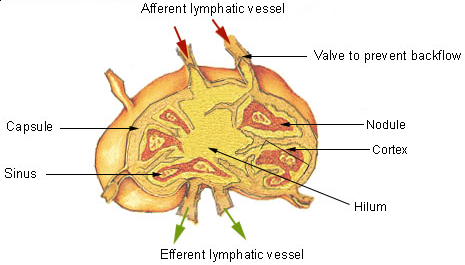
The lymphatic system, or lymphoid system, is an organ system in vertebrates that is part of the immune system, and complementary to the circulatory system. It consists of a large network of lymphatic vessels, lymph nodes, lymphatic or lymphoid organs, and lymphoid tissues. The vessels carry a clear fluid called lymph (the Latin word ''lympha'' refers to the deity of fresh water, "Lympha") back towards the heart, for re-circulation. Unlike the circulatory system that is a closed system, the lymphatic system is open. The human circulatory system processes an average of 20 litres of blood per day through capillary filtration, which removes plasma from the blood. Roughly 17 litres of the filtered blood is reabsorbed directly into the blood vessels, while the remaining three litres are left in the interstitial fluid. One of the main functions of the lymphatic system is to provide an accessory return route to the blood for the surplus three litres. The other main function is that of ... [...More Info...] [...Related Items...] OR: [Wikipedia] [Google] [Baidu] |
Referred Pain
Referred pain, also called reflective pain, is pain perceived at a location other than the site of the painful stimulus. An example is the case of angina pectoris brought on by a myocardial infarction (heart attack), where pain is often felt in the left side of neck, left shoulder, and back rather than in the thorax (chest), the site of the injury. The International Association for the Study of Pain has not officially defined the term; hence several authors have defined it differently. Referred pain is when the pain is located away from or adjacent to the organ involved; for instance, when a person has pain only in their jaw or left arm, but not in the chest. Referred pain has been described since the late 1880s. Despite an increasing amount of literature on the subject, the biological mechanism of referred pain is unknown, although there are several hypotheses. Characteristics * The size of referred pain is related to the intensity and duration of ongoing/evoked pain. * Tempora ... [...More Info...] [...Related Items...] OR: [Wikipedia] [Google] [Baidu] |
Vagus Nerve
The vagus nerve, also known as the tenth cranial nerve, cranial nerve X, or simply CN X, is a cranial nerve that interfaces with the parasympathetic control of the heart, lungs, and digestive tract. It comprises two nerves—the left and right vagus nerves—but they are typically referred to collectively as a single subsystem. The vagus is the longest nerve of the autonomic nervous system in the human body and comprises both sensory and motor fibers. The sensory fibers originate from neurons of the nodose ganglion, whereas the motor fibers come from neurons of the dorsal motor nucleus of the vagus and the nucleus ambiguus. The vagus was also historically called the pneumogastric nerve. Structure Upon leaving the medulla oblongata between the olive and the inferior cerebellar peduncle, the vagus nerve extends through the jugular foramen, then passes into the carotid sheath between the internal carotid artery and the internal jugular vein down to the neck, chest, and abdom ... [...More Info...] [...Related Items...] OR: [Wikipedia] [Google] [Baidu] |
Hepatoduodenal Ligament
The hepatoduodenal ligament is the portion of the lesser omentum extending between the porta hepatis of the liver and the superior part of the duodenum. Running inside it are the following structures collectively known as the portal triad: * hepatic artery proper * portal vein * common bile duct Manual compression of the hepatoduodenal ligament during surgery is known as the Pringle manoeuvre. The cystoduodenal ligament is also found in the lesser omentum and is distinct from both the hepatoduodenal and hepatogastric ligaments. The cystoduodenal ligament is an abnormal peritoneal fold that attaches the duodenum to the gallbladder, representing a rare variation in the anatomy of the lesser sac and its foramen. Another variation sometimes present at the duodenal termination of the hepatoduodenal ligament is the duodenorenal ligament which passes to the front of the right kidney The kidneys are two reddish-brown bean-shaped organs found in vertebrates. They are located on t ... [...More Info...] [...Related Items...] OR: [Wikipedia] [Google] [Baidu] |
Portal Triad
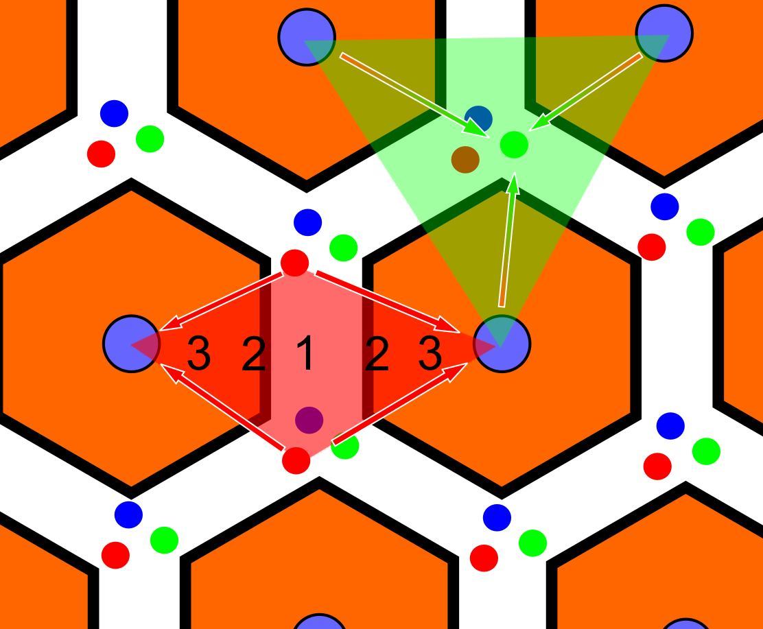
In histology (microscopic anatomy), the lobules of liver, or hepatic lobules, are small divisions of the liver defined at the microscopic scale. The hepatic lobule is a building block of the liver tissue, consisting of a portal triad, hepatocytes arranged in linear cords between a capillary network, and a central vein. Lobules are different from the lobes of liver: they are the smaller divisions of the lobes. The two-dimensional microarchitecture of the liver can be viewed from different perspectives: The term "hepatic lobule", without qualification, typically refers to the classical lobule. Structure The hepatic lobule can be described in terms of metabolic "zones", describing the hepatic acinus (terminal acinus). Each zone is centered on the line connecting two portal triads and extends outwards to the two adjacent central veins. The periportal zone I is nearest to the entering vascular supply and receives the most oxygenated blood, making it least sensitive to ischemic i ... [...More Info...] [...Related Items...] OR: [Wikipedia] [Google] [Baidu] |


