|
Poikilocytosis
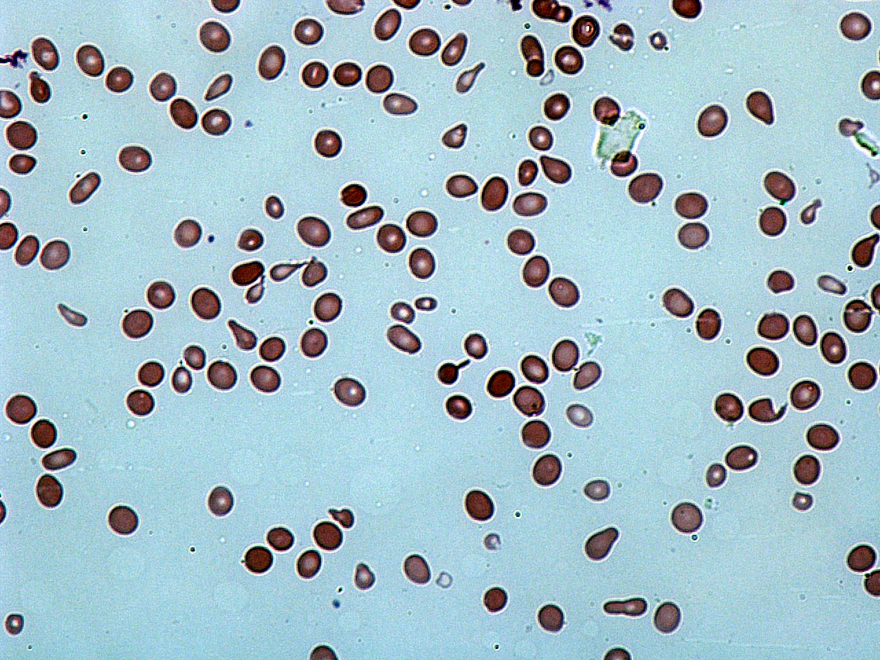
Poikilocytosis is variation in the shapes of red blood cells. Poikilocytes may be oval, teardrop-shaped, sickle-shaped or irregularly contracted. Normal red blood cells are round, flattened disks that are thinner in the middle than at the edges. A ''poikilocyte'' is an abnormally-shaped red blood cell. Generally, poikilocytosis can refer to an increase in abnormal red blood cells of any shape, where they make up 10% or more of the total population of red blood cells. Types Membrane abnormalities # Acanthocytes or Spur/Spike cells # Codocytes or Target cells # Echinocytes and Burr cells # Elliptocytes and Ovalocytes # Spherocytes # Stomatocytes or Mouth cells # Drepanocytes or Sickle Cells # Degmacytes or "bite cells" Trauma # Dacrocytes or Teardrop Cells # Keratocytes # Microspherocytes and Pyropoikilocytes # Schistocytes # Semilunar bodies Diagnosis Poikilocytosis may be diagnosed with a test called a blood smear. During a blood smear, a medical technologist spreads a th ... [...More Info...] [...Related Items...] OR: [Wikipedia] [Google] [Baidu] |
Ovalocyte
Hereditary elliptocytosis, also known as ovalocytosis, is an inherited blood disorder in which an abnormally large number of the person's red blood cells are elliptical rather than the typical biconcave disc shape. Such morphologically distinctive erythrocytes are sometimes referred to as elliptocytes or ovalocytes. It is one of many red-cell membrane defects. In its severe forms, this disorder predisposes to haemolytic anaemia. Although pathological in humans, elliptocytosis is normal in camelids. Presentation RBCs are elleptical in shape rather than normal biconcave shape Most cases are asymptomatic with abnormalities in their peripherial blood film. Pathophysiology Common hereditary elliptocytosis A number of genes have been linked to common hereditary elliptocytosis (many involve the same gene as forms of Hereditary spherocytosis, or HS): These mutations have a common end result; they destabilise the cytoskeletal scaffold of cells. This stability is especially important i ... [...More Info...] [...Related Items...] OR: [Wikipedia] [Google] [Baidu] |
Red Blood Cells
Red blood cells (RBCs), also referred to as red cells, red blood corpuscles (in humans or other animals not having nucleus in red blood cells), haematids, erythroid cells or erythrocytes (from Greek language, Greek ''erythros'' for "red" and ''kytos'' for "hollow vessel", with ''-cyte'' translated as "cell" in modern usage), are the most common type of blood cell and the vertebrate's principal means of delivering oxygen (O2) to the body tissue (biology), tissues—via blood flow through the circulatory system. RBCs take up oxygen in the lungs, or in fish the gills, and release it into tissues while squeezing through the body's capillary, capillaries. The cytoplasm of a red blood cell is rich in hemoglobin, an iron-containing biomolecule that can bind oxygen and is responsible for the red color of the cells and the blood. Each human red blood cell contains approximately 270 million hemoglobin molecules. The cell membrane is composed of proteins and lipids, and this structure ... [...More Info...] [...Related Items...] OR: [Wikipedia] [Google] [Baidu] |
Anisocytosis
Anisocytosis is a medical term meaning that a patient's red blood cells are of unequal size. This is commonly found in anemia and other blood conditions. False diagnostic flagging may be triggered on a complete blood count by an elevated WBC count, agglutinated RBCs, RBC fragments, giant platelets or platelet clumps. In addition, it is a characteristic feature of bovine blood. The red cell distribution width (RDW) is a measurement of anisocytosis and is calculated as a coefficient of variation of the distribution of RBC volumes divided by the mean corpuscular volume ( MCV). Types Anisocytosis is identified by RDW and is classified according to the size of RBC measured by MCV. According to this, it can be divided into *Anisocytosis with microcytosis – Iron deficiency, sickle cell anemia *Anisocytosis with macrocytosis – Folate or vitamin B12 deficiency, autoimmune hemolytic anemia, cytotoxic chemotherapy, chronic liver disease, myelodysplastic syndrome Increased RDW is s ... [...More Info...] [...Related Items...] OR: [Wikipedia] [Google] [Baidu] |
Blood Film
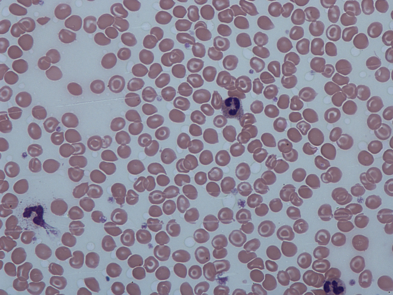
A blood smear, peripheral blood smear or blood film is a thin layer of blood smeared on a glass microscope slide and then stained in such a way as to allow the various blood cells to be examined microscopically. Blood smears are examined in the investigation of hematological (blood) disorders and are routinely employed to look for blood parasites, such as those of malaria and filariasis. Preparation A blood smear is made by placing a drop of blood on one end of a slide, and using a ''spreader slide'' to disperse the blood over the slide's length. The aim is to get a region, called a monolayer, where the cells are spaced far enough apart to be counted and differentiated. The monolayer is found in the "feathered edge" created by the spreader slide as it draws the blood forward. The slide is left to air dry, after which the blood is fixed to the slide by immersing it briefly in methanol. The fixative is essential for good staining and presentation of cellular detail. After fixat ... [...More Info...] [...Related Items...] OR: [Wikipedia] [Google] [Baidu] |
Schistocyte
A schistocyte or schizocyte (from Greek for "divided" and for "hollow" or "cell") is a fragmented part of a red blood cell. Schistocytes are typically irregularly shaped, jagged, and have two pointed ends. Several microangiopathic diseases, including disseminated intravascular coagulation and thrombotic microangiopathies, generate fibrin strands that sever red blood cells as they try to move past a thrombus, creating schistocytes. Schistocytes are often seen in patients with hemolytic anemia. They are frequently a consequence of mechanical artificial heart valves, hemolytic uremic syndrome, and thrombotic thrombocytopenic purpura, among other causes. Excessive schistocytes present in blood can be a sign of microangiopathic hemolytic anemia (MAHA). Appearance Schistocytes are fragmented red blood cells that can take on different shapes. They can be found as triangular, helmet shaped, or comma shaped with pointed edges. Schistocytes are most often found to be microcytic with ... [...More Info...] [...Related Items...] OR: [Wikipedia] [Google] [Baidu] |
Stomatocyte
Hereditary stomatocytosis describes a number of inherited, mostly autosomal dominant human conditions which affect the red blood cell and create the appearance of a slit-like area of central pallor (stomatocyte) among erythrocytes on peripheral blood smear. The erythrocytes' cell membranes may abnormally 'leak' sodium and/or potassium ions, causing abnormalities in cell volume. Hereditary stomatocytosis should be distinguished from acquired causes of stomatocytosis, including dilantin toxicity and alcoholism, as well as artifact from the process of preparing peripheral blood smears. Signs and symptoms Stomatocytosis may present with signs and symptoms consistent with hemolytic anemia as a result of extravascular hemolysis and often intravascular hemolysis. These include fatigue and pallor, as well as signs of jaundice, splenomegaly and gallstone formation from prolonged hemolysis. Certain cases of hereditary stomatocytosis associated with genetic syndromes have additional sympto ... [...More Info...] [...Related Items...] OR: [Wikipedia] [Google] [Baidu] |
Dacrocyte
A dacrocyte (or dacryocyte) is a type of poikilocyte that is shaped like a Tears, teardrop (a "teardrop cell"). A marked increase of dacrocytes is known as dacrocytosis. These tear drop cells are found primarily in diseases with bone marrow fibrosis, such as: primary myelofibrosis, myelodysplastic syndromes during the late course of the disease, rare form of acute leukemias and Myelophthisic anemia, myelophthisis caused by metastatic cancers. Rare causes are myelofibrosis associated with post-irradiation, toxins, autoimmune diseases, metabolic conditions, inborn hemolytic anemias, iron-deficiency anemia or β-thalassemia. Etiology One theory regarding dacrocyte formation is that red blood cells containing various inclusions undergo "pitting" by the spleen to remove these inclusions, and in the process, they can be stretched too far to return to their original shape. It is also thought that this can similarly occur when red blood cells with large inclusions are obstructed from pass ... [...More Info...] [...Related Items...] OR: [Wikipedia] [Google] [Baidu] |
Spherocytosis
Spherocytosis is the presence of spherocytes in the blood, i.e. erythrocytes (red blood cells) that are sphere-shaped rather than bi-concave disk shaped as normal. Spherocytes are found in all hemolytic anemias to some degree. Hereditary spherocytosis and autoimmune hemolytic anemia are characterized by having ''only'' spherocytes. Causes Spherocytes are found in immunologically-mediated hemolytic anemias and in hereditary spherocytosis, but the former would have a positive direct Coombs test and the latter would not. The misshapen but otherwise healthy red blood cells are mistaken by the spleen for old or damaged red blood cells and it thus constantly breaks them down, causing a cycle whereby the body destroys its own blood supply (auto-hemolysis). A complete blood count (CBC) may show increased reticulocytes, a sign of increased red blood cell production, and decreased hemoglobin and hematocrit. The term "non-hereditary spherocytosis" is occasionally used, albeit rarely. Lists ... [...More Info...] [...Related Items...] OR: [Wikipedia] [Google] [Baidu] |
Elliptocyte
Elliptocytes, also known as ovalocytes, are abnormally shaped red blood cells that appear oval or elongated, from slightly egg-shaped to rod or pencil forms. They have normal central pallor with the hemoglobin appearing concentrated at the ends of the elongated cells when viewed through a light microscope. The ends of the cells are blunt and not sharp like sickle cells. __TOC__ Causes Rare elliptocytes (less than 1%) on a peripheral blood smear are a normal finding. These abnormal red blood cells are seen in higher numbers in the blood films of patients with blood disorders such as: * Hereditary elliptocytosis and Southeast Asian ovalocytosis * Thalassemia * Iron deficiency * Myelodysplastic syndrome and myelofibrosis * Megaloblastic anemia Megaloblastic anemia is a type of macrocytic anemia. An anemia is a red blood cell defect that can lead to an undersupply of oxygen. Megaloblastic anemia results from inhibition of DNA replication, DNA synthesis during red blood cell produ ... [...More Info...] [...Related Items...] OR: [Wikipedia] [Google] [Baidu] |
Echinocytes
Echinocyte (from the Greek word ''echinos'', meaning 'hedgehog' or 'sea urchin'), in human biology and medicine, refers to a form of red blood cell that has an abnormal cell membrane characterized by many small, evenly spaced thorny projections.Mentzer WC. Spiculated cells (echinocytes and acanthocytes) and target cells. UpToDate (release: 20.12- C21.4/ref> A more common term for these cells is burr cells. Physiology Echinocytes are frequently confused with acanthocytes, but the mechanism of cell membrane alteration is different. Echinocytosis is a reversible condition of red blood cells that is often merely an artifact produced by EDTA, which is used as an anticoagulant in sampled blood. Echinocytes can be distinguished from acanthocytes by the shape of the projections, which are smaller and more numerous than in acanthocytes and are evenly spaced. Echinocytes also exhibit central pallor, or lightening of color in the center of the cell under Wright staining. Causes In addition t ... [...More Info...] [...Related Items...] OR: [Wikipedia] [Google] [Baidu] |
Codocyte
Codocytes, also known as target cells, are red blood cells that have the appearance of a shooting target with a bullseye. In optical microscopy these cells appear to have a dark center (a central, hemoglobinized area) surrounded by a white ring (an area of relative pallor), followed by dark outer (peripheral) second ring containing a band of hemoglobin. However, in electron microscopy they appear very thin and bell shaped (hence the name codo-: bell). Because of their thinness they are referred to as leptocytes. On routine smear morphology, some people like to make a distinction between leptocytes and codocytes- suggesting that in leptocytes the central spot is not completely detached from the peripheral ring, i.e. the pallor is in a C shape rather than a full ring. These cells are characterized by a disproportional increase in the ratio of surface membrane area to volume. This is also described as a "relative membrane excess." It is due to either increased red cell surface area ... [...More Info...] [...Related Items...] OR: [Wikipedia] [Google] [Baidu] |
Acanthocyte
Acanthocyte (from the Greek word ἄκανθα ''acantha'', meaning 'thorn'), in biology and medicine, refers to an abnormal form of red blood cell that has a spiked cell membrane, due to thorny projections. A similar term is spur cells. Often they may be confused with echinocytes or schistocytes. Acanthocytes have coarse, irregularly spaced, variably sized crenations, resembling many-pointed stars. They are seen on blood films in abetalipoproteinemia, liver disease, chorea acanthocytosis, McLeod syndrome, and several inherited neurological and other disorders such as neuroacanthocytosis, anorexia nervosa, infantile pyknocytosis, hypothyroidism, idiopathic neonatal hepatitis, alcoholism, congestive splenomegaly, Zieve syndrome, and chronic granulomatous disease. Usage Spur cells may refer synonymously to acanthocytes, or may refer in some sources to a specific subset of 'extreme acanthocytes' that have undergone splenic modification whereby additional cell membrane loss has b ... [...More Info...] [...Related Items...] OR: [Wikipedia] [Google] [Baidu] |


_of_echinocytes.png)

.jpg)