|
Pituitary
In vertebrate anatomy, the pituitary gland, or hypophysis, is an endocrine gland, about the size of a chickpea and weighing, on average, in humans. It is a protrusion off the bottom of the hypothalamus at the base of the brain. The hypophysis rests upon the hypophyseal fossa of the sphenoid bone in the center of the middle cranial fossa and is surrounded by a small bony cavity (sella turcica) covered by a dural fold (diaphragma sellae). The anterior pituitary (or adenohypophysis) is a lobe of the gland that regulates several physiological processes including stress, growth, reproduction, and lactation. The intermediate lobe synthesizes and secretes melanocyte-stimulating hormone. The posterior pituitary (or neurohypophysis) is a lobe of the gland that is functionally connected to the hypothalamus by the median eminence via a small tube called the pituitary stalk (also called the infundibular stalk or the infundibulum). Hormones secreted from the pituitary gland help t ... [...More Info...] [...Related Items...] OR: [Wikipedia] [Google] [Baidu] |
Anterior Pituitary
A major organ of the endocrine system, the anterior pituitary (also called the adenohypophysis or pars anterior) is the glandular, anterior lobe that together with the posterior lobe (posterior pituitary, or the neurohypophysis) makes up the pituitary gland (hypophysis). The anterior pituitary regulates several physiological processes, including stress, growth, reproduction, and lactation. Proper functioning of the anterior pituitary and of the organs it regulates can often be ascertained via blood tests that measure hormone levels. Structure The pituitary gland sits in a protective bony enclosure called the sella turcica (''Turkish chair/saddle''). It is composed of three lobes: the anterior, intermediate, and posterior lobes. In many animals, these lobes are distinct. However, in humans, the intermediate lobe is but a few cell layers thick and indistinct; as a result, it is often considered part of the anterior pituitary. In all animals, the fleshy, glandular anterior pitui ... [...More Info...] [...Related Items...] OR: [Wikipedia] [Google] [Baidu] |
Hypothalamus
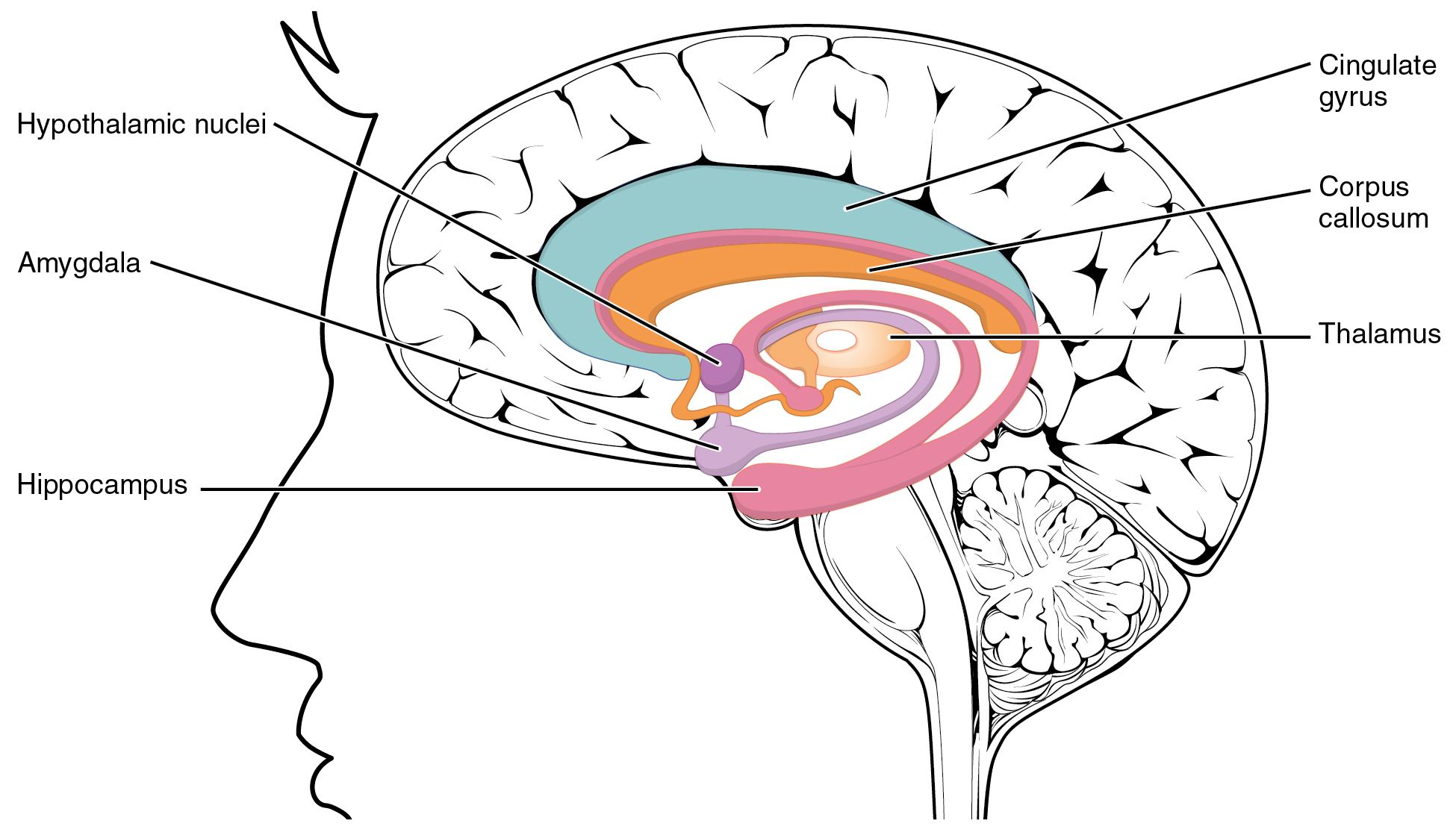
The hypothalamus () is a part of the brain that contains a number of small nuclei with a variety of functions. One of the most important functions is to link the nervous system to the endocrine system via the pituitary gland. The hypothalamus is located below the thalamus and is part of the limbic system. In the terminology of neuroanatomy, it forms the ventral part of the diencephalon. All vertebrate brains contain a hypothalamus. In humans, it is the size of an almond. The hypothalamus is responsible for regulating certain metabolic processes and other activities of the autonomic nervous system. It synthesizes and secretes certain neurohormones, called releasing hormones or hypothalamic hormones, and these in turn stimulate or inhibit the secretion of hormones from the pituitary gland. The hypothalamus controls body temperature, hunger, important aspects of parenting and maternal attachment behaviours, thirst, fatigue, sleep, and circadian rhythms. Structure T ... [...More Info...] [...Related Items...] OR: [Wikipedia] [Google] [Baidu] |
Posterior Pituitary
The posterior pituitary (or neurohypophysis) is the posterior lobe of the pituitary gland which is part of the endocrine system. The posterior pituitary is not glandular as is the anterior pituitary. Instead, it is largely a collection of axonal projections from the hypothalamus that terminate behind the anterior pituitary, and serve as a site for the secretion of neurohypophysial hormones (oxytocin and vasopressin) directly into the blood. The hypothalamic–neurohypophyseal system is composed of the hypothalamus (the paraventricular nucleus and supraoptic nucleus), posterior pituitary, and these axonal projections. Structure The posterior pituitary consists mainly of neuronal projections (axons) of magnocellular neurosecretory cells extending from the supraoptic and paraventricular nuclei of the hypothalamus. These axons store and release neurohypophysial hormones oxytocin and vasopressin into the neurohypophyseal capillaries, from there they get into the systemic circulation ... [...More Info...] [...Related Items...] OR: [Wikipedia] [Google] [Baidu] |
Endocrine Gland
Endocrine glands are ductless glands of the endocrine system that secrete their products, hormones, directly into the blood. The major glands of the endocrine system include the pineal gland, pituitary gland, pancreas, ovaries, testes, thyroid gland, parathyroid gland, hypothalamus and adrenal glands. The hypothalamus and pituitary glands are neuroendocrine organs. Pituitary gland The pituitary gland hangs from the base of the brain by the pituitary stalk, and is enclosed by bone. It consists of a hormone-producing glandular portion of the anterior pituitary and a neural portion of the posterior pituitary, which is an extension of the hypothalamus. The hypothalamus regulates the hormonal output of the anterior pituitary and creates two hormones that it exports to the posterior pituitary for storage and later release. Four of the six anterior pituitary hormones are tropic hormones that regulate the function of other endocrine organs. Most anterior pituitary hormones exhibit ... [...More Info...] [...Related Items...] OR: [Wikipedia] [Google] [Baidu] |
Lactation
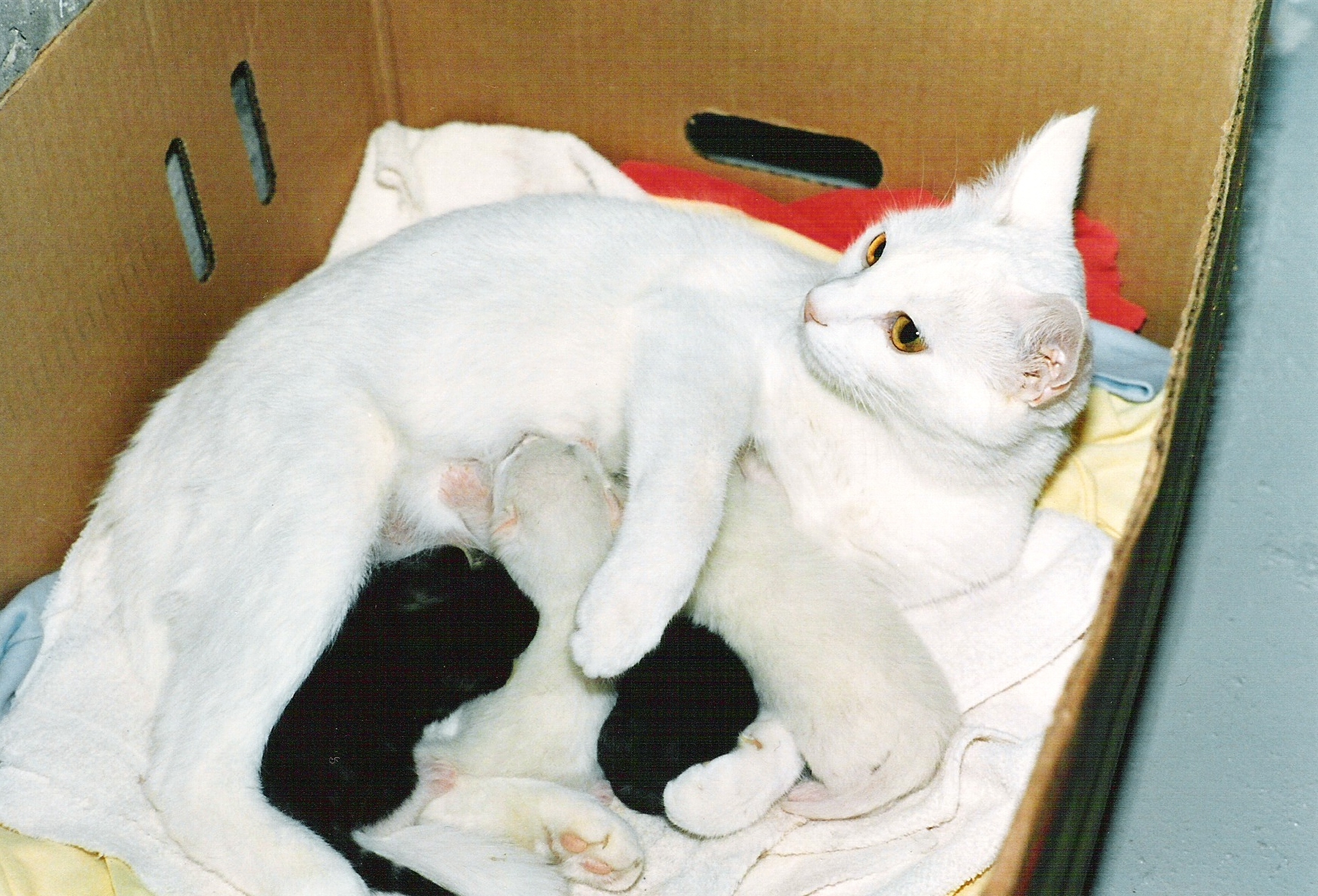
Lactation describes the secretion of milk from the mammary glands and the period of time that a mother lactates to feed her young. The process naturally occurs with all sexually mature female mammals, although it may predate mammals. The process of feeding milk in all animals (including humans) is called ''nursing'', and in humans it is also called ''breastfeeding''. Newborn infants often produce some milk from their own breast tissue, known colloquially as witch's milk. In most species, lactation is a sign that the female has been pregnant at some point in her life, although it can happen without pregnancy. Nearly every species of mammal has nipples; except for monotremes, egg-laying mammals, which instead release milk through ducts in the abdomen. In only one species of mammal, the Dayak fruit bat from Southeast Asia, is milk production a normal male function. ''Galactopoiesis'' is the maintenance of milk production. This stage requires prolactin. Oxytocin is critical for t ... [...More Info...] [...Related Items...] OR: [Wikipedia] [Google] [Baidu] |
Superior Hypophyseal Artery
The superior hypophyseal artery is an artery supplying the pars tuberalis, the infundibulum of the pituitary gland, and the median eminence. It is a branch of the cerebral part of the internal carotid artery The internal carotid artery (Latin: arteria carotis interna) is an artery in the neck which supplies the anterior circulation of the brain. In human anatomy, the internal and external carotids arise from the common carotid arteries, where these b .... References Arteries of the head and neck {{circulatory-stub ... [...More Info...] [...Related Items...] OR: [Wikipedia] [Google] [Baidu] |
Hypophyseal Fossa
The sella turcica (Latin for 'Turkish saddle') is a saddle-shaped depression in the body of the sphenoid bone of the human skull and of the skulls of other hominids including chimpanzees, gorillas and orangutans. It serves as a cephalometric landmark. The pituitary gland or hypophysis is located within the most inferior aspect of the sella turcica, the hypophyseal fossa. Structure The sella turcica is located in the sphenoid bone behind the chiasmatic groove and the tuberculum sellae. It belongs to the middle cranial fossa. The sella turcica's most inferior portion is known as the hypophyseal fossa (the "seat of the saddle"), and contains the pituitary gland (hypophysis). In front of the hypophyseal fossa is the tuberculum sellae. Completing the formation of the saddle posteriorly is the dorsum sellae, which is continuous with the clivus, inferoposteriorly. The dorsum sellae is terminated laterally by the posterior clinoid processes. Development It is widely believed that ... [...More Info...] [...Related Items...] OR: [Wikipedia] [Google] [Baidu] |
Sella Turcica
The sella turcica (Latin for 'Turkish saddle') is a saddle-shaped depression in the body of the sphenoid bone of the human skull and of the skulls of other hominids including chimpanzees, gorillas and orangutans. It serves as a cephalometric landmark. The pituitary gland or hypophysis is located within the most inferior aspect of the sella turcica, the hypophyseal fossa. Structure The sella turcica is located in the sphenoid bone behind the chiasmatic groove and the tuberculum sellae. It belongs to the middle cranial fossa. The sella turcica's most inferior portion is known as the hypophyseal fossa (the "seat of the saddle"), and contains the pituitary gland (hypophysis). In front of the hypophyseal fossa is the tuberculum sellae. Completing the formation of the saddle posteriorly is the dorsum sellae, which is continuous with the clivus, inferoposteriorly. The dorsum sellae is terminated laterally by the posterior clinoid processes. Development It is widely believed that t ... [...More Info...] [...Related Items...] OR: [Wikipedia] [Google] [Baidu] |
Melanocyte-stimulating Hormone
The melanocyte-stimulating hormones, known collectively as MSH, also known as melanotropins or intermedins, are a family of peptide hormones and neuropeptides consisting of α-melanocyte-stimulating hormone (α-MSH), β-melanocyte-stimulating hormone (β-MSH), and γ-melanocyte-stimulating hormone (γ-MSH) that are produced by cells in the pars intermedia of the anterior lobe of the pituitary gland. Synthetic analogues of α-MSH, such as afamelanotide (melanotan I; Scenesse), melanotan II, and bremelanotide (PT-141), have been developed and researched. Biosynthesis The various forms of MSH are generated from different cleavages of the proopiomelanocortin protein, which also yields other important neuropeptides like adrenocorticotropic hormone. Melanocytes in skin make and secrete MSH in response to ultraviolet light, where it increases synthesis of melanin. Some neurons in arcuate nucleus of the hypothalamus make and secrete α-MSH in response to leptin; α-MSH is also m ... [...More Info...] [...Related Items...] OR: [Wikipedia] [Google] [Baidu] |
Intermediate Lobe
Pars intermedia is the boundary between the anterior and posterior lobes of the pituitary. It contains colloid-filled cysts and two types of cells - basophils and chromophobes. The cysts are the remainder of Rathke’s pouch. In human fetal A fetus or foetus (; plural fetuses, feti, foetuses, or foeti) is the unborn offspring that develops from an animal embryo. Following embryonic development the fetal stage of development takes place. In human prenatal development, fetal develo ... life, this area produces melanocyte stimulating hormone or MSH which causes the release of melanin pigment in skin melanocytes (pigment cells). However, the pars intermedia is normally either very small or entirely absent in adulthood. In lower vertebrates (fish, amphibians) MSH from the pars intermedia is responsible for darkening of the skin, often in response to changes in background color. This color change is due to MSH stimulating the dispersion of melanin pigment in dermal (skin) melan ... [...More Info...] [...Related Items...] OR: [Wikipedia] [Google] [Baidu] |
Middle Cranial Fossa
The middle cranial fossa, deeper than the anterior cranial fossa, is narrow medially and widens laterally to the sides of the skull. It is separated from the posterior fossa by the clivus and the petrous crest. It is bounded in front by the posterior margins of the lesser wings of the sphenoid bone, the anterior clinoid processes, and the ridge forming the anterior margin of the chiasmatic groove; behind, by the superior angles of the petrous portions of the temporal bones and the dorsum sellæ; laterally by the temporal squamæ, sphenoidal angles of the parietals, and greater wings of the sphenoid. It is traversed by the squamosal, sphenoparietal, sphenosquamosal, and sphenopetrosal sutures. It houses the temporal lobes of the brain and the pituitary gland. A middle fossa craniotomy is one means to surgically remove acoustic neuromas (vestibular schwannoma) growing within the internal auditory canal of the temporal bone. Middle part The middle part of the fossa presents, i ... [...More Info...] [...Related Items...] OR: [Wikipedia] [Google] [Baidu] |


