|
Photoacoustic Flow Cytometry
Photoacoustic flow cytometry or PAFC is a biomedical imaging modality that utilizes photoacoustic imaging to perform flow cytometry. A flow of cells passes a photoacoustic system producing individual signal response. Each signal is counted to produce a quantitative evaluation of the input sample. Description Traditional flow cytometry uses cells in a Laminar flow, laminar single file stream which then passes through a light source. Using various quantification of light scattering from the cells enables the system to quantify cellular size and complexity which can ultimately be returned in a quantification of cell composition within a sample. Photoacoustic flow cytometry operates on similar principles, but utilizes a photoacoustic signal to differentiate cellular patterns. Furthermore, flow cytometry provides great Ex vivo, ex-vivo analysis, but due to its pure optical source its penetration depth is limited making In vivo, in-vivo analysis limited. Alternatively, photoacoustics ma ... [...More Info...] [...Related Items...] OR: [Wikipedia] [Google] [Baidu] |
Photoacoustic Imaging
Photoacoustic imaging or optoacoustic imaging is a biomedical imaging modality based on the photoacoustic effect. Non-ionizing laser pulses are delivered into biological tissues and part of the energy will be absorbed and converted into heat, leading to transient thermoelastic expansion and thus wideband (i.e. MHz) ultrasonic emission. The generated ultrasonic waves are detected by ultrasonic transducers and then analyzed to produce images. It is known that optical absorption is closely associated with physiological properties, such as hemoglobin concentration and oxygen saturation. As a result, the magnitude of the ultrasonic emission (i.e. photoacoustic signal), which is proportional to the local energy deposition, reveals physiologically specific optical absorption contrast. 2D or 3D images of the targeted areas can then be formed. Biomedical imaging The optical absorption in biological tissues can be due to endogenous molecules such as hemoglobin or melanin, or exogenousl ... [...More Info...] [...Related Items...] OR: [Wikipedia] [Google] [Baidu] |
Methicillin-resistant Staphylococcus Aureus
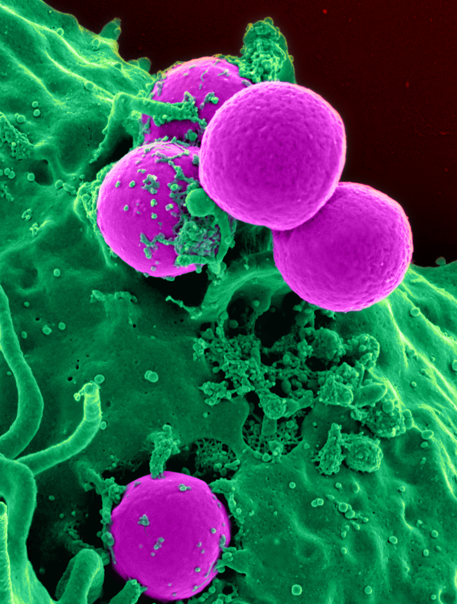
Methicillin-resistant ''Staphylococcus aureus'' (MRSA) is a group of Gram-positive bacteria that are genetically distinct from other strains of ''Staphylococcus aureus''. MRSA is responsible for several difficult-to-treat infections in humans. It caused more than 100,000 deaths attributable to antimicrobial resistance in 2019. MRSA is any strain of ''S. aureus'' that has developed (through natural selection) or acquired (through horizontal gene transfer) a multiple drug resistance to beta-lactam antibiotics. Beta-lactam (β-lactam) antibiotics are a broad-spectrum group that include some penams (penicillin derivatives such as methicillin and oxacillin) and cephems such as the cephalosporins. Strains unable to resist these antibiotics are classified as methicillin-susceptible ''S. aureus'', or MSSA. MRSA is common in hospitals, prisons, and nursing homes, where people with open wounds, invasive devices such as catheters, and weakened immune systems are at greater risk of healt ... [...More Info...] [...Related Items...] OR: [Wikipedia] [Google] [Baidu] |
Jugular Vein
The jugular veins are veins that take deoxygenated blood from the head back to the heart via the superior vena cava. The internal jugular vein descends next to the internal carotid artery and continues posteriorly to the sternocleidomastoid muscle. Structure and Function There are two sets of jugular veins: external and internal. The left and right external jugular veins drain into the subclavian veins. The internal jugular veins join with the subclavian veins more medially to form the brachiocephalic veins. Finally, the left and right brachiocephalic veins join to form the superior vena cava, which delivers deoxygenated blood to the right atrium of the heart. The Jugular veins help carry blood from the heart to and from the brain. An average human brain weighs about 3 pounds, and gets about 15%-20% of the blood that the heart pumps out. It is important for the brain to get enough blood for many reasons. The jugu ... [...More Info...] [...Related Items...] OR: [Wikipedia] [Google] [Baidu] |
Signal-to-noise Ratio
Signal-to-noise ratio (SNR or S/N) is a measure used in science and engineering that compares the level of a desired signal to the level of background noise. SNR is defined as the ratio of signal power to the noise power, often expressed in decibels. A ratio higher than 1:1 (greater than 0 dB) indicates more signal than noise. SNR, bandwidth, and channel capacity of a communication channel are connected by the Shannon–Hartley theorem. Definition Signal-to-noise ratio is defined as the ratio of the power of a signal (meaningful input) to the power of background noise (meaningless or unwanted input): : \mathrm = \frac, where is average power. Both signal and noise power must be measured at the same or equivalent points in a system, and within the same system bandwidth. Depending on whether the signal is a constant () or a random variable (), the signal-to-noise ratio for random noise becomes: : \mathrm = \frac where E refers to the expected value, i.e. in this case ... [...More Info...] [...Related Items...] OR: [Wikipedia] [Google] [Baidu] |
Red Blood Cell
Red blood cells (RBCs), also referred to as red cells, red blood corpuscles (in humans or other animals not having nucleus in red blood cells), haematids, erythroid cells or erythrocytes (from Greek ''erythros'' for "red" and ''kytos'' for "hollow vessel", with ''-cyte'' translated as "cell" in modern usage), are the most common type of blood cell and the vertebrate's principal means of delivering oxygen (O2) to the body tissues—via blood flow through the circulatory system. RBCs take up oxygen in the lungs, or in fish the gills, and release it into tissues while squeezing through the body's capillaries. The cytoplasm of a red blood cell is rich in hemoglobin, an iron-containing biomolecule that can bind oxygen and is responsible for the red color of the cells and the blood. Each human red blood cell contains approximately 270 million hemoglobin molecules. The cell membrane is composed of proteins and lipids, and this structure provides properties essential for physiolo ... [...More Info...] [...Related Items...] OR: [Wikipedia] [Google] [Baidu] |
Hemozoin
Haemozoin is a disposal product formed from the digestion of blood by some blood-feeding parasites. These hematophagous organisms such as malaria parasites (''Plasmodium spp.''), ''Rhodnius'' and ''Schistosoma'' digest haemoglobin and release high quantities of free heme, which is the protein component of haemoglobin. Heme is a prosthetic group consisting of an iron atom contained in the center of a heterocyclic porphyrin ring. Free heme is toxic to cells, so the parasites convert it into an insoluble crystalline form called hemozoin. In malaria parasites, hemozoin is often called ''malaria pigment''. Since the formation of hemozoin is essential to the survival of these parasites, it is an attractive target for developing drugs and is much-studied in ''Plasmodium'' as a way to find drugs to treat malaria (malaria's Achilles' heel). Several currently used antimalarial drugs, such as chloroquine and mefloquine, are thought to kill malaria parasites by inhibiting haemozoin biocrystal ... [...More Info...] [...Related Items...] OR: [Wikipedia] [Google] [Baidu] |
Merozoite
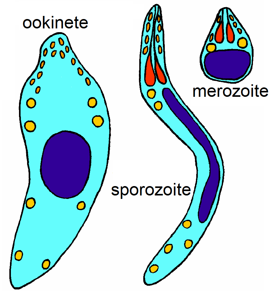
Apicomplexans, a group of intracellular parasites, have life cycle stages that allow them to survive the wide variety of environments they are exposed to during their complex life cycle. Each stage in the life cycle of an apicomplexan organism is typified by a ''cellular variety'' with a distinct morphology and biochemistry. Not all apicomplexa develop all the following cellular varieties and division methods. This presentation is intended as an outline of a hypothetical generalised apicomplexan organism. Methods of asexual replication Apicomplexans (sporozoans) replicate via ways of multiple fission (also known as schizogony). These ways include , and , although the latter is sometimes referred to as schizogony, despite its general meaning. Merogony is an asexually reproductive process of apicomplexa. After infecting a host cell, a trophozoite ( see glossary below) increases in size while repeatedly replicating its nucleus and other organelles. During this process, the org ... [...More Info...] [...Related Items...] OR: [Wikipedia] [Google] [Baidu] |
In Vitro
''In vitro'' (meaning in glass, or ''in the glass'') studies are performed with microorganisms, cells, or biological molecules outside their normal biological context. Colloquially called "test-tube experiments", these studies in biology and its subdisciplines are traditionally done in labware such as test tubes, flasks, Petri dishes, and microtiter plates. Studies conducted using components of an organism that have been isolated from their usual biological surroundings permit a more detailed or more convenient analysis than can be done with whole organisms; however, results obtained from ''in vitro'' experiments may not fully or accurately predict the effects on a whole organism. In contrast to ''in vitro'' experiments, ''in vivo'' studies are those conducted in living organisms, including humans, and whole plants. Definition ''In vitro'' ( la, in glass; often not italicized in English usage) studies are conducted using components of an organism that have been isolated fro ... [...More Info...] [...Related Items...] OR: [Wikipedia] [Google] [Baidu] |
Serology
Serology is the scientific study of Serum (blood), serum and other body fluids. In practice, the term usually refers to the medical diagnosis, diagnostic identification of Antibody, antibodies in the serum. Such antibodies are typically formed in response to an infection (against a given microorganism), against other foreign proteins (in response, for example, to a Acute hemolytic transfusion reaction, mismatched blood transfusion), or to one's own proteins (in instances of autoimmune disease). In either case, the procedure is simple. Serological tests Serological tests are diagnostic methods that are used to identify antibodies and antigens in a patient's sample. Serological tests may be performed to diagnose infections and autoimmune illnesses, to check if a person has immunity (medical), immunity to certain diseases, and in many other situations, such as determining an individual's blood type. Serological tests may also be used in forensic serology to investigate crime scene evid ... [...More Info...] [...Related Items...] OR: [Wikipedia] [Google] [Baidu] |
Blood Film
A blood smear, peripheral blood smear or blood film is a thin layer of blood smeared on a glass microscope slide and then stained in such a way as to allow the various blood cells to be examined microscopically. Blood smears are examined in the investigation of hematological (blood) disorders and are routinely employed to look for blood parasites, such as those of malaria and filariasis. Preparation A blood smear is made by placing a drop of blood on one end of a slide, and using a ''spreader slide'' to disperse the blood over the slide's length. The aim is to get a region, called a monolayer, where the cells are spaced far enough apart to be counted and differentiated. The monolayer is found in the "feathered edge" created by the spreader slide as it draws the blood forward. The slide is left to air dry, after which the blood is fixed to the slide by immersing it briefly in methanol. The fixative is essential for good staining and presentation of cellular detail. After fixat ... [...More Info...] [...Related Items...] OR: [Wikipedia] [Google] [Baidu] |
Malaria
Malaria is a mosquito-borne infectious disease that affects humans and other animals. Malaria causes symptoms that typically include fever, tiredness, vomiting, and headaches. In severe cases, it can cause jaundice, seizures, coma, or death. Symptoms usually begin ten to fifteen days after being bitten by an infected mosquito. If not properly treated, people may have recurrences of the disease months later. In those who have recently survived an infection, reinfection usually causes milder symptoms. This partial resistance disappears over months to years if the person has no continuing exposure to malaria. Malaria is caused by single-celled microorganisms of the ''Plasmodium'' group. It is spread exclusively through bites of infected ''Anopheles'' mosquitoes. The mosquito bite introduces the parasites from the mosquito's saliva into a person's blood. The parasites travel to the liver where they mature and reproduce. Five species of ''Plasmodium'' can infect and be spread by h ... [...More Info...] [...Related Items...] OR: [Wikipedia] [Google] [Baidu] |
Indocyanine Green
Indocyanine green (ICG) is a cyanine dye used in medical diagnostics. It is used for determining cardiac output, hepatic function, liver and gastric blood flow, and for ophthalmic angiography.Definition of indocyanine green National Cancer Institute It has a peak Absorption spectroscopy, spectral absorption at about 800 nm. These infrared frequencies penetrate retinal layers, allowing ICG angiography to image deeper patterns of circulation than fluorescein angiography.Ophthalmic Diagnostic Photography; Indocyanine Green (ICG) Angiography University of Iowa Health Care ICG bi ... [...More Info...] [...Related Items...] OR: [Wikipedia] [Google] [Baidu] |


