|
Pharyngeal (other)
Pharyngeal may refer to: Anatomy * Pharynx, for pharyngeal anatomy * Pharyngeal muscles **Superior pharyngeal constrictor muscle ** Middle pharyngeal constrictor muscle ** Inferior pharyngeal constrictor muscle * Pharyngeal artery * Pharyngeal slit * Pharyngeal tonsil, a mass of lymphoid tissue in the pharynx Other * Pharyngeal consonant A pharyngeal consonant is a consonant that is articulated primarily in the pharynx. Some phoneticians distinguish upper pharyngeal consonants, or "high" pharyngeals, pronounced by retracting the root of the tongue in the mid to upper pharynx, ..., for pharyngeal sounds in phonetics See also * * {{disambiguation ... [...More Info...] [...Related Items...] OR: [Wikipedia] [Google] [Baidu] |
Pharynx
The pharynx (plural: pharynges) is the part of the throat behind the mouth and nasal cavity, and above the oesophagus and trachea (the tubes going down to the stomach and the lungs). It is found in vertebrates and invertebrates, though its structure varies across species. The pharynx carries food and air to the esophagus and larynx respectively. The flap of cartilage called the epiglottis stops food from entering the larynx. In humans, the pharynx is part of the digestive system and the conducting zone of the respiratory system. (The conducting zone—which also includes the nostrils of the nose, the larynx, trachea, bronchi, and bronchioles—filters, warms and moistens air and conducts it into the lungs). The human pharynx is conventionally divided into three sections: the nasopharynx, oropharynx, and laryngopharynx. It is also important in vocalization. In humans, two sets of pharyngeal muscles form the pharynx and determine the shape of its lumen. They are arranged as an ... [...More Info...] [...Related Items...] OR: [Wikipedia] [Google] [Baidu] |
Pharyngeal Muscles
The pharyngeal muscles are a group of muscles that form the pharynx, which is posterior to the oral cavity, determining the shape of its lumen, and affecting its sound properties as the primary resonating cavity. The pharyngeal muscles (involuntary skeletal) push food into the esophagus. There are two muscular layers of the pharynx: the outer circular layer and the inner longitudinal layer. The outer circular layer includes: * Superior constrictor muscle * Middle constrictor muscle * Inferior constrictor muscle During swallowing, these muscles constrict to propel a bolus downwards (an involuntary process). The inner longitudinal layer includes: * Stylopharyngeus muscle * Salpingopharyngeus muscle * Palatopharyngeus muscle During swallowing, these muscles act to shorten and widen the pharynx. They are innervated by the pharyngeal branch of the vagus nerve (CN X) with the exception of the stylopharyngeus muscle which is innervated by the glossopharyngeal nerve The glossophar ... [...More Info...] [...Related Items...] OR: [Wikipedia] [Google] [Baidu] |
Superior Pharyngeal Constrictor Muscle
The superior pharyngeal constrictor muscle is a muscle in the pharynx. It is the highest located muscle of the three pharyngeal constrictors. The muscle is a quadrilateral muscle, thinner and paler than the inferior pharyngeal constrictor muscle and middle pharyngeal constrictor muscle. The muscle is divided into four parts: A pterygopharyngeal, buccopharyngeal, mylopharyngeal and a glossopharyngeal part. Origin and insertion The four parts of this muscle arise from: - the lower third of the posterior margin of the medial pterygoid plate and its hamulus (Pterygopharyngeal part) - from the pterygomandibular raphe (Buccopharyngeal part) - from the alveolar process of the mandible above the posterior end of the mylohyoid line (Mylopharyngeal part) - and by a few fibers from the side of the tongue (Glossopharyngeal part) The fibers curve backward to be inserted into the median raphe, being also prolonged by means of an aponeurosis to the pharyngeal spine on the basilar part of the ... [...More Info...] [...Related Items...] OR: [Wikipedia] [Google] [Baidu] |
Middle Pharyngeal Constrictor Muscle
The middle pharyngeal constrictor is a fan-shaped muscle located in the neck. It is one of three pharyngeal constrictors. Similarly to the superior and inferior pharyngeal constrictor muscles, the middle pharyngeal constrictor is innervated by a branch of the vagus nerve through the pharyngeal plexus. The middle pharyngeal constrictor is smaller than the inferior pharyngeal constrictor muscle. Structure The middle pharyngeal constrictor arises from the whole length of the upper border of the greater cornu of the hyoid bone, from the lesser cornu, and from the stylohyoid ligament. The fibers diverge from their origin: the lower ones descend beneath the constrictor inferior, the middle fibers pass transversely, and the upper fibers ascend and overlap the constrictor superior. It is inserted into the posterior median fibrous raphe, blending in the middle line with the muscle of the opposite side. Function As soon as the bolus of food is received in the pharynx, the elevator mu ... [...More Info...] [...Related Items...] OR: [Wikipedia] [Google] [Baidu] |
Inferior Pharyngeal Constrictor Muscle
The inferior pharyngeal constrictor muscle is a skeletal muscle of the neck. It is the thickest of the three outer pharyngeal muscles. It arises from the sides of the cricoid cartilage and the thyroid cartilage. It is supplied by the vagus nerve (CN X). It is active during swallowing, and partially during breathing and speech. It may be affected by Zenker's diverticulum. Structure The inferior pharyngeal constrictor muscle is composed of two parts. The first part (and more superior) arises from the thyroid cartilage (thyropharyngeal part), and the second part arises from the cricoid cartilage (cricopharyngeal part). * On the ''thyroid cartilage'', it arises from the oblique line on the side of the lamina, from the surface behind this nearly as far as the posterior border and from the inferior horn of the thyroid cartilage. * From the ''cricoid cartilage'', it arises in the interval between the cricothyroid muscle in front, and the articular facet for the inferior horn of the t ... [...More Info...] [...Related Items...] OR: [Wikipedia] [Google] [Baidu] |
Pharyngeal Artery
The ascending pharyngeal artery is an artery in the neck that supplies the pharynx, developing from the proximal part of the embryonic second aortic arch. It is the smallest branch of the external carotid and is a long, slender vessel, deeply seated in the neck, beneath the other branches of the external carotid and under the stylopharyngeus muscle. It lies just superior to the bifurcation of the common carotid arteries. The artery most typically bifurcates into embryologically distinct pharyngeal and neuromeningeal trunks. The pharyngeal trunk usually consists of several branches which supply the middle and inferior pharyngeal constrictor muscles and the stylopharyngeus, ramifying in their substance and in the mucous membranes lining them. These branches are in hemodynamic equilibrium with contributors from the internal maxillary artery. The neuromeningeal trunk classically consists of jugular and hypoglossal divisions, which enter the jugular and hypoglossal foramina to supp ... [...More Info...] [...Related Items...] OR: [Wikipedia] [Google] [Baidu] |
Pharyngeal Slit
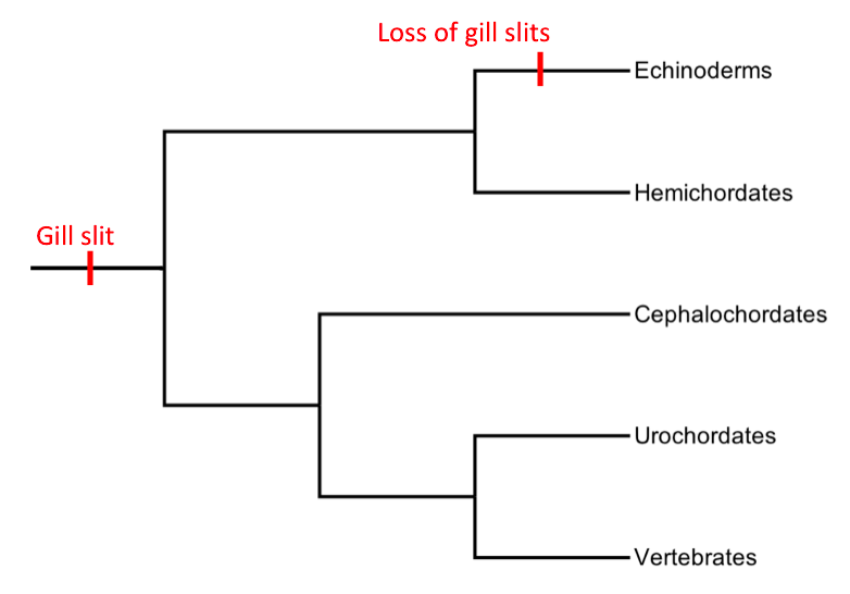
Pharyngeal slits are filter-feeding organs found among deuterostomes. Pharyngeal slits are repeated openings that appear along the pharynx caudal to the mouth. With this position, they allow for the movement of water in the mouth and out the pharyngeal slits. It is postulated that this is how pharyngeal slits first assisted in filter-feeding, and later, with the addition of gills along their walls, aided in respiration of aquatic chordates. These repeated segments are controlled by similar developmental mechanisms. Some hemichordate species can have as many as 200 gill slits. Pharyngeal clefts resembling gill slits are transiently present during the embryonic stages of tetrapod development. The presence of pharyngeal arches and clefts in the neck of the developing human embryo famously led Ernst Haeckel to postulate that "ontogeny recapitulates phylogeny"; this hypothesis, while false, contains elements of truth, as explored by Stephen Jay Gould in ''Ontogeny and Phylogeny''.. A ... [...More Info...] [...Related Items...] OR: [Wikipedia] [Google] [Baidu] |
Pharyngeal Tonsil
In anatomy, the adenoid, also known as the pharyngeal tonsil or nasopharyngeal tonsil, is the superior-most of the tonsils. It is a mass of lymphatic tissue located behind the nasal cavity, in the roof of the nasopharynx, where the nose blends into the throat. In children, it normally forms a soft mound in the roof and back wall of the nasopharynx, just above and behind the uvula. The term ''adenoid'' is also used to represent adenoid hypertrophy, the abnormal growth of the pharyngeal tonsils. Structure The adenoid is a mass of lymphatic tissue located behind the nasal cavity, in the roof of the nasopharynx, where the nose blends into the throat. The adenoid, unlike the palatine tonsils, has pseudostratified epithelium. The adenoids are part of the so-called Waldeyer ring of lymphoid tissue which also includes the palatine tonsils, the lingual tonsils and the tubal tonsils. Development Adenoids develop from a subepithelial infiltration of lymphocytes after the 16th week o ... [...More Info...] [...Related Items...] OR: [Wikipedia] [Google] [Baidu] |


