|
Phantomosaurus Neubigi
''Phantomosaurus'' is an extinct genus of ichthyosaur that lived during the late Anisian stage of the Middle Triassic. Fossils have been found in southern Germany. It was discovered in 1965 and named in 1997 as a species of '' Shastasaurus'' by Sander in the rocks of the Upper Muschelkalk. More recently, in 2005, the braincase was studied by Maisch and Matzke. They found it to be unique among all known ichthyosaurs in terms of braincase morphology. Despite its close relation to many other ichthyosaurs, in particular ''Cymbospondylus, Phantomosaurus'' appears to have a very primitive braincase which resembles other diapsids more than other ichthyosaurs. Morphology ''Phantomosaurus'' is known from a partial skull, the lower jaw, some of the vertebrae and ribs (pictured) and the hind fin. Hind fin The hind fin is very long and slender, with elongated femur, tibia and fibula. These features suggest a long but not particularly powerful limb, which would have generated a reaso ... [...More Info...] [...Related Items...] OR: [Wikipedia] [Google] [Baidu] |
Triassic
The Triassic ( ) is a geologic period and system which spans 50.6 million years from the end of the Permian Period 251.902 million years ago ( Mya), to the beginning of the Jurassic Period 201.36 Mya. The Triassic is the first and shortest period of the Mesozoic Era. Both the start and end of the period are marked by major extinction events. The Triassic Period is subdivided into three epochs: Early Triassic, Middle Triassic and Late Triassic. The Triassic began in the wake of the Permian–Triassic extinction event, which left the Earth's biosphere impoverished; it was well into the middle of the Triassic before life recovered its former diversity. Three categories of organisms can be distinguished in the Triassic record: survivors from the extinction event, new groups that flourished briefly, and other new groups that went on to dominate the Mesozoic Era. Reptiles, especially archosaurs, were the chief terrestrial vertebrates during this time. A specialized subgroup of archo ... [...More Info...] [...Related Items...] OR: [Wikipedia] [Google] [Baidu] |
Fibula
The fibula or calf bone is a leg bone on the lateral side of the tibia, to which it is connected above and below. It is the smaller of the two bones and, in proportion to its length, the most slender of all the long bones. Its upper extremity is small, placed toward the back of the head of the tibia, below the knee joint and excluded from the formation of this joint. Its lower extremity inclines a little forward, so as to be on a plane anterior to that of the upper end; it projects below the tibia and forms the lateral part of the ankle joint. Structure The bone has the following components: * Lateral malleolus * Interosseous membrane connecting the fibula to the tibia, forming a syndesmosis joint * The superior tibiofibular articulation is an arthrodial joint between the lateral condyle of the tibia and the head of the fibula. * The inferior tibiofibular articulation (tibiofibular syndesmosis) is formed by the rough, convex surface of the medial side of the lower end of the f ... [...More Info...] [...Related Items...] OR: [Wikipedia] [Google] [Baidu] |
Skull
The skull is a bone protective cavity for the brain. The skull is composed of four types of bone i.e., cranial bones, facial bones, ear ossicles and hyoid bone. However two parts are more prominent: the cranium and the mandible. In humans, these two parts are the neurocranium and the viscerocranium ( facial skeleton) that includes the mandible as its largest bone. The skull forms the anterior-most portion of the skeleton and is a product of cephalisation—housing the brain, and several sensory structures such as the eyes, ears, nose, and mouth. In humans these sensory structures are part of the facial skeleton. Functions of the skull include protection of the brain, fixing the distance between the eyes to allow stereoscopic vision, and fixing the position of the ears to enable sound localisation of the direction and distance of sounds. In some animals, such as horned ungulates (mammals with hooves), the skull also has a defensive function by providing the mount (on the front ... [...More Info...] [...Related Items...] OR: [Wikipedia] [Google] [Baidu] |
Quadratojugal Bone
The quadratojugal is a skull bone present in many vertebrates, including some living reptiles and amphibians. Anatomy and function In animals with a quadratojugal bone, it is typically found connected to the jugal (cheek) bone from the front and the squamosal bone from above. It is usually positioned at the rear lower corner of the cranium. Many modern tetrapods lack a quadratojugal bone as it has been lost or fused to other bones. Modern examples of tetrapods without a quadratojugal include salamanders, mammals, birds, and squamates (lizards and snakes). In tetrapods with a quadratojugal bone, it often forms a portion of the jaw joint. Developmentally, the quadratojugal bone is a dermal bone in the temporal series, forming the original braincase. The squamosal and quadratojugal bones together form the cheek region and may provide muscular attachments for facial muscles. In reptiles and amphibians In most modern reptiles and amphibians, the quadratojugal is a prominent, strapl ... [...More Info...] [...Related Items...] OR: [Wikipedia] [Google] [Baidu] |
Disarticulation
In medical terminology, disarticulation is the separation of two bones at their joint, either traumatically by way of injury or by a surgeon during arthroplasty or amputation. See also * Joint dislocation * Acrotomophilia * Apotemnophilia * Amputation Amputation is the removal of a limb by trauma, medical illness, or surgery. As a surgical measure, it is used to control pain or a disease process in the affected limb, such as malignancy or gangrene. In some cases, it is carried out on indi ... References Musculoskeletal disorders Amputations {{musculoskeletal-stub ... [...More Info...] [...Related Items...] OR: [Wikipedia] [Google] [Baidu] |
Suprangular
The suprangular or surangular is a jaw bone found in most land vertebrates, except mammals. Usually in the back of the jaw, on the upper edge, it is connected to all other jaw bones: dentary, angular, splenial and articular The articular bone is part of the lower jaw of most vertebrates, including most jawed fish, amphibians, birds and various kinds of reptiles, as well as ancestral mammals. Anatomy In most vertebrates, the articular bone is connected to two oth .... It is often a muscle attachment site. References Vertebrate anatomy {{Vertebrate anatomy-stub ... [...More Info...] [...Related Items...] OR: [Wikipedia] [Google] [Baidu] |
Articular Bone
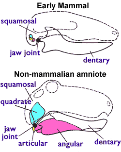
The articular bone is part of the lower jaw of most vertebrates, including most jawed fish, amphibians, birds and various kinds of reptiles, as well as ancestral mammals. Anatomy In most vertebrates, the articular bone is connected to two other lower jaw bones, the suprangular and the angular. Developmentally, it originates from the embryonic mandibular cartilage. The most caudal portion of the mandibular cartilage ossifies to form the articular bone, while the remainder of the mandibular cartilage either remains cartilaginous or disappears. In snakes In snakes, the articular, surangular, and prearticular bones have fused to form the compound bone. The mandible is suspended from the quadrate bone and articulates at this compound bone. Function In amphibians and reptiles In most tetrapods, the articular bone forms the lower portion of the jaw joint. The upper jaw articulates at the quadrate bone. In mammals In mammals, the articular bone evolves to form the malleu ... [...More Info...] [...Related Items...] OR: [Wikipedia] [Google] [Baidu] |
Fish
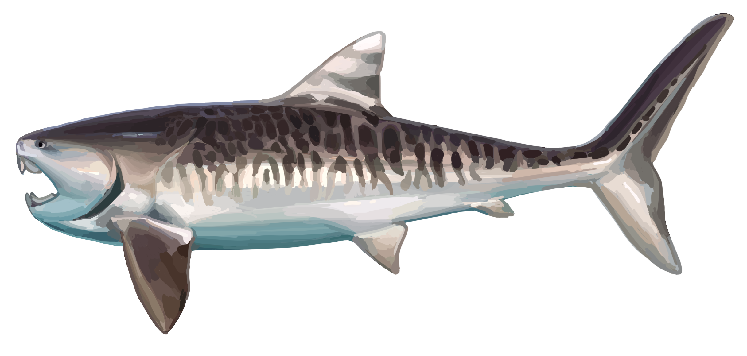
Fish are aquatic, craniate, gill-bearing animals that lack limbs with digits. Included in this definition are the living hagfish, lampreys, and cartilaginous and bony fish as well as various extinct related groups. Approximately 95% of living fish species are ray-finned fish, belonging to the class Actinopterygii, with around 99% of those being teleosts. The earliest organisms that can be classified as fish were soft-bodied chordates that first appeared during the Cambrian period. Although they lacked a true spine, they possessed notochords which allowed them to be more agile than their invertebrate counterparts. Fish would continue to evolve through the Paleozoic era, diversifying into a wide variety of forms. Many fish of the Paleozoic developed external armor that protected them from predators. The first fish with jaws appeared in the Silurian period, after which many (such as sharks) became formidable marine predators rather than just the prey of arthropods. Mos ... [...More Info...] [...Related Items...] OR: [Wikipedia] [Google] [Baidu] |
Tooth
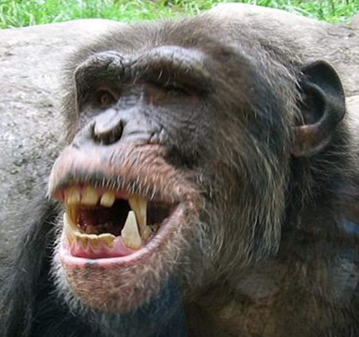
A tooth ( : teeth) is a hard, calcified structure found in the jaws (or mouths) of many vertebrates and used to break down food. Some animals, particularly carnivores and omnivores, also use teeth to help with capturing or wounding prey, tearing food, for defensive purposes, to intimidate other animals often including their own, or to carry prey or their young. The roots of teeth are covered by gums. Teeth are not made of bone, but rather of multiple tissues of varying density and hardness that originate from the embryonic germ layer, the ectoderm. The general structure of teeth is similar across the vertebrates, although there is considerable variation in their form and position. The teeth of mammals have deep roots, and this pattern is also found in some fish, and in crocodilians. In most teleost fish, however, the teeth are attached to the outer surface of the bone, while in lizards they are attached to the inner surface of the jaw by one side. In cartilaginous fish, s ... [...More Info...] [...Related Items...] OR: [Wikipedia] [Google] [Baidu] |
Cone
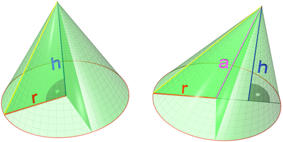
A cone is a three-dimensional geometric shape that tapers smoothly from a flat base (frequently, though not necessarily, circular) to a point called the apex or vertex. A cone is formed by a set of line segments, half-lines, or lines connecting a common point, the apex, to all of the points on a base that is in a plane that does not contain the apex. Depending on the author, the base may be restricted to be a circle, any one-dimensional quadratic form in the plane, any closed one-dimensional figure, or any of the above plus all the enclosed points. If the enclosed points are included in the base, the cone is a solid object; otherwise it is a two-dimensional object in three-dimensional space. In the case of a solid object, the boundary formed by these lines or partial lines is called the ''lateral surface''; if the lateral surface is unbounded, it is a conical surface. In the case of line segments, the cone does not extend beyond the base, while in the case of half-lin ... [...More Info...] [...Related Items...] OR: [Wikipedia] [Google] [Baidu] |
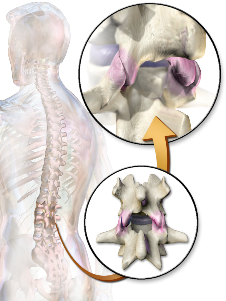
Facet Joint
The facet joints (or zygapophysial joints, zygapophyseal, apophyseal, or Z-joints) are a set of synovial, plane joints between the articular processes of two adjacent vertebrae. There are two facet joints in each spinal motion segment and each facet joint is innervated by the recurrent meningeal nerves. Innervation Innervation to the facet joints vary between segments of the spinal, but they are generally innervated by medial branch nerves that come off the dorsal rami. It is thought that these nerves are for primary sensory input, though there is some evidence that they have some motor input local musculature. Within the cervical spine, most joints are innervated by the medial branch nerve (a branch of the dorsal rami) from the same levels. In other words, the facet joint between C4 and C5 vertebral segments is innervated by the C4 and C5 medial branch nerves. However, there are two exceptions: # The facet joint between C2 and C3 is innervated by the third occipital nerv ... [...More Info...] [...Related Items...] OR: [Wikipedia] [Google] [Baidu] |
Articular Processes
The articular processes or zygapophyses (Greek ζυγον = "yoke" (because it links two vertebrae) + απο = "away" + φυσις = "process") of a vertebra are projections of the vertebra that serve the purpose of fitting with an adjacent vertebra. The actual region of contact is called the ''articular facet''.Moore, Keith L. et al. (2010) ''Clinically Oriented Anatomy'', 6th Ed, p.442 fig. 4.2 Articular processes spring from the junctions of the pedicles and laminæ, and there are two right and left, and two superior and inferior. These stick out of an end of a vertebra to lock with a zygapophysis on the next vertebra, to make the backbone more stable. * The superior processes or prezygapophysis project upward from a lower vertebra, and their articular surfaces are directed more or less backward (oblique coronal plane). * The inferior processes or postzygapophysis project downward from a higher vertebra, and their articular surfaces are directed more or less forward and outwa ... [...More Info...] [...Related Items...] OR: [Wikipedia] [Google] [Baidu] |


