|
Perirhinal
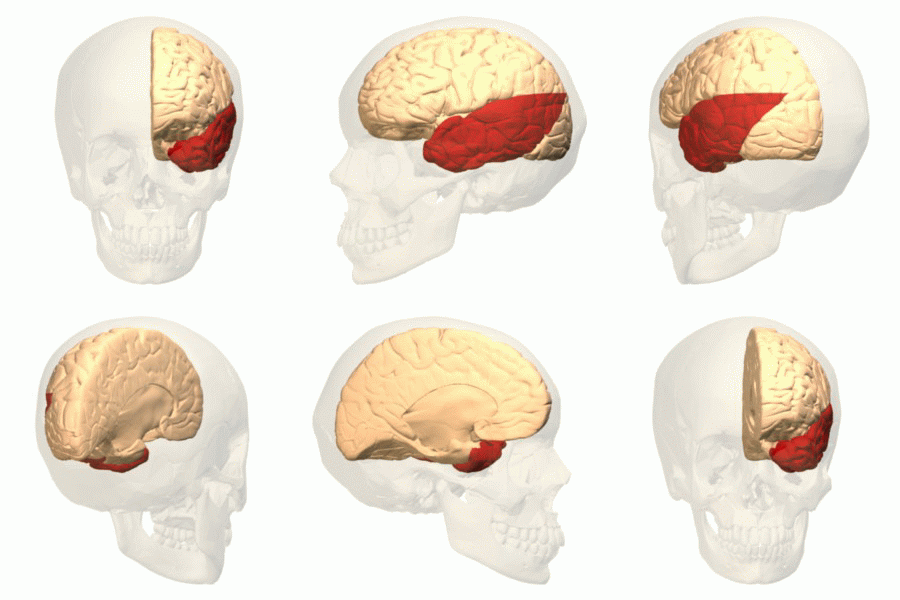
The perirhinal cortex is a cortical region in the medial temporal lobe that is made up of Brodmann areas 35 and 36. It receives highly processed sensory information from all sensory regions, and is generally accepted to be an important region for memory. It is bordered caudally by postrhinal cortex or parahippocampal cortex (homologous regions in rodents and primates, respectively) and ventrally and medially by entorhinal cortex. Structure The perirhinal cortex is composed of two regions: areas 36 and 35. Area 36 is sometimes divided into three subdivisions: 36d is the most rostral and dorsal, 36r ventral and caudal, and 36c the most caudal. Area 35 can be divided in the same manner, into 35d and 35v (for dorsal and ventral, respectively). Area 36 is six-layered, dysgranular, meaning that its layer IV is relatively sparse. Area 35 is agranular cortex (lacking any cells in layer IV). Function The perirhinal cortex is involved in both visual perception and memory; it fac ... [...More Info...] [...Related Items...] OR: [Wikipedia] [Google] [Baidu] |
Medial Temporal Lobe
The temporal lobe is one of the four major lobes of the cerebral cortex in the brain of mammals. The temporal lobe is located beneath the lateral fissure on both cerebral hemispheres of the mammalian brain. The temporal lobe is involved in processing sensory input into derived meanings for the appropriate retention of visual memory, language comprehension, and emotion association. ''Temporal'' refers to the head's temples. Structure The temporal lobe consists of structures that are vital for declarative or long-term memory. Declarative (denotative) or explicit memory is conscious memory divided into semantic memory (facts) and episodic memory (events). Medial temporal lobe structures that are critical for long-term memory include the hippocampus, along with the surrounding hippocampal region consisting of the perirhinal, parahippocampal, and entorhinal neocortical regions. The hippocampus is critical for memory formation, and the surrounding medial temporal cortex is ... [...More Info...] [...Related Items...] OR: [Wikipedia] [Google] [Baidu] |
Brodmann Area
A Brodmann area is a region of the cerebral cortex, in the human or other primate brain, defined by its cytoarchitecture, or histological structure and organization of cells. History Brodmann areas were originally defined and numbered by the German anatomist Korbinian Brodmann based on the cytoarchitectural organization of neurons he observed in the cerebral cortex using the Nissl method of cell staining. Brodmann published his maps of cortical areas in humans, monkeys, and other species in 1909, along with many other findings and observations regarding the general cell types and laminar organization of the mammalian cortex. The same Brodmann area number in different species does not necessarily indicate homologous areas. A similar, but more detailed cortical map was published by Constantin von Economo and Georg N. Koskinas in 1925. Present importance Brodmann areas have been discussed, debated, refined, and renamed exhaustively for nearly a century and remain the ... [...More Info...] [...Related Items...] OR: [Wikipedia] [Google] [Baidu] |
Entorhinal Cortex
The entorhinal cortex (EC) is an area of the brain's allocortex, located in the medial temporal lobe, whose functions include being a widespread network hub for memory, navigation, and the perception of time.Integrating time from experience in the lateral entorhinal cortex Albert Tsao, Jørgen Sugar, Li Lu, Cheng Wang, James J. Knierim, May-Britt Moser & Edvard I. Moser Naturevolume 561, pages57–62 (2018) The EC is the main interface between the hippocampus and neocortex. The EC-hippocampus system plays an important role in declarative (autobiographical/episodic/semantic) memories and in particular spatial memories including memory formation, memory consolidation, and memory optimization in sleep. The EC is also responsible for the pre-processing (familiarity) of the input signals in the reflex nictitating membrane response of classical trace conditioning; the association of impulses from the eye and the ear occurs in the entorhinal cortex. Structure In rodents, th ... [...More Info...] [...Related Items...] OR: [Wikipedia] [Google] [Baidu] |
Cerebral Cortex
The cerebral cortex, also known as the cerebral mantle, is the outer layer of neural tissue of the cerebrum of the brain in humans and other mammals. The cerebral cortex mostly consists of the six-layered neocortex, with just 10% consisting of allocortex. It is separated into two cortices, by the longitudinal fissure that divides the cerebrum into the left and right cerebral hemispheres. The two hemispheres are joined beneath the cortex by the corpus callosum. The cerebral cortex is the largest site of neural integration in the central nervous system. It plays a key role in attention, perception, awareness, thought, memory, language, and consciousness. The cerebral cortex is part of the brain responsible for cognition. In most mammals, apart from small mammals that have small brains, the cerebral cortex is folded, providing a greater surface area in the confined volume of the cranium. Apart from minimising brain and cranial volume, cortical folding is crucial for ... [...More Info...] [...Related Items...] OR: [Wikipedia] [Google] [Baidu] |
Thalamus
The thalamus (from Greek θάλαμος, "chamber") is a large mass of gray matter located in the dorsal part of the diencephalon (a division of the forebrain). Nerve fibers project out of the thalamus to the cerebral cortex in all directions, allowing hub-like exchanges of information. It has several functions, such as the relaying of sensory signals, including motor signals to the cerebral cortex and the regulation of consciousness, sleep, and alertness. Anatomically, it is a paramedian symmetrical structure of two halves (left and right), within the vertebrate brain, situated between the cerebral cortex and the midbrain. It forms during embryonic development as the main product of the diencephalon, as first recognized by the Swiss embryologist and anatomist Wilhelm His Sr. in 1893. Anatomy The thalamus is a paired structure of gray matter located in the forebrain which is superior to the midbrain, near the center of the brain, with nerve fibers projecting out to th ... [...More Info...] [...Related Items...] OR: [Wikipedia] [Google] [Baidu] |
Prefrontal Cortex
In mammalian brain anatomy, the prefrontal cortex (PFC) covers the front part of the frontal lobe of the cerebral cortex. The PFC contains the Brodmann areas BA8, BA9, BA10, BA11, BA12, BA13, BA14, BA24, BA25, BA32, BA44, BA45, BA46, and BA47. The basic activity of this brain region is considered to be orchestration of thoughts and actions in accordance with internal goals. Many authors have indicated an integral link between a person's will to live, personality, and the functions of the prefrontal cortex. This brain region has been implicated in executive functions, such as planning, decision making, short-term memory, personality expression, moderating social behavior and controlling certain aspects of speech and language. Executive function relates to abilities to differentiate among conflicting thoughts, determine good and bad, better and best, same and different, future consequences of current activities, working toward a defined goal, prediction of outc ... [...More Info...] [...Related Items...] OR: [Wikipedia] [Google] [Baidu] |
Prelimbic Cortex
The dorsomedial prefrontal cortex (dmPFC or DMPFC is a section of the prefrontal cortex in some species' brain anatomy. It includes portions of Brodmann areas BA8, BA9, BA10, BA24 and BA32, although some authors identify it specifically with BA8 and BA9 Some notable sub-components include the dorsal anterior cingulate cortex (BA24 and BA32), the prelimbic cortex, and the infralimbic cortex. Functions Evidence shows that the dmPFC plays several roles in humans. The dmPFC is identified to play roles in processing a sense of self, integrating social impressions, theory of mind, morality judgments, empathy, decision making, altruism, fear and anxiety information processing, and top-down motor cortex inhibition The dmPFC also modulates or regulates emotional responses and heart rate in situations of fear or stress and plays a role in long-term memory ). Some argue that the dmPFC is made up of several smaller subregions that are more task-specific. The dmPFC is attributed ... [...More Info...] [...Related Items...] OR: [Wikipedia] [Google] [Baidu] |
Infralimbic Cortex
The infralimbic cortex (IL) is a cortical region in the ventromedial prefrontal cortex which is important in tonic inhibition of subcortical structures and emotional responses, such as fear. Structure Connectivity Primates GABAergic neurons within the amygdala, known as intercalated (ITC) cells, receive a strong projection from the IL medial prefrontal cortex (mPFC) in primates. ITC cells are thought to play a role as the 'off switch' for the amygdala, inhibiting the amygdala's central nucleus output neurons and its basolateral nucleus neurons.Quirk, G.J. & Mueller, D. (2007). Neural mechanisms of extinction learning and retrieval. Neuropsychopharmacology Reviews, 1-17. Further, it has been shown that electrical stimulation of IL reduces conditioned fear and strengthens extinction memory, explaining cortical control over extinction processes, one of the simplest forms of emotional regulation. Rodents Amygdala ITC cells receive strong projection from the IL mPFC in rodents as w ... [...More Info...] [...Related Items...] OR: [Wikipedia] [Google] [Baidu] |
Basal Ganglia
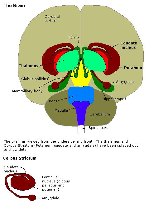
The basal ganglia (BG), or basal nuclei, are a group of subcortical nuclei, of varied origin, in the brains of vertebrates. In humans, and some primates, there are some differences, mainly in the division of the globus pallidus into an external and internal region, and in the division of the striatum. The basal ganglia are situated at the base of the forebrain and top of the midbrain. Basal ganglia are strongly interconnected with the cerebral cortex, thalamus, and brainstem, as well as several other brain areas. The basal ganglia are associated with a variety of functions, including control of voluntary motor movements, procedural learning, habit learning, conditional learning, eye movements, cognition, and emotion. The main components of the basal ganglia – as defined functionally – are the striatum, consisting of both the dorsal striatum ( caudate nucleus and putamen) and the ventral striatum ( nucleus accumbens and olfactory tubercle), the globus pa ... [...More Info...] [...Related Items...] OR: [Wikipedia] [Google] [Baidu] |
Amygdala
The amygdala (; plural: amygdalae or amygdalas; also '; Latin from Greek, , ', 'almond', 'tonsil') is one of two almond-shaped clusters of nuclei located deep and medially within the temporal lobes of the brain's cerebrum in complex vertebrates, including humans. Shown to perform a primary role in the processing of memory, decision making, and emotional responses (including fear, anxiety, and aggression), the amygdalae are considered part of the limbic system. The term "amygdala" was first introduced by Karl Friedrich Burdach in 1822. Structure The regions described as amygdala nuclei encompass several structures of the cerebrum with distinct connectional and functional characteristics in humans and other animals. Among these nuclei are the basolateral complex, the cortical nucleus, the medial nucleus, the central nucleus, and the intercalated cell clusters. The basolateral complex can be further subdivided into the lateral, the basal, and the accessory ba ... [...More Info...] [...Related Items...] OR: [Wikipedia] [Google] [Baidu] |
Basal Forebrain
Part of the human brain, the basal forebrain structures are located in the forebrain to the front of and below the striatum. They include the ventral basal ganglia (including nucleus accumbens and ventral pallidum), nucleus basalis, diagonal band of Broca, substantia innominata, and the medial septal nucleus. These structures are important in the production of acetylcholine, which is then distributed widely throughout the brain. The basal forebrain is considered to be the major cholinergic output of the central nervous system (CNS) centred on the output of the nucleus basalis. The presence of non-cholinergic neurons projecting to the cortex have been found to act with the cholinergic neurons to dynamically modulate activity in the cortex. Function Acetylcholine is known to promote wakefulness in the basal forebrain. Stimulating the basal forebrain gives rise to acetylcholine release, which induces wakefulness and REM sleep, whereas inhibition of acetylcholine release in the ba ... [...More Info...] [...Related Items...] OR: [Wikipedia] [Google] [Baidu] |
Hearing (sense)
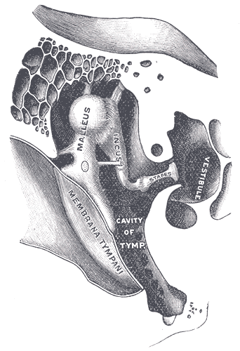
Hearing, or auditory perception, is the ability to perceive sounds through an organ, such as an ear, by detecting vibrations as periodic changes in the pressure of a surrounding medium. The academic field concerned with hearing is auditory science. Sound may be heard through solid, liquid, or gaseous matter. It is one of the traditional five senses. Partial or total inability to hear is called hearing loss. In humans and other vertebrates, hearing is performed primarily by the auditory system: mechanical waves, known as vibrations, are detected by the ear and transduced into nerve impulses that are perceived by the brain (primarily in the temporal lobe). Like touch, audition requires sensitivity to the movement of molecules in the world outside the organism. Both hearing and touch are types of mechanosensation. Hearing mechanism There are three main components of the human auditory system: the outer ear, the middle ear, and the inner ear. Outer ear The outer ... [...More Info...] [...Related Items...] OR: [Wikipedia] [Google] [Baidu] |


