|
Perilymph
Perilymph is an extracellular fluid located within the inner ear. It is found within the scala tympani and scala vestibuli of the cochlea. The ionic composition of perilymph is comparable to that of plasma and cerebrospinal fluid. The major cation in perilymph is sodium, with the values of sodium and potassium concentration in the perilymph being 138 mM and 6.9 mM, respectively. It is also named Cotunnius' liquid and liquor cotunnii for Domenico Cotugno. Structure The inner ear has two major parts, the cochlea and the vestibular organ. They are connected in a series of canals in the temporal bone referred to as the bony labyrinth. The bone canals are separated by the membranes in parallel spaces referred to as the membranous labyrinth. The membranous labyrinth contains endolymph, and is surrounded by perilymph. The perilymph in the bony labyrinth serves as connection to the cerebrospinal fluid of the subarachnoid space via the perilymphatic duct. Composition Peri ... [...More Info...] [...Related Items...] OR: [Wikipedia] [Google] [Baidu] |
Cochlea
The cochlea is the part of the inner ear involved in hearing. It is a spiral-shaped cavity in the bony labyrinth, in humans making 2.75 turns around its axis, the modiolus. A core component of the cochlea is the Organ of Corti, the sensory organ of hearing, which is distributed along the partition separating the fluid chambers in the coiled tapered tube of the cochlea. The name cochlea derives . Structure The cochlea (plural is cochleae) is a spiraled, hollow, conical chamber of bone, in which waves propagate from the base (near the middle ear and the oval window) to the apex (the top or center of the spiral). The spiral canal of the cochlea is a section of the bony labyrinth of the inner ear that is approximately 30 mm long and makes 2 turns about the modiolus. The cochlear structures include: * Three ''scalae'' or chambers: ** the vestibular duct or ''scala vestibuli'' (containing perilymph), which lies superior to the cochlear duct and abuts the oval window ** the ty ... [...More Info...] [...Related Items...] OR: [Wikipedia] [Google] [Baidu] |
Inner Ear
The inner ear (internal ear, auris interna) is the innermost part of the vertebrate ear. In vertebrates, the inner ear is mainly responsible for sound detection and balance. In mammals, it consists of the bony labyrinth, a hollow cavity in the temporal bone of the skull with a system of passages comprising two main functional parts: * The cochlea, dedicated to hearing; converting sound pressure patterns from the outer ear into electrochemical impulses which are passed on to the brain via the auditory nerve. * The vestibular system, dedicated to balance The inner ear is found in all vertebrates, with substantial variations in form and function. The inner ear is innervated by the eighth cranial nerve in all vertebrates. Structure The labyrinth can be divided by layer or by region. Bony and membranous labyrinths The bony labyrinth, or osseous labyrinth, is the network of passages with bony walls lined with periosteum. The three major parts of the bony labyrinth are the ... [...More Info...] [...Related Items...] OR: [Wikipedia] [Google] [Baidu] |
Perilymphatic Duct
In the anatomy of the human ear, the perilymphatic duct is where the perilymphatic space (vestibule of the ear) is connected to the subarachnoid space. This works as a type of shunt to eliminate excess perilymph fluid from the perilymphatic space around the cochlea of the ear. Perilymph is continuous with cerebrospinal fluid (CSF) in the subarachnoid space. CSF pressure abnormalities do not generally have clinical impact on the inner ear which is explained physically by the bore diameter and length of the perilymphatic duct. This duct goes through the skull The skull is a bone protective cavity for the brain. The skull is composed of four types of bone i.e., cranial bones, facial bones, ear ossicles and hyoid bone. However two parts are more prominent: the cranium and the mandible. In humans, t ... and is parallel with but not directly associated with the endolymphatic duct. The duct is lined by an epithelium. References Ear {{anatomy-stub ... [...More Info...] [...Related Items...] OR: [Wikipedia] [Google] [Baidu] |
Cerebrospinal Fluid
Cerebrospinal fluid (CSF) is a clear, colorless body fluid found within the tissue that surrounds the brain and spinal cord of all vertebrates. CSF is produced by specialised ependymal cells in the choroid plexus of the ventricles of the brain, and absorbed in the arachnoid granulations. There is about 125 mL of CSF at any one time, and about 500 mL is generated every day. CSF acts as a shock absorber, cushion or buffer, providing basic mechanical and immunological protection to the brain inside the skull. CSF also serves a vital function in the cerebral autoregulation of cerebral blood flow. CSF occupies the subarachnoid space (between the arachnoid mater and the pia mater) and the ventricular system around and inside the brain and spinal cord. It fills the ventricles of the brain, cisterns, and sulci, as well as the central canal of the spinal cord. There is also a connection from the subarachnoid space to the bony labyrinth of the inner ear via the per ... [...More Info...] [...Related Items...] OR: [Wikipedia] [Google] [Baidu] |
Endolymph
Endolymph is the fluid contained in the membranous labyrinth of the inner ear. The major cation in endolymph is potassium, with the values of sodium and potassium concentration in the endolymph being 0.91 mM and 154 mM, respectively. It is also called ''Scarpa's fluid'', after Antonio Scarpa. Structure The inner ear has two parts: the bony labyrinth and the membranous labyrinth. The membranous labyrinth is contained within the bony labyrinth, and within the membranous labyrinth is a fluid called endolymph. Between the outer wall of the membranous labyrinth and the wall of the bony labyrinth is the location of perilymph. Composition Perilymph and endolymph have unique ionic compositions suited to their functions in regulating electrochemical impulses of hair cells. The electric potential of endolymph is ~80-90 mV more positive than perilymph due to a higher concentration of K compared to Na. The main component of this unique extracellular fluid is potassium, wh ... [...More Info...] [...Related Items...] OR: [Wikipedia] [Google] [Baidu] |
Scala Tympani
The tympanic duct or scala tympani is one of the perilymph-filled cavities in the inner ear of humans. It is separated from the cochlear duct by the basilar membrane, and it extends from the round window to the helicotrema, where it continues as vestibular duct. The purpose of the perilymph-filled tympanic duct and vestibular duct is to transduce the movement of air that causes the tympanic membrane and the ossicles to vibrate, to movement of liquid and the basilar membrane. This movement is conveyed to the organ of Corti inside the cochlear duct, composed of hair cells attached to the basilar membrane and their stereocilia embedded in the tectorial membrane. The movement of the basilar membrane compared to the tectorial membrane causes the stereocilia to bend. They then depolarise and send impulses to the brain via the cochlear nerve. This produces the sensation of sound. Additional images File:Gray921.png, Interior of right osseous labyrinth. (Scala tympani labeled ... [...More Info...] [...Related Items...] OR: [Wikipedia] [Google] [Baidu] |
Hair Cells
Hair cells are the sensory receptors of both the auditory system and the vestibular system in the ears of all vertebrates, and in the lateral line organ of fishes. Through mechanotransduction, hair cells detect movement in their environment. In mammals, the auditory hair cells are located within the spiral organ of Corti on the thin basilar membrane in the cochlea of the inner ear. They derive their name from the tufts of stereocilia called ''hair bundles'' that protrude from the apical surface of the cell into the fluid-filled cochlear duct. The stereocilia number from 50-100 in each cell while being tightly packed together and decrease in size the further away they are located from the kinocilium. The hair bundles are arranged as stiff columns that move at their base in response to stimuli applied to the tips. Mammalian cochlear hair cells are of two anatomically and functionally distinct types, known as outer, and inner hair cells. Damage to these hair cells results i ... [...More Info...] [...Related Items...] OR: [Wikipedia] [Google] [Baidu] |
Scala Vestibuli
The vestibular duct or scala vestibuli is a perilymph-filled cavity inside the cochlea of the inner ear that conducts sound vibrations to the cochlear duct. It is separated from the cochlear duct by Reissner's membrane and extends from the vestibule of the ear to the helicotrema where it joins the tympanic duct. Additional images Image:Gray923.png, The cochlea and vestibule, viewed from above. Image:Gray903.png, Transverse section of the cochlear duct of a fetal cat. Image:Gray921.png, Interior of right osseous labyrinth. Image:Gray928.png, Diagrammatic longitudinal section of the cochlea. See also * Tympanic duct References internal websites Slidefrom University of Kansas Diagramat Indiana University – Purdue University Indianapolis Imageat University of New England (United States) The University of New England (UNE) is a private research university in Maine with campuses in Portland and Biddeford, as well as a study abroad campus in Tangier, Morocco. Dur ... [...More Info...] [...Related Items...] OR: [Wikipedia] [Google] [Baidu] |
Endocochlear Potential
The endocochlear potential (EP; also called endolymphatic potential) is the positive voltage of 80-100mV seen in the cochlear endolymphatic spaces. Within the cochlea the EP varies in the magnitude all along its length. When a sound is presented, the endocochlear potential changes either positive or negative in the endolymph, depending on the stimulus. The change in the potential is called the summating potential. With the movement of the basilar membrane, a shear force is created and a small potential is generated due to a difference in potential between the endolymph (scala media Scala or SCALA may refer to: Automobiles * Renault Scala, multiple automobile models * Škoda Scala, a Czech compact hatchback Music * Scala (band), an English electronic music group * Escala (group), an electronic string quartet formerly k ..., +80 mV) and the perilymph (vestibular and tympanic ducts, 0 mV). EP is highest in the basal turn of the cochlea (95 mV in mice) and decreases in the magn ... [...More Info...] [...Related Items...] OR: [Wikipedia] [Google] [Baidu] |
Membranous Labyrinth
The membranous labyrinth is a collection of fluid filled tubes and chambers which contain the receptors for the senses of equilibrium and hearing. It is lodged within the bony labyrinth in the inner ear and has the same general form; it is, however, considerably smaller and is partly separated from the bony walls by a quantity of fluid, the perilymph. In certain places, it is fixed to the walls of the cavity. The membranous labyrinth contains fluid called endolymph. The walls of the membranous labyrinth are lined with distributions of the cochlear nerve, one of the two branches of the vestibulocochlear nerve. The other branch is the vestibular nerve. Within the vestibule, the membranous labyrinth does not quite preserve the form of the bony labyrinth, but consists of two membranous sacs, the utricle, and the saccule The saccule is a bed of sensory cells in the inner ear The inner ear (internal ear, auris interna) is the innermost part of the vertebrate ear. In verteb ... [...More Info...] [...Related Items...] OR: [Wikipedia] [Google] [Baidu] |
Bony Labyrinth
The bony labyrinth (also osseous labyrinth or otic capsule) is the rigid, bony outer wall of the inner ear in the temporal bone. It consists of three parts: the vestibule, semicircular canals, and cochlea. These are cavities hollowed out of the substance of the bone, and lined by periosteum. They contain a clear fluid, the perilymph, in which the membranous labyrinth is situated. A fracture classification system in which temporal bone fractures detected by computed tomography are delineated based on disruption of the otic capsule has been found to be predictive for complications of temporal bone trauma such as facial nerve injury, sensorineural deafness and cerebrospinal fluid otorrhea. On radiographic images, the otic capsule is the densest portion of the temporal bone. In otospongiosis, a leading cause of adult-onset hearing loss, the otic capsule is exclusively affected. This area normally undergoes no remodeling in adult life and is extremely dense. With otospong ... [...More Info...] [...Related Items...] OR: [Wikipedia] [Google] [Baidu] |
Extracellular Fluid
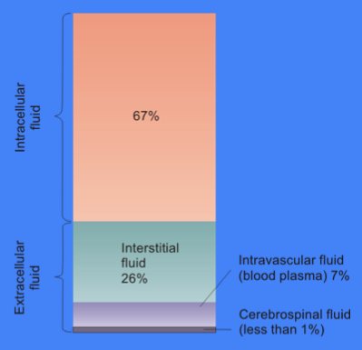
In cell biology, extracellular fluid (ECF) denotes all body fluid outside the cells of any multicellular organism. Total body water in healthy adults is about 60% (range 45 to 75%) of total body weight; women and the obese typically have a lower percentage than lean men. Extracellular fluid makes up about one-third of body fluid, the remaining two-thirds is intracellular fluid within cells. The main component of the extracellular fluid is the interstitial fluid that surrounds cells. Extracellular fluid is the internal environment of all multicellular animals, and in those animals with a blood circulatory system, a proportion of this fluid is blood plasma. Plasma and interstitial fluid are the two components that make up at least 97% of the ECF. Lymph makes up a small percentage of the interstitial fluid. The remaining small portion of the ECF includes the transcellular fluid (about 2.5%). The ECF can also be seen as having two components – plasma and lymph as a delivery sy ... [...More Info...] [...Related Items...] OR: [Wikipedia] [Google] [Baidu] |


