|
Pelvimetry
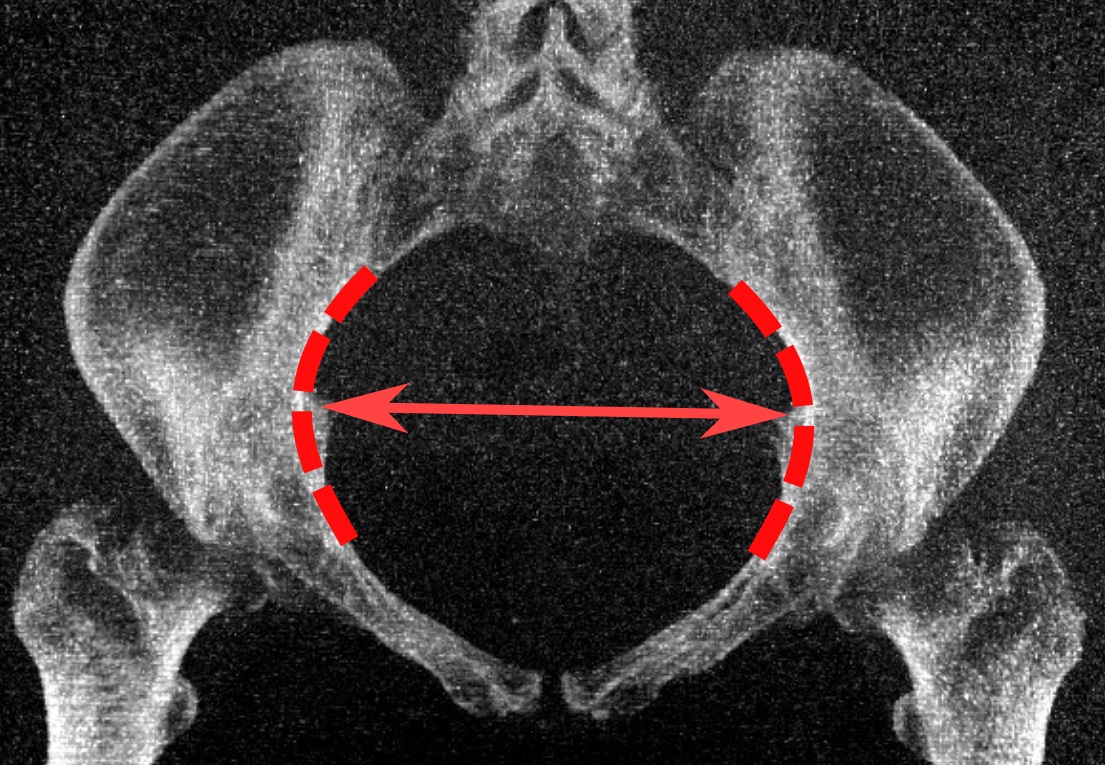
Pelvimetry is the measurement of the female pelvis. It can theoretically identify cephalo-pelvic disproportion, which is when the capacity of the pelvis is inadequate to allow the fetus to negotiate the birth canal. However, clinical evidence indicate that all pregnant women should be allowed a trial of labor regardless of pelvimetry results. Indication Theoretically, pelvimetry may identify cephalo-pelvic disproportion, which is when the capacity of the pelvis is inadequate to allow the fetus to negotiate the birth canal. However, a woman's pelvis loosens up before birth (with the help of hormones). A Cochrane review in 2017 found that there was too little evidence to show whether pelvimetry is beneficial and safe when the baby is in cephalic presentation. A review in 2003 came to the conclusion that pelvimetry does not change the management of pregnant women, and recommended that all women should be allowed a trial of labor regardless of pelvimetry results. It considered rout ... [...More Info...] [...Related Items...] OR: [Wikipedia] [Google] [Baidu] |
Cephalo-pelvic Disproportion
Cephalopelvic disproportion exists when the capacity of the pelvis is inadequate to allow the fetus to negotiate the birth canal. This may be due to a small pelvis, a nongynecoid pelvic formation, a large fetus, an unfavorable orientation of the fetus, or a combination of these factors. Certain medical conditions may distort pelvic bones, such as rickets or a pelvic fracture, and lead to CPD. Transverse diagonal measurement has been proposed as a predictive method. Causes A large fetus can be one cause of CPD. A large fetus can be caused by gestational diabetes, postterm pregnancy, genetic factors, and multiparity. The shape of the pelvis can also be a cause of CPD. The pelvis may be too small, or the shape of the pelvis may be malformed. Shorter women are more likely to have CPD as are adolescents. Diagnosis Diagnosis of CPD may be made when there is failure to progress, but not all cases of prolonged labour are the result of CPD. Use of ultrasound to measure the size of t ... [...More Info...] [...Related Items...] OR: [Wikipedia] [Google] [Baidu] |
Pelvic Inlet
The pelvic inlet or superior aperture of the pelvis is a planar surface which defines the boundary between the pelvic cavity and the abdominal cavity (or, according to some authors, between two parts of the pelvic cavity, called lesser pelvis and greater pelvis). It is a major target of measurements of pelvimetry. Its position and orientation relative to the skeleton of the pelvis is anatomically defined by its edge, the pelvic brim. The pelvic brim is an approximately apple-shaped line passing through the prominence of the sacrum, the arcuate and pectineal lines, and the upper margin of the pubic symphysis. Occasionally, the terms pelvic inlet and pelvic brim are used interchangeably. Boundaries The edge of the pelvic inlet (pelvic brim) is formed as follows: Diameters The diameters or conjugates of the pelvis are measured at the pelvic inlet and outlet and as oblique diameters. Two diameters may be measured from the outside of the body using a pelvimeter Additional ... [...More Info...] [...Related Items...] OR: [Wikipedia] [Google] [Baidu] |
Pelvic Outlet
The lower circumference of the lesser pelvis is very irregular; the space enclosed by it is named the inferior aperture or pelvic outlet. It is an important component of pelvimetry. Boundaries It has the following boundaries: * anteriorly: the pubic arch * laterally: the ischial tuberosities * posterolaterally: the inferior margin of the sacrotuberous ligament * posteriorly: the anterior border of the middle of the coccyx. Notches These eminences are separated by three notches: * one in front, the pubic arch, formed by the convergence of the inferior rami of the ischium and pubis on either side. * The other notches, one on either side, are formed by the sacrum and coccyx behind, the ischium in front, and the ilium above; they are called the sciatic notches; in the natural state they are converted into foramina by the sacrotuberous and sacrospinous ligaments. In situ When the ligaments are in situ, the inferior aperture of the pelvis is lozenge-shaped, bounded as follows: * ... [...More Info...] [...Related Items...] OR: [Wikipedia] [Google] [Baidu] |
Pelvic Inlet
The pelvic inlet or superior aperture of the pelvis is a planar surface which defines the boundary between the pelvic cavity and the abdominal cavity (or, according to some authors, between two parts of the pelvic cavity, called lesser pelvis and greater pelvis). It is a major target of measurements of pelvimetry. Its position and orientation relative to the skeleton of the pelvis is anatomically defined by its edge, the pelvic brim. The pelvic brim is an approximately apple-shaped line passing through the prominence of the sacrum, the arcuate and pectineal lines, and the upper margin of the pubic symphysis. Occasionally, the terms pelvic inlet and pelvic brim are used interchangeably. Boundaries The edge of the pelvic inlet (pelvic brim) is formed as follows: Diameters The diameters or conjugates of the pelvis are measured at the pelvic inlet and outlet and as oblique diameters. Two diameters may be measured from the outside of the body using a pelvimeter Additional ... [...More Info...] [...Related Items...] OR: [Wikipedia] [Google] [Baidu] |
Human Pelvis
The pelvis (plural pelves or pelvises) is the lower part of the Trunk (anatomy), trunk, between the human abdomen, abdomen and the thighs (sometimes also called pelvic region), together with its embedded skeleton (sometimes also called bony pelvis, or pelvic skeleton). The pelvic region of the trunk includes the bony pelvis, the pelvic cavity (the space enclosed by the bony pelvis), the pelvic floor, below the pelvic cavity, and the perineum, below the pelvic floor. The pelvic skeleton is formed in the area of the back, by the sacrum and the coccyx and anteriorly and to the left and right sides, by a pair of hip bones. The two hip bones connect the spine with the lower limbs. They are attached to the sacrum posteriorly, connected to each other anteriorly, and joined with the two femurs at the hip joints. The gap enclosed by the bony pelvis, called the pelvic cavity, is the section of the body underneath the abdomen and mainly consists of the reproductive organs (sex organs) and ... [...More Info...] [...Related Items...] OR: [Wikipedia] [Google] [Baidu] |
Joseph C
Joseph is a common male given name, derived from the Hebrew Yosef (יוֹסֵף). "Joseph" is used, along with "Josef", mostly in English, French and partially German languages. This spelling is also found as a variant in the languages of the modern-day Nordic countries. In Portuguese and Spanish, the name is "José". In Arabic, including in the Quran, the name is spelled '' Yūsuf''. In Persian, the name is "Yousef". The name has enjoyed significant popularity in its many forms in numerous countries, and ''Joseph'' was one of the two names, along with ''Robert'', to have remained in the top 10 boys' names list in the US from 1925 to 1972. It is especially common in contemporary Israel, as either "Yossi" or "Yossef", and in Italy, where the name "Giuseppe" was the most common male name in the 20th century. In the first century CE, Joseph was the second most popular male name for Palestine Jews. In the Book of Genesis Joseph is Jacob's eleventh son and Rachel's first son, and k ... [...More Info...] [...Related Items...] OR: [Wikipedia] [Google] [Baidu] |
Coccyx
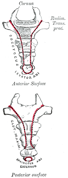
The coccyx ( : coccyges or coccyxes), commonly referred to as the tailbone, is the final segment of the vertebral column in all apes, and analogous structures in certain other mammals such as horses. In tailless primates (e.g. humans and other great apes) since ''Nacholapithecus'' (a Miocene hominoid),Nakatsukasa 2004, ''Acquisition of bipedalism'' (SeFig. 5entitled ''First coccygeal/caudal vertebra in short-tailed or tailless primates.''.) the coccyx is the remnant of a vestigial tail. In animals with bony tails, it is known as ''tailhead'' or ''dock'', in bird anatomy as ''tailfan''. It comprises three to five separate or fused coccygeal vertebrae below the sacrum, attached to the sacrum by a fibrocartilaginous joint, the sacrococcygeal symphysis, which permits limited movement between the sacrum and the coccyx. Structure The coccyx is formed of three, four or five rudimentary vertebrae. It articulates superiorly with the sacrum. In each of the first three segments may ... [...More Info...] [...Related Items...] OR: [Wikipedia] [Google] [Baidu] |
Sacrococcygeal Joint
The sacrococcygeal symphysis (sacrococcygeal articulation, articulation of the sacrum and coccyx) is an amphiarthrodial joint, formed between the oval surface at the apex of the sacrum, and the base of the coccyx. It is a slightly moveable jointMorris (2005), p 59 which is frequently, partially or completely, obliterated in old age,Palastanga (2006), p 334 homologous with the joints between the bodies of the vertebrae. Disc The sacrococcygeal disc or interosseus ligamentHuijbregts (2001), p 13 is similar to the intervertebral discs but thinner, thicker in front and behind than at the sides, and with a firmer texture. The articular surfaces are elliptical with longer transversal axes. The surface on the sacrum is convex and that on the coccyx concave. Occasionally the coccyx is freely movable on the sacrum, most notably during pregnancy; in such cases a synovial membrane is present. Ligaments The joint is strengthened by a series of ligaments: * The ventral or anterior sac ... [...More Info...] [...Related Items...] OR: [Wikipedia] [Google] [Baidu] |


