|
Pelvic Digit
A pelvic digit, pelvic finger, or pelvic rib is a rare congenital abnormality in humans, in which Osseous tissue, bone tissue develops in the soft tissue near the human pelvis, pelvis, resembling a rib or finger and often divided into one or more segments with Articulation (anatomy), pseudo-articulations. Pelvic digits are typically benign tumor, benign and asymptomatic, and are usually discovered accidentally. Approximately 41 cases have been reported. The pelvic digit was first reported by D. Sullivan and W.S. Cornwell in 1974. Pelvic digits may be located at any level of the pelvis, the lower ribs, or even the anterior abdominal wall. It is theorized that pelvic digit anomalies arise during the mesenchymal stage of bone growth, within the first six weeks of embryogenesis. Their formation may result from a failure of the primordium of the coccyx to fuse to the vertebral column, leading to the independent development of a proto-rib structure. See also *Supernumerary body part Re ... [...More Info...] [...Related Items...] OR: [Wikipedia] [Google] [Baidu] |
Mesenchymal
Mesenchyme () is a type of loosely organized animal embryonic connective tissue of undifferentiated cells that give rise to most tissues, such as skin, blood or bone. The interactions between mesenchyme and epithelium help to form nearly every organ in the developing embryo. Vertebrates Structure Mesenchyme is characterized morphologically by a prominent ground substance matrix containing a loose aggregate of reticular fibers and unspecialized mesenchymal stem cells. Mesenchymal cells can migrate easily (in contrast to epithelial cells, which lack mobility), are organized into closely adherent sheets, and are polarized in an apical-basal orientation. Development The mesenchyme originates from the mesoderm. From the mesoderm, the mesenchyme appears as an embryologically primitive "soup". This "soup" exists as a combination of the mesenchymal cells plus serous fluid plus the many different tissue proteins. Serous fluid is typically stocked with the many serous elements, such a ... [...More Info...] [...Related Items...] OR: [Wikipedia] [Google] [Baidu] |
Congenital Disorders
A birth defect, also known as a congenital disorder, is an abnormal condition that is present at birth regardless of its cause. Birth defects may result in disabilities that may be physical, intellectual, or developmental. The disabilities can range from mild to severe. Birth defects are divided into two main types: structural disorders in which problems are seen with the shape of a body part and functional disorders in which problems exist with how a body part works. Functional disorders include metabolic and degenerative disorders. Some birth defects include both structural and functional disorders. Birth defects may result from genetic or chromosomal disorders, exposure to certain medications or chemicals, or certain infections during pregnancy. Risk factors include folate deficiency, drinking alcohol or smoking during pregnancy, poorly controlled diabetes, and a mother over the age of 35 years old. Many are believed to involve multiple factors. Birth defects may be vi ... [...More Info...] [...Related Items...] OR: [Wikipedia] [Google] [Baidu] |
Supernumerary Body Part
Supernumerary body parts are most commonly a congenital disorder involving the growth of an additional part of the body and a deviation from the body plan. Body parts may be easily visible or hidden away, such as internal organs. Many additional body parts form by the same process as conjoined twins: the zygote begins to split but fails to completely separate. This condition may also be a symptom of repeated occurrences of continuous inbreeding in a genetic line. Specific types of occurrence Specific types of additional body parts include: *Accessory breast – one or more additional breastsOnline Mendelian Inheritance in Man. Nipples, Supernumerary. Accessed 10 July 2006. * Accessory spleen – one or more additional spleens *Cervical rib – an additional rib *Diphallia – having two penes/penises *Hermaphroditism – having both male and female sex organs *Hyperdontia – additional teeth * Pelvic digit – a bony growth in the soft tissue of the pelvic region *Polycephaly ... [...More Info...] [...Related Items...] OR: [Wikipedia] [Google] [Baidu] |
Vertebral Column
The vertebral column, also known as the backbone or spine, is part of the axial skeleton. The vertebral column is the defining characteristic of a vertebrate in which the notochord (a flexible rod of uniform composition) found in all chordata, chordates has been replaced by a segmented series of bone: vertebrae separated by intervertebral discs. Individual vertebrae are named according to their region and position, and can be used as anatomical landmarks in order to guide procedures such as Lumbar puncture, lumbar punctures. The vertebral column houses the spinal canal, a cavity that encloses and protects the spinal cord. There are about 50,000 species of animals that have a vertebral column. The human vertebral column is one of the most-studied examples. Many different diseases in humans can affect the spine, with spina bifida and scoliosis being recognisable examples. The general structure of human vertebrae is fairly typical of that found in mammals, reptiles, and birds. Th ... [...More Info...] [...Related Items...] OR: [Wikipedia] [Google] [Baidu] |
Coccyx
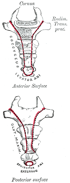
The coccyx ( : coccyges or coccyxes), commonly referred to as the tailbone, is the final segment of the vertebral column in all apes, and analogous structures in certain other mammals such as horses. In tailless primates (e.g. humans and other great apes) since ''Nacholapithecus'' (a Miocene hominoid),Nakatsukasa 2004, ''Acquisition of bipedalism'' (SeFig. 5entitled ''First coccygeal/caudal vertebra in short-tailed or tailless primates.''.) the coccyx is the remnant of a vestigial tail. In animals with bony tails, it is known as ''tailhead'' or ''dock'', in bird anatomy as ''tailfan''. It comprises three to five separate or fused coccygeal vertebrae below the sacrum, attached to the sacrum by a fibrocartilaginous joint, the sacrococcygeal symphysis, which permits limited movement between the sacrum and the coccyx. Structure The coccyx is formed of three, four or five rudimentary vertebrae. It articulates superiorly with the sacrum. In each of the first three segments may ... [...More Info...] [...Related Items...] OR: [Wikipedia] [Google] [Baidu] |
Primordium
A primordium (; plural: primordia; synonym: anlage) in embryology, is an organ or tissue in its earliest recognizable stage of development. Cells of the primordium are called primordial cells. A primordium is the simplest set of cells capable of triggering growth of the would-be organ and the initial foundation from which an organ is able to grow. In flowering plants, a floral primordium gives rise to a flower. Although it is a frequently used term in plant biology, the word is used in describing the biology of all multicellular organisms (for example: a tooth primordium in animals, a leaf primordium in plants or a sporophore primordium in fungi.) Primordium development in plants Plants produce both leaf and flower primordia cells at the shoot apical meristem (SAM). Primordium development in plants is critical to the proper positioning and development of plant organs and cells. The process of primordium development is intricately regulated by a set of genes that affect the pos ... [...More Info...] [...Related Items...] OR: [Wikipedia] [Google] [Baidu] |
Embryogenesis
An embryo is an initial stage of development of a multicellular organism. In organisms that reproduce sexually, embryonic development is the part of the life cycle that begins just after fertilization of the female egg cell by the male sperm cell. The resulting fusion of these two cells produces a single-celled zygote that undergoes many cell divisions that produce cells known as blastomeres. The blastomeres are arranged as a solid ball that when reaching a certain size, called a morula, takes in fluid to create a cavity called a blastocoel. The structure is then termed a blastula, or a blastocyst in mammals. The mammalian blastocyst hatches before implantating into the endometrial lining of the womb. Once implanted the embryo will continue its development through the next stages of gastrulation, neurulation, and organogenesis. Gastrulation is the formation of the three germ layers that will form all of the different parts of the body. Neurulation forms the nervous sys ... [...More Info...] [...Related Items...] OR: [Wikipedia] [Google] [Baidu] |
British Journal Of Radiology
''The British Journal of Radiology'' is a monthly peer-reviewed medical journal covering radiology. It is published by the British Institute of Radiology and the editors-in-chief are Simon Jackson (University Hospitals Plymouth NHS Trust) and Andrew Nisbet (University College London). According to the ''Journal Citation Reports'', the journal has a 2021 impact factor The impact factor (IF) or journal impact factor (JIF) of an academic journal is a scientometric index calculated by Clarivate that reflects the yearly mean number of citations of articles published in the last two years in a given journal, as ... of 3.629. History The journal's forerunner, the ''Archives of Clinical Skiagraphy'' was established by Sydney Domville Rowland in May 1896. In July 1897 it was renamed the ''Archives of the Roentgen Ray'' and reported that it would keep a "record fthe proceedings of the recently formed Roentgen Society, and will consist of original communications, notes, and correspo ... [...More Info...] [...Related Items...] OR: [Wikipedia] [Google] [Baidu] |
Congenital Abnormality
A birth defect, also known as a congenital disorder, is an abnormal condition that is present at birth regardless of its cause. Birth defects may result in disabilities that may be physical, intellectual, or developmental. The disabilities can range from mild to severe. Birth defects are divided into two main types: structural disorders in which problems are seen with the shape of a body part and functional disorders in which problems exist with how a body part works. Functional disorders include metabolic and degenerative disorders. Some birth defects include both structural and functional disorders. Birth defects may result from genetic or chromosomal disorders, exposure to certain medications or chemicals, or certain infections during pregnancy. Risk factors include folate deficiency, drinking alcohol or smoking during pregnancy, poorly controlled diabetes, and a mother over the age of 35 years old. Many are believed to involve multiple factors. Birth defects may be visib ... [...More Info...] [...Related Items...] OR: [Wikipedia] [Google] [Baidu] |
Benign Tumor
A benign tumor is a mass of cells (tumor) that does not invade neighboring tissue or metastasize (spread throughout the body). Compared to malignant (cancerous) tumors, benign tumors generally have a slower growth rate. Benign tumors have relatively well differentiated cells. They are often surrounded by an outer surface (fibrous sheath of connective tissue) or stay contained within the epithelium. Common examples of benign tumors include moles and uterine fibroids. Some forms of benign tumors may be harmful to health. Benign tumor growth causes a mass effect that can compress neighboring tissues. This can lead to nerve damage, blood flow reduction ( ischemia), tissue death (necrosis), or organ damage. The health effects of benign tumor growth may be more prominent if the tumor is contained within an enclosed space such as the cranium, respiratory tract, sinus, or bones. For example, unlike most benign tumors elsewhere in the body, benign brain tumors can be life-threateni ... [...More Info...] [...Related Items...] OR: [Wikipedia] [Google] [Baidu] |
Articulation (anatomy)
A joint or articulation (or articular surface) is the connection made between bones, ossicles, or other hard structures in the body which link an animal's skeletal system into a functional whole.Saladin, Ken. Anatomy & Physiology. 7th ed. McGraw-Hill Connect. Webp.274/ref> They are constructed to allow for different degrees and types of movement. Some joints, such as the knee, elbow, and shoulder, are self-lubricating, almost frictionless, and are able to withstand compression and maintain heavy loads while still executing smooth and precise movements. Other joints such as sutures between the bones of the skull permit very little movement (only during birth) in order to protect the brain and the sense organs. The connection between a tooth and the jawbone is also called a joint, and is described as a fibrous joint known as a gomphosis. Joints are classified both structurally and functionally. Classification The number of joints depends on if sesamoids are included, age of the h ... [...More Info...] [...Related Items...] OR: [Wikipedia] [Google] [Baidu] |


