|
Paratenial Nucleus
The paratenial nucleus, or parataenial nucleus ( la, nucleus parataenialis), is a component of the midline nuclear group in the thalamus. It is sometimes subdivided into the nucleus parataenialis interstitialis and nucleus parataenialis parvocellularis (Hassler). It is located above the bordering paraventricular nucleus of thalamus and below the anterodorsal nucleus. The paratenial nucleus, like other midline nuclei, receives inputs from a large number of regions in the brainstem, hypothalamus and limbic system. It projects back to an equally wide range, but in a fairly specific manner (in the past, the midline nuclei have often been described as "nonspecific" because of their global effects). Particular targets include medial frontal polar cortex, the anterior cingulate, insula, the piriform and entorhinal cortices, the ventral subiculum, claustrum, the core and shell of nucleus accumbens, the medial striatum, the bed nucleus of stria terminalis, and caudal parts of the centr ... [...More Info...] [...Related Items...] OR: [Wikipedia] [Google] [Baidu] |
Midline Nuclear Group
The midline nuclear group (or midline thalamic nuclei) is a region of the thalamus consisting of the following nuclei: * paraventricular nucleus of thalamus (''nucleus paraventricularis thalami'') - not to be confused with paraventricular nucleus of hypothalamus * paratenial nucleus The paratenial nucleus, or parataenial nucleus ( la, nucleus parataenialis), is a component of the midline nuclear group in the thalamus. It is sometimes subdivided into the nucleus parataenialis interstitialis and nucleus parataenialis parvocellu ... (''nucleus parataenialis'') * nucleus reuniens * rhomboid nucleus (''nucleus commissuralis rhomboidalis'') * subfascicular nucleus (''nucleus subfascicularis'') The midline nuclei are often called "nonspecific" in that they project widely to the cortex and elsewhere. This has led to the assumption that they may be involved in general functions such as alerting. However, anatomical connections might suggest more specific functions, with the paraventr ... [...More Info...] [...Related Items...] OR: [Wikipedia] [Google] [Baidu] |
Claustrum
The claustrum (Latin, meaning "to close" or "to shut") is a thin, bilateral collection of neurons and supporting glial cells, that connects to cortical (e.g., the pre-frontal cortex) and subcortical regions (e.g., the thalamus) of the brain. It is located between the insula medially and the putamen laterally, separated by the extreme and external capsules respectively. The blood supply to the claustrum is fulfilled via the middle cerebral artery. It is considered to be the most densely connected structure in the brain, allowing for integration of various cortical inputs (e.g., colour, sound and touch) into one experience rather than singular events. The claustrum is difficult to study given the limited number of individuals with claustral lesions and the poor resolution of neuroimaging. The claustrum is made up of various cell types differing in size, shape and neurochemical composition. Five cell types exist, and a majority of these cells resemble pyramidal neurons found in ... [...More Info...] [...Related Items...] OR: [Wikipedia] [Google] [Baidu] |
Fear Conditioning
Pavlovian fear conditioning is a behavioral paradigm in which organisms learn to predict aversive events. It is a form of learning in which an aversive stimulus (e.g. an electrical shock) is associated with a particular neutral context (e.g., a room) or neutral stimulus (e.g., a tone), resulting in the expression of fear responses to the originally neutral stimulus or context. This can be done by pairing the neutral stimulus with an aversive stimulus (e.g., an electric shock, loud noise, or unpleasant odor). Eventually, the neutral stimulus alone can elicit the state of fear. In the vocabulary of classical conditioning, the neutral stimulus or context is the "conditional stimulus" (CS), the aversive stimulus is the "unconditional stimulus" (US), and the fear is the "conditional response" (CR). Fear conditioning has been studied in numerous species, from snails to humans. In humans, conditioned fear is often measured with verbal report and galvanic skin response. In other animals ... [...More Info...] [...Related Items...] OR: [Wikipedia] [Google] [Baidu] |
Memory Consolidation
Memory consolidation is a category of processes that stabilize a memory trace after its initial acquisition. A memory trace is a change in the nervous system caused by memorizing something. Consolidation is distinguished into two specific processes. The first, synaptic consolidation, which is thought to correspond to late-phase long-term potentiation, occurs on a small scale in the synaptic connections and neural circuits within the first few hours after learning. The second process is systems consolidation, occurring on a much larger scale in the brain, rendering hippocampus-dependent memories independent of the hippocampus over a period of weeks to years. Recently, a third process has become the focus of research, reconsolidation, in which previously consolidated memories can be made labile again through reactivation of the memory trace. History Memory consolidation was first referred to in the writings of the renowned Roman teacher of rhetoric Quintillian. He noted the "cu ... [...More Info...] [...Related Items...] OR: [Wikipedia] [Google] [Baidu] |
Hunger (motivational State)
Hunger is a sensation that motivates the consumption of food. The sensation of hunger typically manifests after only a few hours without eating and is generally considered to be unpleasant. Satiety occurs between 5 and 20 minutes after eating. There are several theories about how the feeling of hunger arises. The desire to eat food, or appetite, is another sensation experienced with regards to eating. The term ''hunger'' is also the most commonly used in social science and policy discussions to describe the condition of people who suffer from a chronic lack of sufficient food and constantly or frequently experience the sensation of hunger, and can lead to malnutrition. A healthy, well-nourished individual can survive for weeks without food intake (see fasting), with claims ranging from three to ten weeks. Satiety is the opposite of hunger; it is the sensation of feeling full. Hunger pangs The physical sensation of hunger is related to contractions of the stomach muscles. Th ... [...More Info...] [...Related Items...] OR: [Wikipedia] [Google] [Baidu] |
Amygdala
The amygdala (; plural: amygdalae or amygdalas; also '; Latin from Greek, , ', 'almond', 'tonsil') is one of two almond-shaped clusters of nuclei located deep and medially within the temporal lobes of the brain's cerebrum in complex vertebrates, including humans. Shown to perform a primary role in the processing of memory, decision making, and emotional responses (including fear, anxiety, and aggression), the amygdalae are considered part of the limbic system. The term "amygdala" was first introduced by Karl Friedrich Burdach in 1822. Structure The regions described as amygdala nuclei encompass several structures of the cerebrum with distinct connectional and functional characteristics in humans and other animals. Among these nuclei are the basolateral complex, the cortical nucleus, the medial nucleus, the central nucleus, and the intercalated cell clusters. The basolateral complex can be further subdivided into the lateral, the basal, and the accessory basal nucle ... [...More Info...] [...Related Items...] OR: [Wikipedia] [Google] [Baidu] |
Central Nucleus Of The Amygdala
The central nucleus of the amygdala (CeA or aCeN) is a nucleus within the amygdala. It "serves as the major output nucleus of the amygdala and participates in receiving and processing pain information." CeA "connects with brainstem areas that control the expression of innate behaviors and associated physiological responses." CeA is responsible for "autonomic components of emotions (e.g., changes in heart rate, blood pressure, and respiration) primarily through output pathways to the lateral hypothalamus and brain stem." The CeA is also responsible for "conscious perception of emotion primarily through the ventral amygdalofugal output pathway to the anterior cingulate cortex, orbitofrontal cortex, and prefrontal cortex." Amygdala subdividisions and outputs The regions described as amygdala nuclei encompass several structures with distinct connectional and functional characteristics in humans and other animals. Among these nuclei are the basolateral complex, the cortical nucleu ... [...More Info...] [...Related Items...] OR: [Wikipedia] [Google] [Baidu] |
Stria Terminalis
The stria terminalis (or terminal stria) is a structure in the brain consisting of a band of fibers running along the lateral margin of the ventricular surface of the thalamus. Serving as a major output pathway of the amygdala, the stria terminalis runs from its centromedial division to the ventromedial nucleus of the hypothalamus. Anatomy The stria terminalis covers the superior thalamostriate vein, marking a line of separation between the thalamus and the caudate nucleus as seen upon gross dissection of the ventricles of the brain, viewed from the superior aspect. The stria terminalis extends from the region of the interventricular foramina to the temporal horn of the lateral ventricle, carrying fibers from the amygdala to the septal nuclei, hypothalamic, and thalamic areas of the brain. It also carries fibers projecting from these areas back to the amygdala. Bed nucleus of the stria terminalis (BNST) The activity of the bed nucleus of the stria terminalis correlates with ... [...More Info...] [...Related Items...] OR: [Wikipedia] [Google] [Baidu] |
Striatum
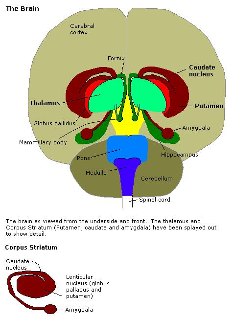
The striatum, or corpus striatum (also called the striate nucleus), is a nucleus (a cluster of neurons) in the subcortical basal ganglia of the forebrain. The striatum is a critical component of the motor and reward systems; receives glutamatergic and dopaminergic inputs from different sources; and serves as the primary input to the rest of the basal ganglia. Functionally, the striatum coordinates multiple aspects of cognition, including both motor and action planning, decision-making, motivation, reinforcement, and reward perception. The striatum is made up of the caudate nucleus and the lentiform nucleus. The lentiform nucleus is made up of the larger putamen, and the smaller globus pallidus. Strictly speaking the globus pallidus is part of the striatum. It is common practice, however, to implicitly exclude the globus pallidus when referring to striatal structures. In primates, the striatum is divided into a ventral striatum, and a dorsal striatum, subdivisions that are ... [...More Info...] [...Related Items...] OR: [Wikipedia] [Google] [Baidu] |
Nucleus Accumbens
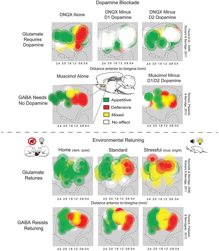
The nucleus accumbens (NAc or NAcc; also known as the accumbens nucleus, or formerly as the ''nucleus accumbens septi'', Latin for "nucleus adjacent to the septum") is a region in the basal forebrain rostral to the preoptic area of the hypothalamus. The nucleus accumbens and the olfactory tubercle collectively form the ventral striatum. The ventral striatum and dorsal striatum collectively form the striatum, which is the main component of the basal ganglia. The dopaminergic neurons of the mesolimbic pathway project onto the GABAergic medium spiny neurons of the nucleus accumbens and olfactory tubercle. Each cerebral hemisphere has its own nucleus accumbens, which can be divided into two structures: the nucleus accumbens core and the nucleus accumbens shell. These substructures have different morphology and functions. Different NAcc subregions (core vs shell) and neuron subpopulations within each region (D1-type vs D2-type medium spiny neurons) are responsible for different ... [...More Info...] [...Related Items...] OR: [Wikipedia] [Google] [Baidu] |
Nucleus Accumbens Shell
The nucleus accumbens (NAc or NAcc; also known as the accumbens nucleus, or formerly as the ''nucleus accumbens septi'', Latin for "nucleus adjacent to the septum") is a region in the basal forebrain rostral to the preoptic area of the hypothalamus. The nucleus accumbens and the olfactory tubercle collectively form the ventral striatum. The ventral striatum and dorsal striatum collectively form the striatum, which is the main component of the basal ganglia. The dopaminergic neurons of the mesolimbic pathway project onto the GABAergic medium spiny neurons of the nucleus accumbens and olfactory tubercle. Each cerebral hemisphere has its own nucleus accumbens, which can be divided into two structures: the nucleus accumbens core and the nucleus accumbens shell. These substructures have different morphology and functions. Different NAcc subregions (core vs shell) and neuron subpopulations within each region (D1-type vs D2-type medium spiny neurons) are responsible for different co ... [...More Info...] [...Related Items...] OR: [Wikipedia] [Google] [Baidu] |
Nucleus Accumbens Core
The nucleus accumbens (NAc or NAcc; also known as the accumbens nucleus, or formerly as the ''nucleus accumbens septi'', Latin for "nucleus adjacent to the septum") is a region in the basal forebrain rostral to the preoptic area of the hypothalamus. The nucleus accumbens and the olfactory tubercle collectively form the ventral striatum. The ventral striatum and dorsal striatum collectively form the striatum, which is the main component of the basal ganglia. The dopaminergic neurons of the mesolimbic pathway project onto the GABAergic medium spiny neurons of the nucleus accumbens and olfactory tubercle. Each cerebral hemisphere has its own nucleus accumbens, which can be divided into two structures: the nucleus accumbens core and the nucleus accumbens shell. These substructures have different morphology and functions. Different NAcc subregions (core vs shell) and neuron subpopulations within each region (D1-type vs D2-type medium spiny neurons) are responsible for different ... [...More Info...] [...Related Items...] OR: [Wikipedia] [Google] [Baidu] |
_(18167720666).jpg)


