|
Parapsicephalus
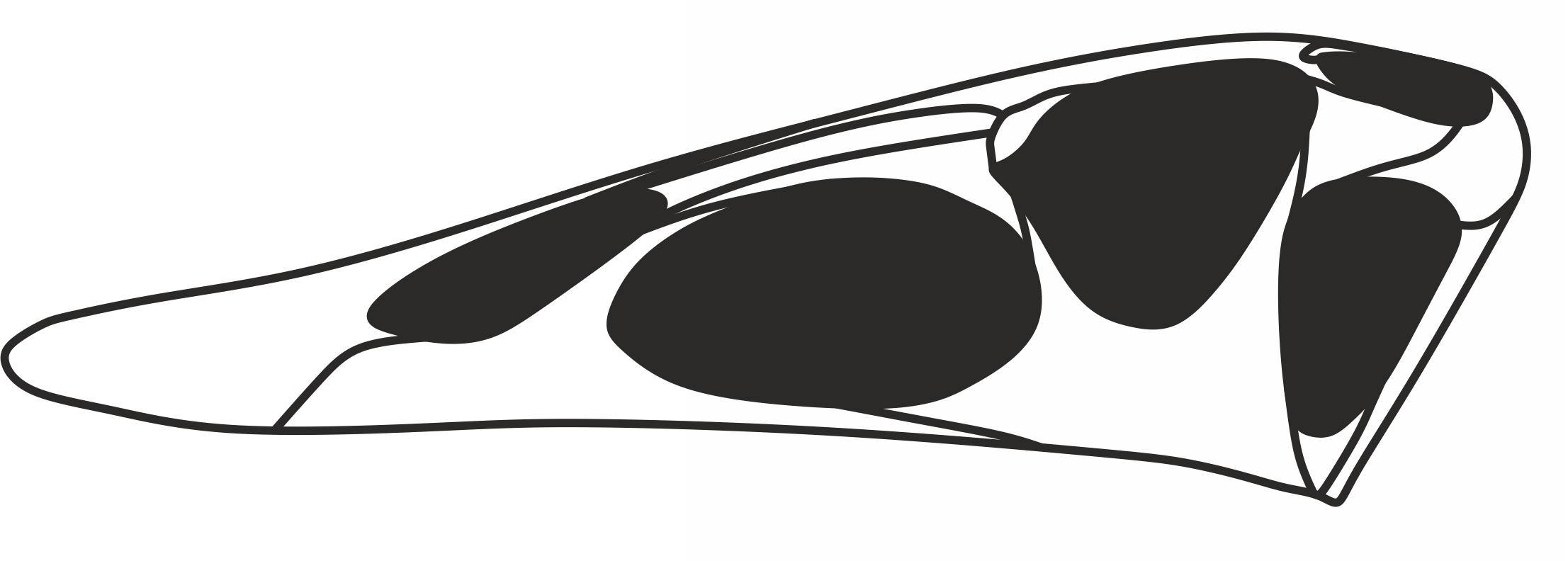
''Parapsicephalus'' (meaning "beside arch head") is a genus of long-tailed rhamphorhynchid pterosaurs from the Lower Jurassic Whitby, Yorkshire, England. It contains a single species, ''P. purdoni'', named initially as a species of the related rhamphorhynchid ''Scaphognathus'' in 1888 but moved to its own genus in 1919 on account of a unique combination of characteristics. In particular, the top surface of the skull of ''Parapsicephalus'' is convex, which is otherwise only seen in dimorphodontians. This has been the basis of its referral to the Dimorphodontia by some researchers, but it is generally agreed upon that ''Parapsicephalus'' probably represents a rhamphorhynchid. Within the Rhamphorhynchidae, ''Parapsicephalus'' has been synonymized with the roughly contemporary ''Dorygnathus''; this, however, is not likely given the many differences between the two taxa, including the aforementioned convex top surface of the skull. ''Parapsicephalus'' has been tentatively referred to t ... [...More Info...] [...Related Items...] OR: [Wikipedia] [Google] [Baidu] |
Dimorphodontia
Dimorphodontidae (or dimorphodontids) is a group of early "rhamphorhynchoid" pterosaurs named after '' Dimorphodon'', that lived in the Late Triassic to Early Jurassic. While fossils that can be definitively referred to the group are rare, dimorphodontids may have had a broad distribution, with fossils known from the UK, the southwest United States, and possibly Antarctica. Dimorphodontidae was named in 1870 by Harry Govier Seeley (as "Dimorphodontae") with ''Dimorphodon'' as the only known member. In 2003, David Unwin defined a clade Dimorphodontidae, as the group consisting of the last common ancestor of ''Dimorphodon macronyx'' and ''Peteinosaurus zambellii'', and all its descendants.Unwin, D. M. 2003. "On the phylogeny and evolutionary history of pterosaurs". In: Buffetaut, E. & Mazin, J.-M. (eds), ''Evolution and Palaeobiology of Pterosaurs''. Geological Society, London, Special Publications 217: 139-190 However, later studies found that ''Dimorphodon'' may not be closely re ... [...More Info...] [...Related Items...] OR: [Wikipedia] [Google] [Baidu] |
Rhamphorhynchidae
Rhamphorhynchidae is a group of early pterosaurs named after ''Rhamphorhynchus'', that lived in the Late Jurassic. The family Rhamphorhynchidae was named in 1870 by Harry Govier Seeley.Seeley, H.G. (1870). "The Orithosauria: An Elementary Study of the Bones of Pterodactyles." Cambridge, 135 p. Members of the group possess no more than 11 pairs of teeth in the rostrum, a deltopectoral crest that is constricted at the base but expanded at the distal end, and a bent phalange on the fifth toe. Rhamphorhynchidae traditionally contains two subfamilies: the Rhamphorhynchinae and the Scaphognathinae. While not recovered as distinct clades by all analyses, there do appear to be traits uniting members of each group. Rhamphorhynchines are more common, were lightly built, and had jaws ending in pointed tips that contained more teeth, which are often procumbent (pointed forward). Scaphognathines are comparatively quite rare, were more robust skeletally, and had shorter wing proportions. The b ... [...More Info...] [...Related Items...] OR: [Wikipedia] [Google] [Baidu] |
Rhamphorhynchid
Rhamphorhynchidae is a group of early pterosaurs named after ''Rhamphorhynchus'', that lived in the Late Jurassic. The family Rhamphorhynchidae was named in 1870 by Harry Govier Seeley.Seeley, H.G. (1870). "The Orithosauria: An Elementary Study of the Bones of Pterodactyles." Cambridge, 135 p. Members of the group possess no more than 11 pairs of teeth in the rostrum, a deltopectoral crest that is constricted at the base but expanded at the distal end, and a bent phalange on the fifth toe. Rhamphorhynchidae traditionally contains two subfamilies: the Rhamphorhynchinae and the Scaphognathinae. While not recovered as distinct clades by all analyses, there do appear to be traits uniting members of each group. Rhamphorhynchines are more common, were lightly built, and had jaws ending in pointed tips that contained more teeth, which are often procumbent (pointed forward). Scaphognathines are comparatively quite rare, were more robust skeletally, and had shorter wing proportions. The br ... [...More Info...] [...Related Items...] OR: [Wikipedia] [Google] [Baidu] |
Toarcian
The Toarcian is, in the ICS' geologic timescale, an age and stage in the Early or Lower Jurassic. It spans the time between 182.7 Ma (million years ago) and 174.1 Ma. It follows the Pliensbachian and is followed by the Aalenian. The Toarcian Age began with the Toarcian turnover, the extinction event that sets its fossil faunas apart from the previous Pliensbachian age. It is believed to have ended with a global cooling event known as the Comptum Cooling Event, although whether it represented a worldwide event is controversial. Stratigraphic definitions The Toarcian takes its name from the city of Thouars, just south of Saumur in the Loire Valley of France. The stage was introduced by French palaeontologist Alcide d'Orbigny in 1842, after examining rock strata of this age in a quarry near Thouars. In Europe this period is represented by the upper part of the Lias. The base of the Toarcian is defined as the place in the stratigraphic record where the ammonite genus '' Eoda ... [...More Info...] [...Related Items...] OR: [Wikipedia] [Google] [Baidu] |
Pterygoid Bone
The pterygoid is a paired bone forming part of the palate of many vertebrates, behind the palatine bone In anatomy, the palatine bones () are two irregular bones of the facial skeleton in many animal species, located above the uvula in the throat. Together with the maxillae, they comprise the hard palate. (''Palate'' is derived from the Latin ''pa ...s. It is a flat and thin lamina, united to the medial side of the pterygoid process of the sphenoid bone, and to the perpendicular lamina of the palatine bone. Bones of the head and neck {{musculoskeletal-stub ... [...More Info...] [...Related Items...] OR: [Wikipedia] [Google] [Baidu] |
Nasal Bone
The nasal bones are two small oblong bones, varying in size and form in different individuals; they are placed side by side at the middle and upper part of the face and by their junction, form the bridge of the upper one third of the nose. Each has two surfaces and four borders. Structure The two nasal bones are joined at the midline internasal suture and make up the bridge of the nose. Surfaces The ''outer surface'' is concavo-convex from above downward, convex from side to side; it is covered by the procerus and nasalis muscles, and perforated about its center by a foramen, for the transmission of a small vein. The ''inner surface'' is concave from side to side, and is traversed from above downward, by a groove for the passage of a branch of the nasociliary nerve. Articulations The nasal articulates with four bones: two of the cranium, the frontal and ethmoid, and two of the face, the opposite nasal and the maxilla. Other animals In primitive bony fish and tetrapod ... [...More Info...] [...Related Items...] OR: [Wikipedia] [Google] [Baidu] |
Lacrimal Bone
The lacrimal bone is a small and fragile bone of the facial skeleton; it is roughly the size of the little fingernail. It is situated at the front part of the medial wall of the orbit. It has two surfaces and four borders. Several bony landmarks of the lacrimal bone function in the process of lacrimation or crying. Specifically, the lacrimal bone helps form the nasolacrimal canal necessary for tear translocation. A depression on the anterior inferior portion of the bone, the lacrimal fossa, houses the membranous lacrimal sac. Tears or lacrimal fluid, from the lacrimal glands, collect in this sac during excessive lacrimation. The fluid then flows through the nasolacrimal duct and into the nasopharynx. This drainage results in what is commonly referred to a runny nose during excessive crying or tear production. Injury or fracture of the lacrimal bone can result in posttraumatic obstruction of the lacrimal pathways. Structure Lateral or orbital surface The lateral or orbital surface i ... [...More Info...] [...Related Items...] OR: [Wikipedia] [Google] [Baidu] |
Vomer
The vomer (; lat, vomer, lit=ploughshare) is one of the unpaired facial bones of the skull. It is located in the midsagittal line, and articulates with the sphenoid, the ethmoid, the left and right palatine bones, and the left and right maxillary bones. The vomer forms the inferior part of the nasal septum in humans, with the superior part formed by the perpendicular plate of the ethmoid bone. The name is derived from the Latin word for a ploughshare and the shape of the bone. In humans The vomer is situated in the median plane, but its anterior portion is frequently bent to one side. It is thin, somewhat quadrilateral in shape, and forms the hinder and lower part of the nasal septum; it has two surfaces and four borders. The surfaces are marked by small furrows for blood vessels, and on each is the nasopalatine groove, which runs obliquely downward and forward, and lodges the nasopalatine nerve and vessels. Borders The ''superior border'', the thickest, presents a dee ... [...More Info...] [...Related Items...] OR: [Wikipedia] [Google] [Baidu] |
Quadratojugal Bone
The quadratojugal is a skull bone present in many vertebrates, including some living reptiles and amphibians. Anatomy and function In animals with a quadratojugal bone, it is typically found connected to the jugal (cheek) bone from the front and the squamosal bone from above. It is usually positioned at the rear lower corner of the cranium. Many modern tetrapods lack a quadratojugal bone as it has been lost or fused to other bones. Modern examples of tetrapods without a quadratojugal include salamanders, mammals, birds, and squamates (lizards and snakes). In tetrapods with a quadratojugal bone, it often forms a portion of the jaw joint. Developmentally, the quadratojugal bone is a dermal bone in the temporal series, forming the original braincase. The squamosal and quadratojugal bones together form the cheek region and may provide muscular attachments for facial muscles. In reptiles and amphibians In most modern reptiles and amphibians, the quadratojugal is a prominent, strapl ... [...More Info...] [...Related Items...] OR: [Wikipedia] [Google] [Baidu] |
Jugal Bone
The jugal is a skull bone found in most reptiles, amphibians and birds. In mammals, the jugal is often called the malar or zygomatic. It is connected to the quadratojugal and maxilla, as well as other bones, which may vary by species. Anatomy The jugal bone is located on either side of the skull in the circumorbital region. It is the origin of several masticatory muscles in the skull. The jugal and lacrimal bones are the only two remaining from the ancestral circumorbital series: the prefrontal, postfrontal, postorbital, jugal, and lacrimal bones. During development, the jugal bone originates from dermal bone. In dinosaurs This bone is considered key in the determination of general traits in cases in which the entire skull has not been found intact (for instance, as with dinosaurs in paleontology). In some dinosaur genera the jugal also forms part of the lower margin of either the antorbital fenestra or the infratemporal fenestra, or both. Most commonly, this bone articu ... [...More Info...] [...Related Items...] OR: [Wikipedia] [Google] [Baidu] |
Maxilla
The maxilla (plural: ''maxillae'' ) in vertebrates is the upper fixed (not fixed in Neopterygii) bone of the jaw formed from the fusion of two maxillary bones. In humans, the upper jaw includes the hard palate in the front of the mouth. The two maxillary bones are fused at the intermaxillary suture, forming the anterior nasal spine. This is similar to the mandible (lower jaw), which is also a fusion of two mandibular bones at the mandibular symphysis. The mandible is the movable part of the jaw. Structure In humans, the maxilla consists of: * The body of the maxilla * Four processes ** the zygomatic process ** the frontal process of maxilla ** the alveolar process ** the palatine process * three surfaces – anterior, posterior, medial * the Infraorbital foramen * the maxillary sinus * the incisive foramen Articulations Each maxilla articulates with nine bones: * two of the cranium: the frontal and ethmoid * seven of the face: the nasal, zygomatic, lacrimal, inferior n ... [...More Info...] [...Related Items...] OR: [Wikipedia] [Google] [Baidu] |
Quadrate Bone
The quadrate bone is a skull bone in most tetrapods, including amphibians, sauropsids (reptiles, birds), and early synapsids. In most tetrapods, the quadrate bone connects to the quadratojugal and squamosal bones in the skull, and forms upper part of the jaw joint. The lower jaw articulates at the articular bone, located at the rear end of the lower jaw. The quadrate bone forms the lower jaw articulation in all classes except mammals. Evolutionarily, it is derived from the hindmost part of the primitive cartilaginous upper jaw. Function in reptiles In certain extinct reptiles, the variation and stability of the morphology of the quadrate bone has helped paleontologists in the species-level taxonomy and identification of mosasaur squamates and spinosaurine dinosaurs. In some lizards and dinosaurs, the quadrate is articulated at both ends and movable. In snakes, the quadrate bone has become elongated and very mobile, and contributes greatly to their ability to swallow very ... [...More Info...] [...Related Items...] OR: [Wikipedia] [Google] [Baidu] |

.jpg)

