|
Paracytophagy
Paracytophagy () is the cellular process whereby a cell engulfs a protrusion which extends from a neighboring cell. This protrusion may contain material which is actively transferred between the cells. The process of paracytophagy was first described as a crucial step during cell-to-cell spread of the intracellular bacterial pathogen ''Listeria monocytogenes'', and is also commonly observed in ''Shigella flexneri''. Paracytophagy allows these intracellular pathogens to spread directly from cell to cell, thus escaping immune detection and destruction. Studies of this process have contributed significantly to our understanding of the role of the actin cytoskeleton in eukaryotic cells. Actin cytoskeleton Actin is one of the main cytoskeletal proteins in eukaryotic cells. The polymerization of actin filaments is responsible for the formation of pseudopods, filopodia and lamellipodia during cell motility. Cells actively build actin microfilaments that push the cell membrane towards ... [...More Info...] [...Related Items...] OR: [Wikipedia] [Google] [Baidu] |
Trogocytosis
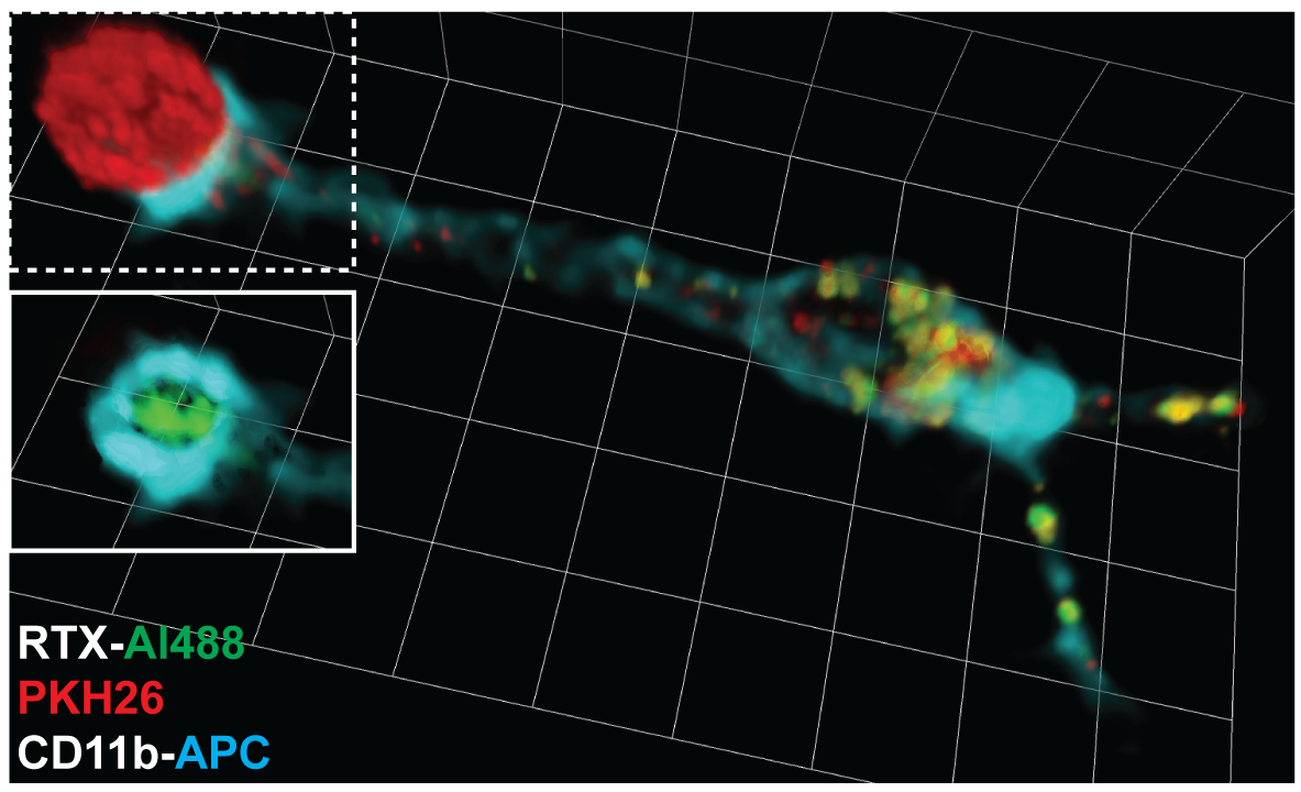
Trogocytosis ( gr, trogo; ''gnaw'') is when a cell nibbles another cell. It is a process whereby lymphocytes (B cell, B, T cell, T and Natural killer cell, NK cell (biology), cells) conjugated to antigen-presenting cells extract Cell surface molecule, surface molecules from these cells and express them on their own surface. The molecular reorganization occurring at the interface between the lymphocyte and the antigen-presenting cell during conjugation is also called "immunological synapse". Steps in the discovery of trogocytosis First indication for the existence of this process dates back late 70s when several research groups reported on the presence of unexpected molecules such as Major Histocompatibility complex molecules (MHC) on T cells. The notion that membrane fragments, and not isolated molecules, could be captured by T cells on antigen-presenting cells was suggested by the capture of MHC molecules fused to the green fluorescent protein (GFP) in their intracellular porti ... [...More Info...] [...Related Items...] OR: [Wikipedia] [Google] [Baidu] |
Cell (biology)
The cell is the basic structural and functional unit of life forms. Every cell consists of a cytoplasm enclosed within a membrane, and contains many biomolecules such as proteins, DNA and RNA, as well as many small molecules of nutrients and metabolites.Cell Movements and the Shaping of the Vertebrate Body in Chapter 21 of Molecular Biology of the Cell '' fourth edition, edited by Bruce Alberts (2002) published by Garland Science. The Alberts text discusses how the "cellular building blocks" move to shape developing embryos. It is also common to describe small molecules such as ... [...More Info...] [...Related Items...] OR: [Wikipedia] [Google] [Baidu] |
Secretion
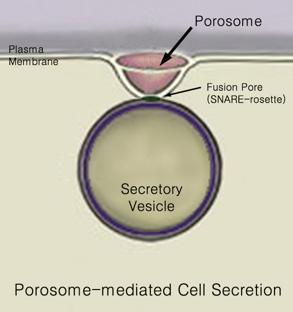
440px Secretion is the movement of material from one point to another, such as a secreted chemical substance from a cell or gland. In contrast, excretion is the removal of certain substances or waste products from a cell or organism. The classical mechanism of cell secretion is via secretory portals at the plasma membrane called porosomes. Porosomes are permanent cup-shaped lipoprotein structures embedded in the cell membrane, where secretory vesicles transiently dock and fuse to release intra-vesicular contents from the cell. Secretion in bacterial species means the transport or translocation of effector molecules for example: proteins, enzymes or toxins (such as cholera toxin in pathogenic bacteria e.g. ''Vibrio cholerae'') from across the interior (cytoplasm or cytosol) of a bacterial cell to its exterior. Secretion is a very important mechanism in bacterial functioning and operation in their natural surrounding environment for adaptation and survival. In eukaryotic cells ... [...More Info...] [...Related Items...] OR: [Wikipedia] [Google] [Baidu] |
Ezrin
Ezrin also known as cytovillin or villin-2 is a protein that in humans is encoded by the ''EZR'' gene. Structure The N-terminus of ezrin contains a FERM domain which is further subdivided into three subdomains. The C-terminus contain an ERM domain. Function The cytoplasmic peripheral protein encoded by this gene can be phosphorylated by protein-tyrosine kinase in microvilli and is a member of the ERM protein family. This protein serves as a linker between plasma membrane and actin cytoskeleton. It plays a key role in cell surface structure adhesion, migration, and organization. The N-terminal domain (also called FERM domain) binds sodium-hydrogen exchanger regulatory factor (NHERF) protein (involving long-range allostery). This binding can happen only when ezrin is in its active state. The activation of ezrin occurs in synergism of the two factors: 1) binding of the N-terminal domain to phosphatidylinositol(4,5)bis-phosphate (PIP2) and 2) phosphorylation of threonine T5 ... [...More Info...] [...Related Items...] OR: [Wikipedia] [Google] [Baidu] |
Cell Membrane
The cell membrane (also known as the plasma membrane (PM) or cytoplasmic membrane, and historically referred to as the plasmalemma) is a biological membrane that separates and protects the interior of all cells from the outside environment (the extracellular space). The cell membrane consists of a lipid bilayer, made up of two layers of phospholipids with cholesterols (a lipid component) interspersed between them, maintaining appropriate membrane fluidity at various temperatures. The membrane also contains membrane proteins, including integral proteins that span the membrane and serve as membrane transporters, and peripheral proteins that loosely attach to the outer (peripheral) side of the cell membrane, acting as enzymes to facilitate interaction with the cell's environment. Glycolipids embedded in the outer lipid layer serve a similar purpose. The cell membrane controls the movement of substances in and out of cells and organelles, being selectively permeable to ions a ... [...More Info...] [...Related Items...] OR: [Wikipedia] [Google] [Baidu] |
Cytoplasm
In cell biology, the cytoplasm is all of the material within a eukaryotic cell, enclosed by the cell membrane, except for the cell nucleus. The material inside the nucleus and contained within the nuclear membrane is termed the nucleoplasm. The main components of the cytoplasm are cytosol (a gel-like substance), the organelles (the cell's internal sub-structures), and various cytoplasmic inclusions. The cytoplasm is about 80% water and is usually colorless. The submicroscopic ground cell substance or cytoplasmic matrix which remains after exclusion of the cell organelles and particles is groundplasm. It is the hyaloplasm of light microscopy, a highly complex, polyphasic system in which all resolvable cytoplasmic elements are suspended, including the larger organelles such as the ribosomes, mitochondria, the plant plastids, lipid droplets, and vacuoles. Most cellular activities take place within the cytoplasm, such as many metabolic pathways including glycolysis, and proces ... [...More Info...] [...Related Items...] OR: [Wikipedia] [Google] [Baidu] |
Electron Microscopy
An electron microscope is a microscope that uses a beam of accelerated electrons as a source of illumination. As the wavelength of an electron can be up to 100,000 times shorter than that of visible light photons, electron microscopes have a higher resolving power than light microscopes and can reveal the structure of smaller objects. A scanning transmission electron microscope has achieved better than 50 pm resolution in annular dark-field imaging mode and magnifications of up to about 10,000,000× whereas most light microscopes are limited by diffraction to about 200 nm resolution and useful magnifications below 2000×. Electron microscopes use shaped magnetic fields to form electron optical lens systems that are analogous to the glass lenses of an optical light microscope. Electron microscopes are used to investigate the ultrastructure of a wide range of biological and inorganic specimens including microorganisms, cells, large molecules, biopsy samples, ... [...More Info...] [...Related Items...] OR: [Wikipedia] [Google] [Baidu] |
J Cell Biol 2002 Aug 158(3) 409-14, Figure 1
J, or j, is the tenth letter in the Latin alphabet, used in the modern English alphabet, the alphabets of other western European languages and others worldwide. Its usual name in English is ''jay'' (pronounced ), with a now-uncommon variant ''jy'' ."J", ''Oxford English Dictionary,'' 2nd edition (1989) When used in the International Phonetic Alphabet for the ''y'' sound, it may be called ''yod'' or ''jod'' (pronounced or ). History The letter ''J'' used to be used as the swash letter ''I'', used for the letter I at the end of Roman numerals when following another I, as in XXIIJ or xxiij instead of XXIII or xxiii for the Roman numeral twenty-three. A distinctive usage emerged in Middle High German. Gian Giorgio Trissino (1478–1550) was the first to explicitly distinguish I and J as representing separate sounds, in his ''Ɛpistola del Trissino de le lettere nuωvamente aggiunte ne la lingua italiana'' ("Trissino's epistle about the letters recently added in the Ital ... [...More Info...] [...Related Items...] OR: [Wikipedia] [Google] [Baidu] |
Drosophila Melanogaster
''Drosophila melanogaster'' is a species of fly (the taxonomic order Diptera) in the family Drosophilidae. The species is often referred to as the fruit fly or lesser fruit fly, or less commonly the "vinegar fly" or "pomace fly". Starting with Charles W. Woodworth's 1901 proposal of the use of this species as a model organism, ''D. melanogaster'' continues to be widely used for biological research in genetics, physiology, microbial pathogenesis, and life history evolution. As of 2017, five Nobel Prizes have been awarded to drosophilists for their work using the insect. ''D. melanogaster'' is typically used in research owing to its rapid life cycle, relatively simple genetics with only four pairs of chromosomes, and large number of offspring per generation. It was originally an African species, with all non-African lineages having a common origin. Its geographic range includes all continents, including islands. ''D. melanogaster'' is a common pest in homes, restaurants, and othe ... [...More Info...] [...Related Items...] OR: [Wikipedia] [Google] [Baidu] |
MHCII
MHC Class II molecules are a class of major histocompatibility complex (MHC) molecules normally found only on professional antigen-presenting cells such as dendritic cells, mononuclear phagocytes, some endothelial cells, thymic epithelial cells, and B cells. These cells are important in initiating immune responses. The antigens presented by class II peptides are derived from extracellular proteins (not cytosolic as in MHC class I). Loading of a MHC class II molecule occurs by phagocytosis; extracellular proteins are endocytosed, digested in lysosomes, and the resulting epitopic peptide fragments are loaded onto MHC class II molecules prior to their migration to the cell surface. In humans, the MHC class II protein complex is encoded by the human leukocyte antigen gene complex (HLA). HLAs corresponding to MHC class II are HLA-DP, HLA-DM, HLA-DOA, HLA-DOB, HLA-DQ, and HLA-DR. Mutations in the HLA gene complex can lead to bare lymphocyte syndrome (BLS), which is a typ ... [...More Info...] [...Related Items...] OR: [Wikipedia] [Google] [Baidu] |
Cross-presentation
Cross-presentation is the ability of certain professional antigen-presenting cells (mostly dendritic cells) to take up, process and present ''extracellular'' antigens with MHC class I molecules to CD8 T cells (cytotoxic T cells). Cross-priming, the result of this process, describes the stimulation of naive cytotoxic CD8+ T cells into activated cytotoxic CD8+ T cells. This process is necessary for immunity against most tumors and against viruses that infect dendritic cells and sabotage their presentation of virus antigens. Cross presentation is also required for the induction of cytotoxic immunity by vaccination with protein antigens, for example, tumour vaccination. Cross-presentation is of particular importance, because it permits the presentation of exogenous antigens, which are normally presented by MHC II on the surface of dendritic cells, to also be presented through the MHC I pathway. The MHC I pathway is normally used to present endogenous antigens that have infected a parti ... [...More Info...] [...Related Items...] OR: [Wikipedia] [Google] [Baidu] |
Hematopoietic Stem Cell
Hematopoietic stem cells (HSCs) are the stem cells that give rise to other blood cells. This process is called haematopoiesis. In vertebrates, the very first definitive HSCs arise from the ventral endothelial wall of the embryonic aorta within the (midgestational) aorta-gonad-mesonephros region, through a process known as endothelial-to-hematopoietic transition. In adults, haematopoiesis occurs in the red bone marrow, in the core of most bones. The red bone marrow is derived from the layer of the embryo called the mesoderm. Haematopoiesis is the process by which all mature blood cells are produced. It must balance enormous production needs (the average person produces more than 500 billion blood cells every day) with the need to regulate the number of each blood cell type in the circulation. In vertebrates, the vast majority of hematopoiesis occurs in the bone marrow and is derived from a limited number of hematopoietic stem cells that are multipotent and capable of extensive se ... [...More Info...] [...Related Items...] OR: [Wikipedia] [Google] [Baidu] |
_409-14%2C_Figure_1.png)


