|
Parabrachial Nuclei
The parabrachial nuclei, also known as the parabrachial complex, are a group of nuclei in the dorsolateral pons that surrounds the superior cerebellar peduncle as it enters the brainstem from the cerebellum. They are named from the Latin term for the superior cerebellar peduncle, the ''brachium conjunctivum''. In the human brain, the expansion of the superior cerebellar peduncle expands the parabrachial nuclei, which form a thin strip of grey matter over most of the peduncle. The parabrachial nuclei are typically divided along the lines suggested by Baxter and Olszewski in humans, into a medial parabrachial nucleus and lateral parabrachial nucleus. These have in turn been subdivided into a dozen subnuclei: the superior, dorsal, ventral, internal, external and extreme lateral subnuclei; the lateral crescent and subparabrachial nucleus (Kolliker-Fuse nucleus) along the ventrolateral margin of the lateral parabrachial complex; and the medial and external medial subnuclei Component ... [...More Info...] [...Related Items...] OR: [Wikipedia] [Google] [Baidu] |
Brainstem
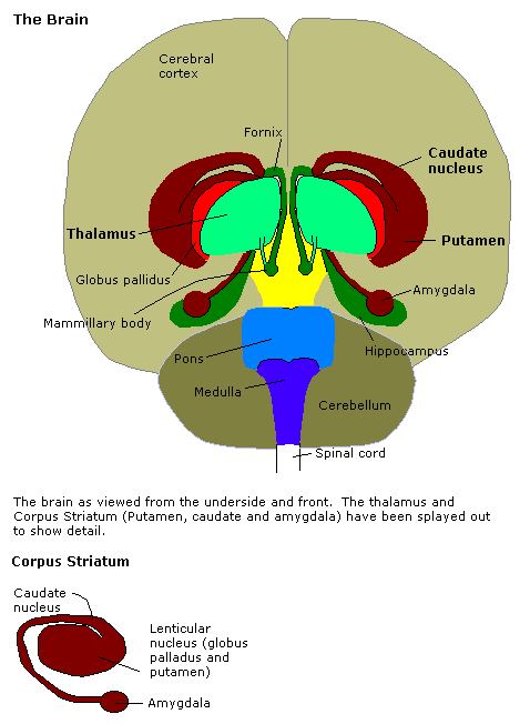
The brainstem (or brain stem) is the posterior stalk-like part of the brain that connects the cerebrum with the spinal cord. In the human brain the brainstem is composed of the midbrain, the pons, and the medulla oblongata. The midbrain is continuous with the thalamus of the diencephalon through the tentorial notch, and sometimes the diencephalon is included in the brainstem. The brainstem is very small, making up around only 2.6 percent of the brain's total weight. It has the critical roles of regulating cardiac, and respiratory function, helping to control heart rate and breathing rate. It also provides the main motor and sensory nerve supply to the face and neck via the cranial nerves. Ten pairs of cranial nerves come from the brainstem. Other roles include the regulation of the central nervous system and the body's sleep cycle. It is also of prime importance in the conveyance of motor and sensory pathways from the rest of the brain to the body, and from the body back to t ... [...More Info...] [...Related Items...] OR: [Wikipedia] [Google] [Baidu] |
Solitary Tract
The solitary tract (tractus solitarius, or fasciculus solitarius), is a compact fiber bundle that extends longitudinally through the posterolateral region of the medulla oblongata. The solitary tract is surrounded by the solitary nucleus, and descends to the upper cervical segments of the spinal cord. It was first named by Theodor Meynert in 1872. Composition The solitary tract is made up of primary sensory fibers and descending fibers of the vagus, glossopharyngeal, and facial nerves. Function The solitary tract conveys afferent information from stretch receptors and chemoreceptors in the walls of the cardiovascular, respiratory, and intestinal tracts. Afferent fibers from cranial nerves 7, 9 and 10 convey taste ( SVA) in its rostral portion, and general visceral sense ( general visceral afferent fibers, GVA) in its caudal part. Taste buds in the mucosa of the tongue can also generate impulses in the rostral regions of the solitary tract. The efferent fibers are distrib ... [...More Info...] [...Related Items...] OR: [Wikipedia] [Google] [Baidu] |
Calcitonin Gene-related Peptide
Calcitonin gene-related peptide (CGRP) is a member of the calcitonin family of peptides consisting of calcitonin, amylin, adrenomedullin, adrenomedullin 2 ( intermedin) and calcitonin‑receptor‑stimulating peptide. Calcitonin is mainly produced by thyroid C cells whilst CGRP is secreted and stored in the nervous system. This peptide, in humans, exists in two forms: CGRP alpha (α-CGRP or CGRP I), and CGRP beta (β-CGRP or CGRP II). α-CGRP is a 37-amino acid neuropeptide and is formed by alternative splicing of the calcitonin/CGRP gene located on chromosome 11. β-CGRP is less studied. In humans, β-CGRP differs from α-CGRP by three amino acids and is encoded in a separate, nearby gene. The CGRP family includes calcitonin (CT), adrenomedullin (AM), and amylin (AMY). Function CGRP is produced in both peripheral and central neurons. It is a potent peptide vasodilator and can function in the transmission of nociception. In the spinal cord, the function and expression of CGRP ... [...More Info...] [...Related Items...] OR: [Wikipedia] [Google] [Baidu] |
Neuropeptide
Neuropeptides are chemical messengers made up of small chains of amino acids that are synthesized and released by neurons. Neuropeptides typically bind to G protein-coupled receptors (GPCRs) to modulate neural activity and other tissues like the gut, muscles, and heart. There are over 100 known neuropeptides, representing the largest and most diverse class of signaling molecules in the nervous system. Neuropeptides are synthesized from large precursor proteins which are cleaved and post-translationally processed then packaged into dense core vesicles. Neuropeptides are often co-released with other neuropeptides and neurotransmitters in a single neuron, yielding a multitude of effects. Once released, neuropeptides can diffuse widely to affect a broad range of targets. Synthesis Neuropeptides are synthesized from large, inactive precursor proteins called prepropeptides. Prepropeptides contain sequences for a family of distinct peptides and often contain repeated copies of the sa ... [...More Info...] [...Related Items...] OR: [Wikipedia] [Google] [Baidu] |
Forebrain
In the anatomy of the brain of vertebrates, the forebrain or prosencephalon is the Anatomical terms of location#Directional terms, rostral (forward-most) portion of the brain. The forebrain (prosencephalon), the midbrain (mesencephalon), and hindbrain (rhombencephalon) are the three Brain vesicle, primary brain vesicles during the early development of the nervous system. The forebrain controls body temperature, reproductive functions, eating, sleeping, and the display of emotions. At the five-vesicle stage, the forebrain separates into the diencephalon (thalamus, hypothalamus, subthalamus, and epithalamus) and the telencephalon which develops into the cerebrum. The cerebrum consists of the cerebral cortex, underlying white matter, and the basal ganglia. In humans, by 5 weeks in utero it is visible as a single portion toward the front of the fetus. At 8 weeks in utero, the forebrain splits into the left and right cerebral hemispheres. When the embryonic forebrain fails to divide ... [...More Info...] [...Related Items...] OR: [Wikipedia] [Google] [Baidu] |
Ventrolateral Medulla
The ventrolateral medulla, part of the medulla oblongata of the brainstem, plays a major role in regulating arterial blood pressure and breathing. It regulates blood pressure by regulating the activity of the sympathetic nerves that target the heart and peripheral blood vessels. The ventrolateral medulla consists of a rostral ventrolateral medulla (RVLM) and a caudal ventrolateral medulla (CVLM). Neurons in the RVLM project directly to preganglionic neurons in the spinal cord The spinal cord is a long, thin, tubular structure made up of nervous tissue, which extends from the medulla oblongata in the brainstem to the lumbar region of the vertebral column (backbone). The backbone encloses the central canal of the spi ... and maintain tonic activity in the sympathetic vasomotor nerves. This activity is inhibited by GABA output from the CVLM. References Sympathetic nervous system Reflexes Medulla oblongata Cardiovascular physiology {{neuroanatomy-stub ... [...More Info...] [...Related Items...] OR: [Wikipedia] [Google] [Baidu] |
Intralaminar Thalamic Nuclei
The intralaminar thalamic nuclei (ITN) are collections of neurons in the internal medullary lamina of the thalamus that are generally divided in two groups as follows:Mancall, E., Brock, D. & Gray, H. (2011). Gray's clinical neuroanatomy the anatomic basis for clinical neuroscience. Philadelphia: Elsevier/Saunders. * anterior (rostral) group ** central medial nucleus ** paracentral nucleus ** central lateral nucleus * posterior (caudal) intralaminar group ** centromedian nucleus ** parafascicular nucleus Some sources also include a "central dorsal" nucleus. Degeneration of this area can be associated with progressive supranuclear palsy and Parkinson's disease. See also * Central tegmental tract * Output of the Reticular activating system, ARAS References External links Diagram at University of Florida Thalamic nuclei {{neuroanatomy-stub ... [...More Info...] [...Related Items...] OR: [Wikipedia] [Google] [Baidu] |
Taste
The gustatory system or sense of taste is the sensory system that is partially responsible for the perception of taste (flavor). Taste is the perception produced or stimulated when a substance in the mouth reacts chemically with taste receptor cells located on taste buds in the oral cavity, mostly on the tongue. Taste, along with olfaction and trigeminal nerve stimulation (registering texture, pain, and temperature), determines flavors of food and other substances. Humans have taste receptors on taste buds and other areas, including the upper surface of the tongue and the epiglottis. The gustatory cortex is responsible for the perception of taste. The tongue is covered with thousands of small bumps called papillae, which are visible to the naked eye. Within each papilla are hundreds of taste buds. The exception to this is the filiform papillae that do not contain taste buds. There are between 2000 and 5000Boron, W.F., E.L. Boulpaep. 2003. Medical Physiology. 1st ed. Elsevier ... [...More Info...] [...Related Items...] OR: [Wikipedia] [Google] [Baidu] |
Special Visceral Afferent
A Special visceral afferent fibers (SVA) is a afferent fiber that develop in association with the gastrointestinal tract. They carry the special senses of smell (olfaction) and taste (gustation). The cranial nerves containing SVA fibers are the olfactory nerve (I), the facial nerve (VII), the glossopharyngeal nerve (IX), and the vagus nerve (X). The facial nerve receives taste from the anterior 2/3 of the tongue The tongue is a muscular organ in the mouth of a typical tetrapod. It manipulates food for mastication and swallowing as part of the digestive process, and is the primary organ of taste. The tongue's upper surface (dorsum) is covered by taste ...; the glossopharyngeal from the posterior 1/3, and the vagus nerve from the epiglottis. The sensory processes, using their primary cell bodies from the inferior ganglion, send projections to the medulla, from which they travel in the tractus solitarius, later terminating at the rostral nucleus solitarius.Bhatnagar C. Subhash. '' ... [...More Info...] [...Related Items...] OR: [Wikipedia] [Google] [Baidu] |
Amygdala
The amygdala (; plural: amygdalae or amygdalas; also '; Latin from Greek, , ', 'almond', 'tonsil') is one of two almond-shaped clusters of nuclei located deep and medially within the temporal lobes of the brain's cerebrum in complex vertebrates, including humans. Shown to perform a primary role in the processing of memory, decision making, and emotional responses (including fear, anxiety, and aggression), the amygdalae are considered part of the limbic system. The term "amygdala" was first introduced by Karl Friedrich Burdach in 1822. Structure The regions described as amygdala nuclei encompass several structures of the cerebrum with distinct connectional and functional characteristics in humans and other animals. Among these nuclei are the basolateral complex, the cortical nucleus, the medial nucleus, the central nucleus, and the intercalated cell clusters. The basolateral complex can be further subdivided into the lateral, the basal, and the accessory basal nucle ... [...More Info...] [...Related Items...] OR: [Wikipedia] [Google] [Baidu] |
Spinal Cord
The spinal cord is a long, thin, tubular structure made up of nervous tissue, which extends from the medulla oblongata in the brainstem to the lumbar region of the vertebral column (backbone). The backbone encloses the central canal of the spinal cord, which contains cerebrospinal fluid. The brain and spinal cord together make up the central nervous system (CNS). In humans, the spinal cord begins at the occipital bone, passing through the foramen magnum and then enters the spinal canal at the beginning of the cervical vertebrae. The spinal cord extends down to between the first and second lumbar vertebrae, where it ends. The enclosing bony vertebral column protects the relatively shorter spinal cord. It is around long in adult men and around long in adult women. The diameter of the spinal cord ranges from in the cervical and lumbar regions to in the thoracic area. The spinal cord functions primarily in the transmission of nerve signals from the motor cortex to the body, ... [...More Info...] [...Related Items...] OR: [Wikipedia] [Google] [Baidu] |
Medulla Oblongata
The medulla oblongata or simply medulla is a long stem-like structure which makes up the lower part of the brainstem. It is anterior and partially inferior to the cerebellum. It is a cone-shaped neuronal mass responsible for autonomic (involuntary) functions, ranging from vomiting to sneezing. The medulla contains the cardiac, respiratory, vomiting and vasomotor centers, and therefore deals with the autonomic functions of breathing, heart rate and blood pressure as well as the sleep–wake cycle. During embryonic development, the medulla oblongata develops from the myelencephalon. The myelencephalon is a secondary vesicle which forms during the maturation of the rhombencephalon, also referred to as the hindbrain. The bulb is an archaic term for the medulla oblongata. In modern clinical usage, the word bulbar (as in bulbar palsy) is retained for terms that relate to the medulla oblongata, particularly in reference to medical conditions. The word bulbar can refer to the nerves ... [...More Info...] [...Related Items...] OR: [Wikipedia] [Google] [Baidu] |


